Superior sagittal sinus dural arteriovenous fistula caused by treatment of meningioma masquerades as sinus thrombosis
- Affiliations
-
- 1Department of Neurosurgery, Penn State Health Milton S Hershey Medical Center, PA, USA
- KMID: 2520886
- DOI: http://doi.org/10.7461/jcen.2021.E2021.01.002
Abstract
- Dural arteriovenous fistulas (DAVF) are rare acquired lesions resulting from abnormal shunting between intracranial dural arteries and venous system. Typically arising from structural weakness of the dura and a coinciding trigger factor, DAVFs can present with similar clinical and imaging characteristics to sinus thrombosis. A 61-year-old male with a history of meningioma previously managed with subtotal resection and stereotactic radiosurgery presented with progressive right-sided vision loss and bilateral papilledema. Initial imaging suggested possible sinus occlusion. Catheter angiogram revealed a Borden-Shucart grade III DAVF of the superior sagittal sinus and elevated venous pressures and the patient subsequently underwent endovascular transarterial intervention twice. We report on the first case of a superior sagittal sinus DAVF occurring after surgical resection of a parasagittal meningioma.
Figure
-
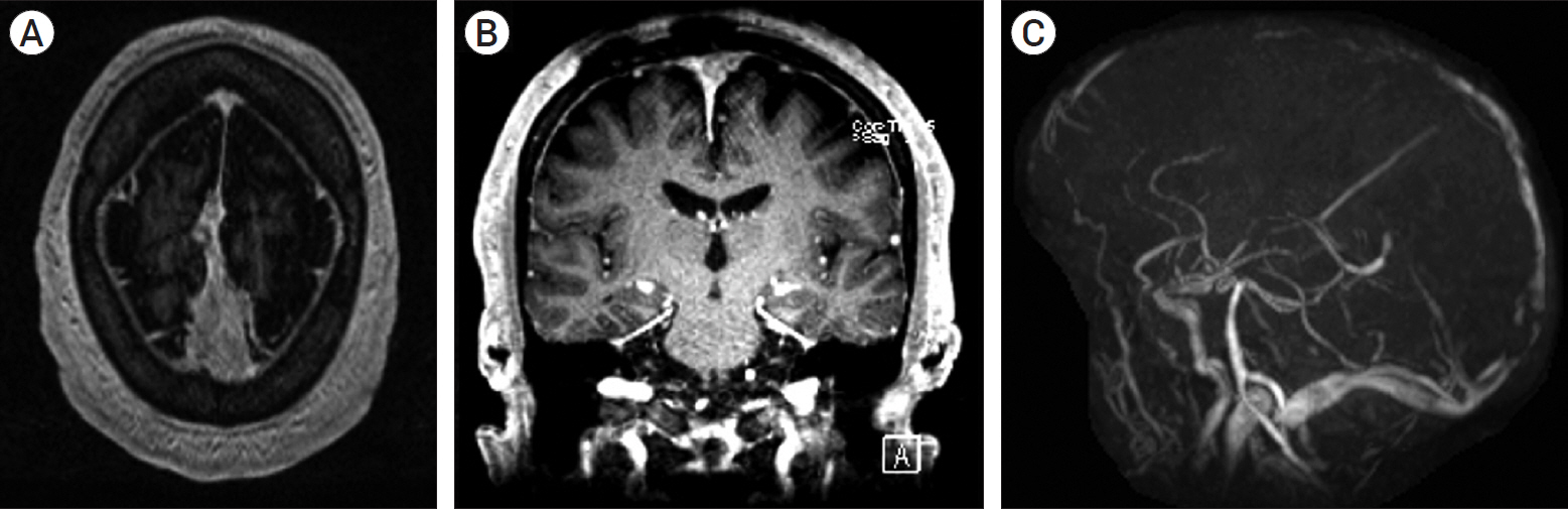
Fig. 1. Magnetic resonance imaging (MRI). (A), (B) Axial and coronal view of parafalcine meningioma with tumor extension into the superior sagittal sinus. (C) MRI venogram revealing anterior-to-mid superior sagittal sinus occlusion as a result of tumor invasion of the sinus.
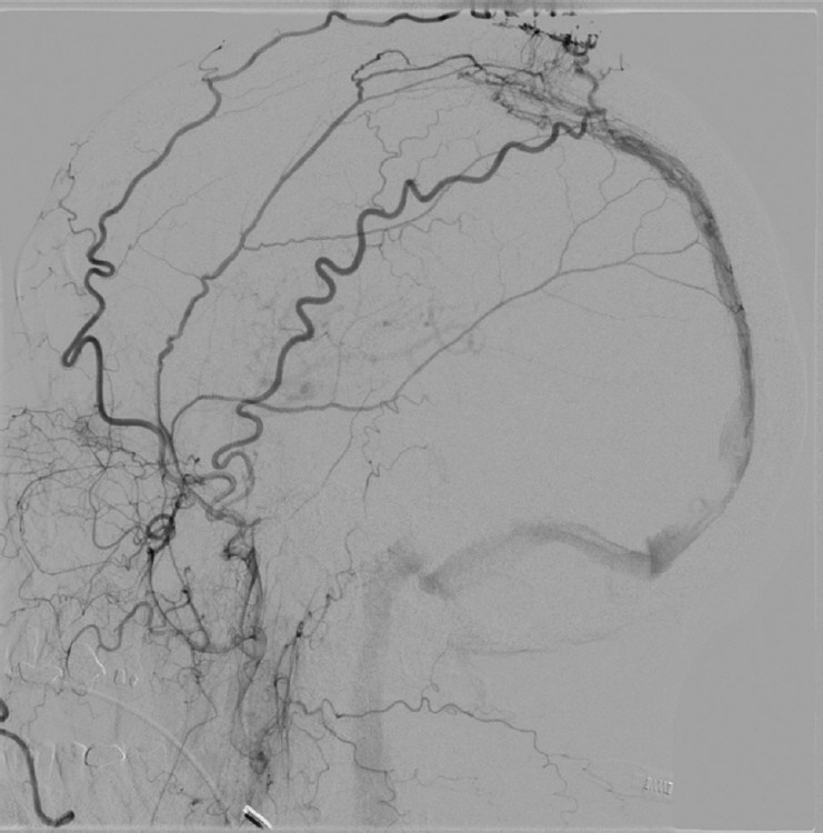
Fig. 2. Digital subtraction angiography (DSA), lateral view. Superior sagittal sinus dural arteriovenous fistula with the bilateral superficial temporal, bilateral meningeal, and right occipital arterial feeding vessels and retrograde cortical venous drainage (Borden-Shucart grade III; Cognard grade III).
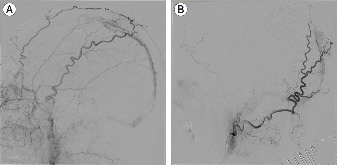
Fig. 3. Digital subtraction angiography (DSA), lateral view. First embolization leading to complete obliteration of the left meningeal feeding artery and right anterior branch of the middle meningeal artery as well as slowing of flow (A). Follow-up embolization 2 weeks later was performed on the right posterior branch of the middle meningeal and right occipital artery revealing residual dural arteriovenous fistula with continued right-sided feeding from the left via the occipital artery (B).

Fig. 4. Digital subtraction angiography (DSA), lateral view in (A) arterial phase from external carotid artery injection, (B) arterial phase from internal carotid artery injection, and (C) venous phase following internal carotid artery injection. Follow-up angiography 3 months after initial embolization demonstrates complete obliteration of the dural arteriovenous fistula.
Reference
-
1. Arnautović KI, Al-Mefty O, Angtuaco E, Phares LJ. Dural arteriovenous malformations of the transverse/sigmoid sinus acquired from dominant sinus occlusion by a tumor: report of two cases. Neurosurgery. 1998; Feb. 42(2):383–8.2. Awad IA, Little JR, Akarawi WP, Ahl J. Intracranial dural arteriovenous malformations: factors predisposing to an aggressive neurological course. J Neurosurg. 1990; Jun. 72(6):839–50.
Article3. Borden JA, Wu JK, Shucart WA. A proposed classification for spinal and cranial dural arteriovenous fistulous malformations and implications for treatment. J Neurosurg. 1995; Feb. 82(2):166–79.
Article4. Chandra RV, Leslie-Mazwi TM, Mehta BP, Yoo AJ, Rabinov JD, Pryor JC, et al. Transarterial onyx embolization of cranial dural arteriovenous fistulas: long-term follow-up. AJNR Am J Neuroradiol. 2014; Sep. 35(9):1793–7.
Article5. Cognard C, Casasco A, Toevi M, Houdart E, Chiras J, Merland JJ. Dural arteriovenous fistulas as a cause of intracranial hypertension due to impairment of cranial venous outflow. J Neurol Neurosurg Psychiatry. 1998; Sep. 65(3):308–16.
Article6. Deguchi J, Yamada M, Kobata H, Kuroiwa T. Regional cerebral blood flow after acetazolamide challenge in patients with dural arteriovenous fistula: simple way to evaluate intracranial venous hypertension. AJNR Am J Neuroradiol. 2005; May. 26(5):1101–6.7. Fukai J, Terada T, Kuwata T, Hyotani G, Raimura M, Nakagawa M, et al. Transarterial intravenous coil embolization of dural arteriovenous fistula involving the superior sagittal sinus. Surg Neurol. 2001; Jun. 55(6):353–8.
Article8. Halbach VV, Higashida RT, Hieshima GB, Rosenblum M, Cahan L. Treatment of dural arteriovenous malformations involving the superior sagittal sinus. AJNR Am J Neuroradiol. 1988; Mar-Apr. 9(2):337–43.9. Horinaka N, Nonaka Y, Nakayama T, Mori K, Wada R, Maeda M. Dural arteriovenous fistula of the transverse sinus with concomitant ipsilateral meningioma. Acta Neurochir (Wien). 2003; Jun. 145(6):501–4. discussion 504.
Article10. Houser OW, Baker HL Jr, Rhoton AL Jr, Okazaki H. Intracranial dural arteriovenous malformations. Radiology. 1972; Oct. 105(1):55–64.
Article11. Ilyas A, Chen CJ, Ding D, Buell TJ, Raper DMS, Lee CC, et al. Radiation-induced changes after stereotactic radiosurgery for brain arteriovenous malformations: A systematic review and meta-analysis. Neurosurgery. 2018; Sep. 83(3):365–76.
Article12. Kim B, Jeon P, Kim K, Kim S, Kim H, Byun HS, Jo KI. Predictive Factors for Response of Intracranial Dural Arteriovenous Fistulas to Transarterial Onyx Embolization: Angiographic Subgroup Analysis of Treatment Outcomes. World Neurosurg. 2016; Apr. 88:609–18.
Article13. Kurl S, Saari T, Vanninen R, Hernesniemi J. Dural arteriovenous fistulas of superior sagittal sinus: case report and review of literature. Surg Neurol. 1996; Mar. 45(3):250–5.
Article14. Murphy ES, Xie H, Merchant TE, Yu JS, Chao ST, Suh JH. Review of cranial radiotherapy-induced vasculopathy. J Neurooncol. 2015; May. 122(3):421–9.
Article15. Narayanan S. Endovascular management of intracranial dural arteriovenous fistulas. Neurol Clin. 2010; Nov. 28(4):899–911.
Article16. Natarajan SK, Ghodke B, Kim LJ, Hallam DK, Britz GW, Sekhar LN. Multimodality treatment of intracranial dural arteriovenous fistulas in the Onyx era: a single center experience. World Neurosurg. 2010; Apr. 73(4):365–79.
Article17. Oh SH, Choi JH, Kim BS, Lee KS, Shin YS. Treatment outcomes according to various treatment modalities for intracranial dural arteriovenous fistulas in the Onyx era. A 10-year single-center experience. World Neurosurg. 2019; Jun. 126:e825–34.
Article18. Quigg M, Yen CP, Chatman M, Quigg AH, McNeill IT, Przybylowski CJ, et al. Risks of history of diabetes mellitus, hypertension, and other factors related to radiation-induced changes following Gamma Knife surgery for cerebral arteriovenous malformations. J Neurosurg. 2012; Dec. 117 Suppl:144–9.
Article19. Simon S, Yao T, Ulm AJ, Rosenbaum BP, Mericle RA. Dural arteriovenous fistulas masquerading as dural sinus thrombosis. J Neurosurg. 2009; Mar. 110(3):514–7.
Article20. Song W, Sun H, Liu J, Liu L, Liu J. Spontaneous resolution of venous aneurysms after transarterial embolization of a variant superior sagittal sinus dural arteriovenous fistula: case report and literature review. Neurologist. 2017; Sep. 22(5):186–95.21. Vanlandingham M, Fox B, Hoit D, Elijovich L, Arthur AS. Endovascular treatment of intracranial dural arteriovenous fistulas. Neurosurgery. 2014; Feb. 74 Suppl 1:S42–9.
Article
- Full Text Links
-
- Actions
-
Cited
- CITED
-
- Close
- Share
-
- Similar articles
-
- Dural Arteriovenous Malformation Associated with Meningioma: Spontaneous Disappearance after Tumor Removal: Case Report
- Transient Neurologic Deterioration after Total Removal of Parasagittal Meningioma Including Completely Occluding Superior Sagittal Sinus
- Treatment of Superior Sagittal Sinus Dural Arteriovenous Fistula
- Contralateral Transverse Sinus Occlusion After Treatment of Transverse-Sigmoid Sinus Dural Arteriovenous Fistula: A Case Report
- A Case of Dural Arteriovenous Fistula of Superior Sagittal Sinus presented with ReVersible Dementia
