Source Image Based New 3D Rotational Angiography for Differential Diagnosis between the Infundibulum and an Internal Carotid Artery Aneurysm : Pilot Study
- Affiliations
-
- 1Department of Neurosurgery, International St. Mary’s Hospital, College of Medicine, Catholic Kwandong University, Incheon, Korea
- 2Department of Radiology, Ajou University Hospital, College of Medicine, Ajou University, Suwon, Korea
- 3Department of Radiology, College of Medicine, Kangwon National University, Chuncheon, Korea
- 4Department of Radiology, Bundang Jesaeng General Hospital, Sungnam, Korea
- 5Department of Neurosurgery, Ewha Womans University Seoul Hospital, College of Medicine, Ewha Womans University, Seoul, Korea
- 6Department of Neurosurgery, Yongin Severance Hospital, College of Medicine, Yonsei University, Yongin, Korea
- KMID: 2519699
- DOI: http://doi.org/10.3340/jkns.2020.0332
Abstract
Objective
: Distinguishing between an infundibulum and a true aneurysm is clinically important. This study aimed to evaluate whether using source image based new three-dimensional rotational angiography (S-n3DRA) can increase the rate of aneurysm detection and improve distinction between a true aneurysm and an infundibulum.
Methods
: Twenty-two consecutive patients with 23 lesions, were evaluated by time-of-flight (TOF) magnetic resonance angiography (MRA), S-n3DRA, and digital subtraction angiography (DSA). The data were retrospectively and independently reviewed by two neurointerventionists, and the diagnoses based on TOF MRA, S-n3DRA, and DSA were compared. The diagnostic efficacy (interobserver agreement and diagnostic performance) of S-n3DRA was compared with that of TOF MRA.
Results
: S-n3DRA showed higher interobserver agreement (κ=0.923) than TOF MRA (κ=0.465) and significantly higher accuracy than MRA in distinguishing an aneurysm from an infundibulum (p=0.0039).
Conclusion
: Compared to MRA, S-n3DRA could provide better screening accuracy and information for distinguishing an aneurysm from an infundibulum. Therefore, S-n3DRA has the potential to reduce the need for DSA.
Keyword
Figure
-
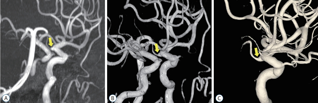
Fig. 1. A 74-year-old woman was admitted for the treatment of an unruptured aneurysm. A : On time-of-flight magnetic resonance angiography three-dimensional (3D) reconstruction images, a saccular dilatation, possibly an aneurysm (arrow), was suspected near the posterior communicating artery (PcomA). B : Source image based new 3D rotational angiography showed that the PcomA originated from the wedge-shaped infundibulum (arrow). C : This lesion was confirmed as an infundibulum (arrow) on 3D rotational angiogram.

Fig. 2. A 62-year-old man underwent magnetic resonance angiography (MRA) for a medical check-up. A : On time-of-flight MRA three-dimensional (3D) reconstruction images, a saccular dilatation (arrow) was identified in the anterior communicating artery (AcomA). B : Source image based new 3D rotational angiography showed that the median artery of the corpus callosum originated from the wedge-shaped infundibulum (arrow). C : This lesion was confirmed as an infundibulum (arrow) on 3D rotational angiogram.

Fig. 3. A 56-year-old woman underwent magnetic resonance angiography (MRA) for a headache. A : On time-of-flight MRA three-dimensional (3D) reconstruction images, a bulbous dilatation (arrow) was identified in the right middle cerebral artery (MCA). B : Source image based new 3D rotational angiography showed that the MCA branch originated from the infundibulum (arrow). C : This lesion was confirmed as an infundibulum (arrow) on 3D rotational angiogram.
Reference
-
References
1. Adams WM, Laitt RD, Jackson A. The role of MR angiography in the pretreatment assessment of intracranial aneurysms: a comparative study. AJNR Am J Neuroradiol. 21:1618–1628. 2000.2. Bederson JB, Connolly ES Jr, Batjer HH, Dacey RG, Dion JE, Diringer MN, et al. Guidelines for the management of aneurysmal subarachnoid hemorrhage: a statement for healthcare professionals from a special writing group of the Stroke Council, American Heart Association. Stroke. 40:994–1025. 2009.
Article3. Brisman JL, Song JK, Newell DW. Cerebral aneurysms. N Engl J Med. 355:928–939. 2006.
Article4. Fifi JT, Meyers PM, Lavine SD, Cox V, Silverberg L, Mangla S, et al. Complications of modern diagnostic cerebral angiography in an academic medical center. J Vasc Interv Radiol. 20:442–447. 2009.
Article5. Kaufmann TJ, Huston J 3rd, Mandrekar JN, Schleck CD, Thielen KR, Kallmes DF. Complications of diagnostic cerebral angiography: evaluation of 19,826 consecutive patients. Radiology. 243:812–819. 2007.
Article6. Menke J, Larsen J, Kallenberg K. Diagnosing cerebral aneurysms by computed tomographic angiography: meta-analysis. Ann Neurol. 69:646–654. 2011.
Article7. Numminen J, Tarkiainen A, Niemelä M, Porras M, Hernesniemi J, Kangasniemi M. Detection of unruptured cerebral artery aneurysms by MRA at 3.0 tesla: comparison with multislice helical computed tomographic angiography. Acta Radiol. 52:670–674. 2011.
Article8. Okahara M, Kiyosue H, Yamashita M, Nagatomi H, Hata H, Saginoya T, et al. Diagnostic accuracy of magnetic resonance angiography for cerebral aneurysms in correlation with 3D-digital subtraction angiographic images: a study of 133 aneurysms. Stroke. 33:1803–1808. 2002.
Article9. Sailer AM, Wagemans BA, Nelemans PJ, de Graaf R, van Zwam WH. Diagnosing intracranial aneurysms with MR angiography: systematic review and meta-analysis. Stroke. 45:119–126. 2014.
Article10. Satoh T, Omi M, Ohsako C, Fujiwara K, Tsuno K, Sasahara W, et al. Differential diagnosis of the infundibular dilation and aneurysm of internal carotid artery: assessment with fusion imaging of 3D MR cisternography/angiography. AJNR Am J Neuroradiol. 27:306–312. 2006.11. Satoh T, Onoda K, Tsuchimoto S. Visualization of intraaneurysmal flow patterns with transluminal flow images of 3D MR angiograms in conjunction with aneurysmal configurations. AJNR Am J Neuroradiol. 24:1436–1445. 2003.12. van Gijn J, Kerr RS, Rinkel GJ. Subarachnoid haemorrhage. Lancet. 369:306–318. 2007.
Article13. van Rooij WJ, Sprengers ME, de Gast AN, Peluso JP, Sluzewski M. 3D rotational angiography: the new gold standard in the detection of additional intracranial aneurysms. AJNR Am J Neuroradiol. 29:976–979. 2008.
Article14. Wermer MJ, van Walderveen MA, Garpebring A, van Osch MJ, Versluis MJ. 7Tesla MRA for the differentiation between intracranial aneurysms and infundibula. Magn Reson Imaging. 37:16–20. 2017.
Article15. Willinsky RA, Taylor SM, TerBrugge K, Farb RI, Tomlinson G, Montanera W. Neurologic complications of cerebral angiography: prospective analysis of 2,899 procedures and review of the literature. Radiology. 227:522–528. 2003.
Article
- Full Text Links
-
- Actions
-
Cited
- CITED
-
- Close
- Share
-
- Similar articles
-
- Supraclinoid Internal Carotid Artery Fenestration Harboring an Unruptured Aneurysm and Another Remote Ruptured Aneurysm: Case Report and Review of the Literature
- Clinical Usefulness of Virtual Angioscopy Using 3D MR Angiography DICOM Images
- Ruptured Aneurysm of the Ophthalmic Artery
- Imaging of Distal Dural Ring Plane and Paraclinoid Internal Carotid Artery Aneurysms with 3D Rotational Angiography
- A Pitfall in the Use of Three Dimensional Computed Tomographic Angiography for Early Surgery of Ruptured Cerebral Aneurysm: Case Report
