Ann Rehabil Med.
2021 Aug;45(4):325-330. 10.5535/arm.21101.
Is Palmar Cutaneous Branch of the Median Nerve More Swollen in Carpal Tunnel Syndrome?
- Affiliations
-
- 1Department of Physical Medicine and Rehabilitation, Korea University Guro Hospital, Seoul, Korea
- 2Department of Physical Medicine and Rehabilitation, The Armed Forces Medical Command, Seongnam, Korea
- KMID: 2519639
- DOI: http://doi.org/10.5535/arm.21101
Abstract
Objective
To investigate the characteristics of the palmar cutaneous branch of the median nerve (PCBMN) in patient with carpal tunnel syndrome (CTS) using high-resolution ultrasound.
Methods
Fourteen healthy volunteers (17 wrists) and 31 patients with CTS (41 wrists) were evaluated by high-resolution ultrasound. All patients were classified into three groups based on the electrophysiologic CTS impairment severity: mild, moderate, and severe. Using high-resolution ultrasound, the cross-sectional areas (CSAs) of the PCBMN were measured at the proximal wrist crease, bistyloid line, and distal wrist crease, and the largest CSA was defined as the maximal CSA.
Results
The maximal CSA of the PCBMN of the control, mild, moderate, and severe CTS groups were 0.27±0.08, 0.30±0.07, 0.35±0.10, and 0.47±0.13 mm2, respectively. The maximal CSA of the PCBMN was significantly larger in the severe CTS group than in the other groups.
Conclusion
The PCBMN could be concomitantly affected in patients with severe CTS.
Figure
-
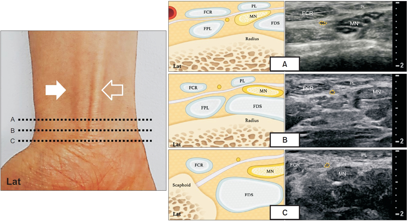
Fig. 1. Scans of the palmar cutaneous branch of the median nerve at three different levels (A, B, C) of the wrist. MN, median nerve; FCR, flexor carpi radialis; PL, palmaris longus; FPL, flexor pollicis longus; FDS, flexor digitorum superficialis.

Fig. 2. Comparisons of the maximal cross-sectional areas (CSAs). (A) Control group versus carpal tunnel syndrome (CTS) group. (B) Between control, mild, moderate, and severe CTS groups. *p<0.05.
Reference
-
1. Kamath J, Jayasheelan N, Mathews R. Compressive neuropathy of the palmar cutaneous branch of the median nerve after a malunited fracture of the distal radius. J Hand Surg Eur Vol. 2016; 41:231–2.
Article2. Chaynes P, Becue J, Vaysse P, Laude M. Relationships of the palmar cutaneous branch of the median nerve: a morphometric study. Surg Radiol Anat. 2004; 26:275–80.
Article3. Gupta SK, Benstead TJ. Symptoms experienced by patients with carpal tunnel syndrome. Can J Neurol Sci. 1997; 24:338–42.
Article4. Sharma V, Wilder-Smith EP. Self-administered hand symptom diagram for carpal tunnel syndrome diagnosis. J Hand Surg Br. 2004; 29:571–4.
Article5. Uluc K, Aktas I, Sunter G, Kahraman Koytak P, Akyuz G, İsak B, et al. Palmar cutaneous nerve conduction in patients with carpal tunnel syndrome. Int J Neurosci. 2015; 125:817–22.
Article6. Imai T, Wada T, Matsumoto H. Entrapment neuropathy of the palmar cutaneous branch of the median nerve in carpal tunnel syndrome. Clin Neurophysiol. 2004; 115:2514–7.
Article7. Jeong YH, Choi JH, Choi HS, Kang S, Yang SN, Yoon JS. Risk assessment of injury to palmar cutaneous branch of the median nerve using high-resolution ultrasound. Ann Rehabil Med. 2019; 43:458–64.
Article8. Cartwright MS, Walker FO. Neuromuscular ultrasound in common entrapment neuropathies. Muscle Nerve. 2013; 48:696–704.
Article9. Hobson-Webb LD. Emerging technologies in neuromuscular ultrasound. Muscle Nerve. 2020; 61:719–25.
Article10. Beekman R, Visser LH. Sonography in the diagnosis of carpal tunnel syndrome: a critical review of the literature. Muscle Nerve. 2003; 27:26–33.
Article11. Tagliafico A, Pugliese F, Bianchi S, Bodner G, Padua L, Rubino M, et al. High-resolution sonography of the palmar cutaneous branch of the median nerve. AJR Am J Roentgenol. 2008; 191:107–14.
Article12. Stevens JC. AAEM minimonograph #26: the electrodiagnosis of carpal tunnel syndrome. Muscle Nerve. 1997; 20:1477–86.
Article13. Lee HJ, Kwon HK. Electrophysiologic classification of severity of carpal tunnel syndrome. J Korean Assoc EMG-Electrodiagn Med. 2004; 6:1–3.14. Cho YS, Lee SH, Kwon HK, Lee HJ. Reappraisal of nerve conduction studies in carpal tunnel syndrome. J Korean Acad Rehabil Med. 1998; 22:861–5.15. Rathakrishnan R, Therimadasamy AK, Chan YH, Wilder-Smith EP. The median palmar cutaneous nerve in normal subjects and CTS. Clin Neurophysiol. 2007; 118:776–80.
Article16. Norbury JW, Nazarian LN. Ultrasound-guided treatment of peripheral entrapment mononeuropathies. Muscle Nerve. 2019; 60:222–31.
Article17. Ginanneschi F, Filippou G, Reale F, Scarselli C, Galeazzi M, Rossi A. Ultrasonographic and functional changes of the ulnar nerve at Guyon’s canal after carpal tunnel release. Clin Neurophysiol. 2010; 121:208–13.
Article
- Full Text Links
-
- Actions
-
Cited
- CITED
-
- Close
- Share
-
- Similar articles
-
- The Abnormal Ultrasonographic Findings of Carpal Tunnel in Carpal Tunnel Syndrome
- Lipofibromatous Hamartoma of Median Nerve with Carpal Tunnel Syndrome: A Case Report
- Median Nerve Block for Treatment of Carpal Tunnel Syndrome: Report of 5 cases
- An Unusual Cause of Carpal Tunnel Syndrome: A Case of Tuberculosis of the Median Nerve
- Carpal Tunnel Syndrome with Recurrent Motor Branch Entrapment: A Case Report
