Clinical Applications of Linked Color Imaging and Blue Laser/Light Imaging in the Screening, Diagnosis, and Treatment of Superficial Colorectal Tumors
- Affiliations
-
- 1Endoscopy Division, National Cancer Center Hospital, Tokyo, Japan
- KMID: 2518850
- DOI: http://doi.org/10.5946/ce.2021.157
Abstract
- Considering its contribution to reducing colorectal cancer morbidity and mortality, the most important task of colonoscopy is to find all existing polyps. Moreover, the accurate detection of existing polyps determines the risk of colorectal cancer morbidity and is an important factor in deciding the appropriate surveillance program for patients. Image-enhanced endoscopy is an easy-to-use modality with improved lesion detection. Linked color imaging (LCI) and blue laser/light imaging (BLI) are useful modalities for improving colonoscopy quality. Each mode has unique optical features; therefore, their intended use differs. LCI contributes to improved polyp detection due to its brightness and high color contrast between the lesion and normal mucosa, while BLI contributes to the characterization of detected polyps by evaluating the vessel and surface patterns of detected lesions. The proper use of these observation modes allows for more efficient endoscopic diagnosis. Moreover, recent developments in artificial intelligence will soon change the clinical practice of colonoscopy and this system will provide an efficient education modality for novice endoscopists.
Figure
-
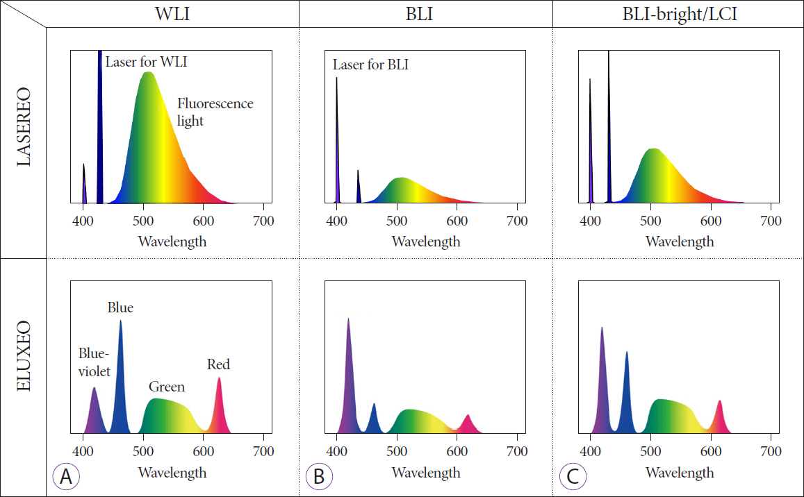
Fig. 1. Wavelength differences among the three observation modes. (A) White light imaging (WLI). (B) Blue laser/light imaging (BLI). (C) Linked color imaging (LCI).

Fig. 2. Differences in brightness among the three observation modes. (A) Linked color imaging (LCI). (B) White light imaging (WLI). (C) Blue laser/light imaging (BLI).

Fig. 3. An example of the clinical application of the three observation modes. (A, B) White light imaging (WLI) or linked color imaging (LCI) for the detection of polyps. (C) Characterization or depth diagnosis was performed by evaluating the vessels and surface pattern using magnifying blue laser/light imaging (BLI). Irregular vessel and surface patterns are shown in the center of the lesion. (D) Chromoendoscopy with crystal violet staining was additionally performed to confirm the depth diagnosis as a final judgment. A severe irregular pit pattern was found at the center of the lesion. From this finding, the depth diagnosis was a deep submucosal invasion.

Fig. 4. CAD-EYE system. (A) Detection of a colorectal polyp with linked color imaging (LCI) and computer-aided detection (CADe system). (B) The polyp was then characterized by blue laser/light imaging (BLI) and computer-aided diagnosis (CADx system).
Reference
-
1. Pan J, Xin L, Ma YF, Hu LH, Li ZS. Colonoscopy reduces colorectal cancer incidence and mortality in patients with non-malignant findings: a meta-analysis. Am J Gastroenterol. 2016; 111:355–365.
Article2. Brand EC, Dik VK, van Oijen MGH, Siersema PD. Missed adenomas with behind-folds visualizing colonoscopy technologies compared with standard colonoscopy: a pooled analysis of 3 randomized back-to-back tandem colonoscopy studies. Gastrointest Endosc. 2017; 86:376–385.e2.
Article3. Osawa H, Miura Y, Takezawa T, et al. Linked color imaging and blue laser imaging for upper gastrointestinal screening. Clin Endosc. 2018; 51:513–526.
Article4. Yoshida N, Dohi O, Inoue K, et al. Blue laser imaging, blue light imaging, and linked color imaging for the detection and characterization of colorectal tumors. Gut Liver. 2019; 13:140–148.
Article5. Min M, Deng P, Zhang W, Sun X, Liu Y, Nong B. Comparison of linked color imaging and white-light colonoscopy for detection of colorectal polyps: a multicenter, randomized, crossover trial. Gastrointest Endosc. 2017; 86:724–730.
Article6. Paggi S, Mogavero G, Amato A, et al. Linked color imaging reduces the miss rate of neoplastic lesions in the right colon: a randomized tandem colonoscopy study. Endoscopy. 2018; 50:396–402.
Article7. Kudo T, Horiuchi A, Kyodo R, et al. Linked colour imaging versus white-light colonoscopy for the detection of flat colorectal lesions: a randomized controlled trial. Colorectal Dis. 2021; 23:1414–1420.
Article8. Shinozaki S, Kobayashi Y, Hayashi Y, et al. Colon polyp detection using linked color imaging compared to white light imaging: systematic review and meta-analysis. Dig Endosc. 2020; 32:874–881.
Article9. Sakamoto T, Tomizawa Y, Cho H, et al. Additional value of linked color imaging in colonoscopy: a retrospective study. Endosc Int Open. 2019; 7:E1448–E1454.
Article10. Kaneko K, Oono Y, Yano T, et al. Effect of novel bright image enhanced endoscopy using blue laser imaging (BLI). Endosc Int Open. 2014; 2:E212–E219.
Article11. Ikematsu H, Sakamoto T, Togashi K, et al. Detectability of colorectal neoplastic lesions using a novel endoscopic system with blue laser imaging: a multicenter randomized controlled trial. Gastrointest Endosc. 2017; 86:386–394.
Article12. Shimoda R, Sakata Y, Fujise T, et al. The adenoma miss rate of blue-laser imaging vs. white-light imaging during colonoscopy: a randomized tandem trial. Endoscopy. 2017; 49:186–190.
Article13. Ang TL, Li JW, Wong YJ, et al. A prospective randomized study of colonoscopy using blue laser imaging and white light imaging in detection and differentiation of colonic polyps. Endosc Int Open. 2019; 7:E1207–E1213.
Article14. Bisschops R, Hassan C, Bhandari P, et al. BASIC (BLI Adenoma Serrated International Classification) classification for colorectal polyp characterization with blue light imaging. Endoscopy. 2018; 50:211–220.
Article15. Subramaniam S, Hayee B, Aepli P, et al. Optical diagnosis of colorectal polyps with blue light imaging using a new international classification. United European Gastroenterol J. 2019; 7:316–325.
Article16. Rondonotti E, Hassan C, Andrealli A, et al. Clinical validation of BASIC classification for the resect and discard strategy for diminutive colorectal polyps. Clin Gastroenterol Hepatol. 2020; 18:2357–2365.e4.
Article17. Lieberman DA, Rex DK, Winawer SJ, Giardiello FM, Johnson DA, Levin TR. Guidelines for colonoscopy surveillance after screening and polypectomy: a consensus update by the US Multi-Society Task Force on Colorectal Cancer. Gastroenterology. 2012; 143:844–857.
Article18. Hassan C, Quintero E, Dumonceau JM, et al. Post-polypectomy colonoscopy surveillance: European Society of Gastrointestinal Endoscopy (ESGE) guideline. Endoscopy. 2013; 45:842–851.
Article19. Desai M, Kennedy K, Aihara H, et al. External validation of blue light imaging (BLI) criteria for the optical characterization of colorectal polyps by endoscopy experts. J Gastroenterol Hepatol. 2021; Apr. 30. [Epub]. https://doi.org/10.1111/jgh.15529.
Article20. Sakamoto T, Nakajima T, Matsuda T, et al. Comparison of the diagnostic performance between magnifying chromoendoscopy and magnifying narrow-band imaging for superficial colorectal neoplasms: an online survey. Gastrointest Endosc. 2018; 87:1318–1323.
Article21. Sakamoto T, Inoki K, Takamaru H, et al. Efficacy of linked colour imaging in magnifying chromoendoscopy with crystal violet staining: a pilot study. Int J Colorectal Dis. 2019; 34:1341–1344.
Article22. Yoshida N, Hisabe T, Inada Y, et al. The ability of a novel blue laser imaging system for the diagnosis of invasion depth of colorectal neoplasms. J Gastroenterol. 2014; 49:73–80.
Article23. Yoshida N, Yagi N, Inada Y, et al. Ability of a novel blue laser imaging system for the diagnosis of colorectal polyps. Dig Endosc. 2014; 26:250–258.
Article24. Nakano A, Hirooka Y, Yamamura T, et al. Comparison of the diagnostic ability of blue laser imaging magnification versus pit pattern analysis for colorectal polyps. Endosc Int Open. 2017; 5:E224–E231.
Article25. Weigt J, Repici A, Antonelli G, et al. Performance of a new integrated computer-assisted system (CADe/CADx) for detection and characterization of colorectal neoplasia. Endoscopy. 2021; 1. 25. [Epub]. https://doi.org/10.1055/a-1372-0419.
Article
- Full Text Links
-
- Actions
-
Cited
- CITED
-
- Close
- Share
-
- Similar articles
-
- Blue Laser Imaging, Blue Light Imaging, and Linked Color Imaging for the Detection and Characterization of Colorectal Tumors
- Detection of Gastrointestinal Cancer using Linked Color Imaging and Blue Light Imaging
- Detecting colorectal lesions with image-enhanced endoscopy: an updated review from clinical trials
- Linked Color Imaging and Blue Laser Imaging for Upper Gastrointestinal Screening
- Advanced Treatment and Imaging in Colonoscopy: The Pocket-Creation Method for Complete Resection and Linked Color Imaging for Better Detection of Early Neoplastic Lesions by Colonoscopy
