Mandibular skeletal posterior anatomic limit for molar distalization in patients with Class III malocclusion with different vertical facial patterns
- Affiliations
-
- 1Department of Orthodontics, Dankook University College of Dentistry, Cheonan, Korea
- KMID: 2518518
- DOI: http://doi.org/10.4041/kjod.2021.51.4.250
Abstract
Objective
The aim of this study was to compare the differences in mandibular posterior anatomic limit (MPAL) distances stratified by vertical patterns in patients with skeletal Class III malocclusion by using cone-beam computed tomography (CBCT).
Methods
CBCT images of 48 patients with skeletal Class III malocclusion (mean age, 22.8 ± 3.1 years) categorized according to the vertical patterns (hypodivergent, normodivergent, and hyperdivergent; n = 16 per group) were analyzed. While parallel to the posterior occlusal line, the shortest linear distances from the distal root of the mandibular second molar to the inner cortex of the mandibular body were measured at depths of 4, 6, and 8 mm from the cementoenamel junction. MPAL distances were compared between the three groups, and their correlations were analyzed.
Results
The mean ages, sex distribution, asymmetry, and crowding in the three groups showed no significant differences. MPAL distance was significantly longer in male (3.8 ± 2.6 mm) than in female (1.8 ± 1.2 mm) at the 8-mm root level. At all root levels, MPAL distances were significantly different in the hypodivergent and hyperdivergent groups (p < 0.001) and between the normodivergent and hyperdivergent groups (p < 0.01). MPAL distances were the shortest in the hyperdivergent group. The mandibular plane angle highly correlated with MPAL distances at all root levels (p < 0.01).
Conclusions
MPAL distances were the shortest in patients with hyperdivergent patterns and showed a decreasing tendency as the mandibular plane angle increased. MPAL distances were significantly shorter (~3.16 mm) at the 8-mm root level.
Figure
-

Figure 1 Cone-beam computed tomography-reconstructed image of the left lingual side of the mandible. Mylohyoid ridge (yellow line) and submandibular fossa (dotted red line).

Figure 2 Measurement of mandibular posterior anatomic limit (MPAL) distance. A, Axial slice at the crown level; dotted red line, posterior occlusal line (POL). B, Axial slice at the root level; orange line, MPAL distance. C, Close-up view of the black box in B; white line, inner cortex; dotted white line, outer cortex of the mandibular body.
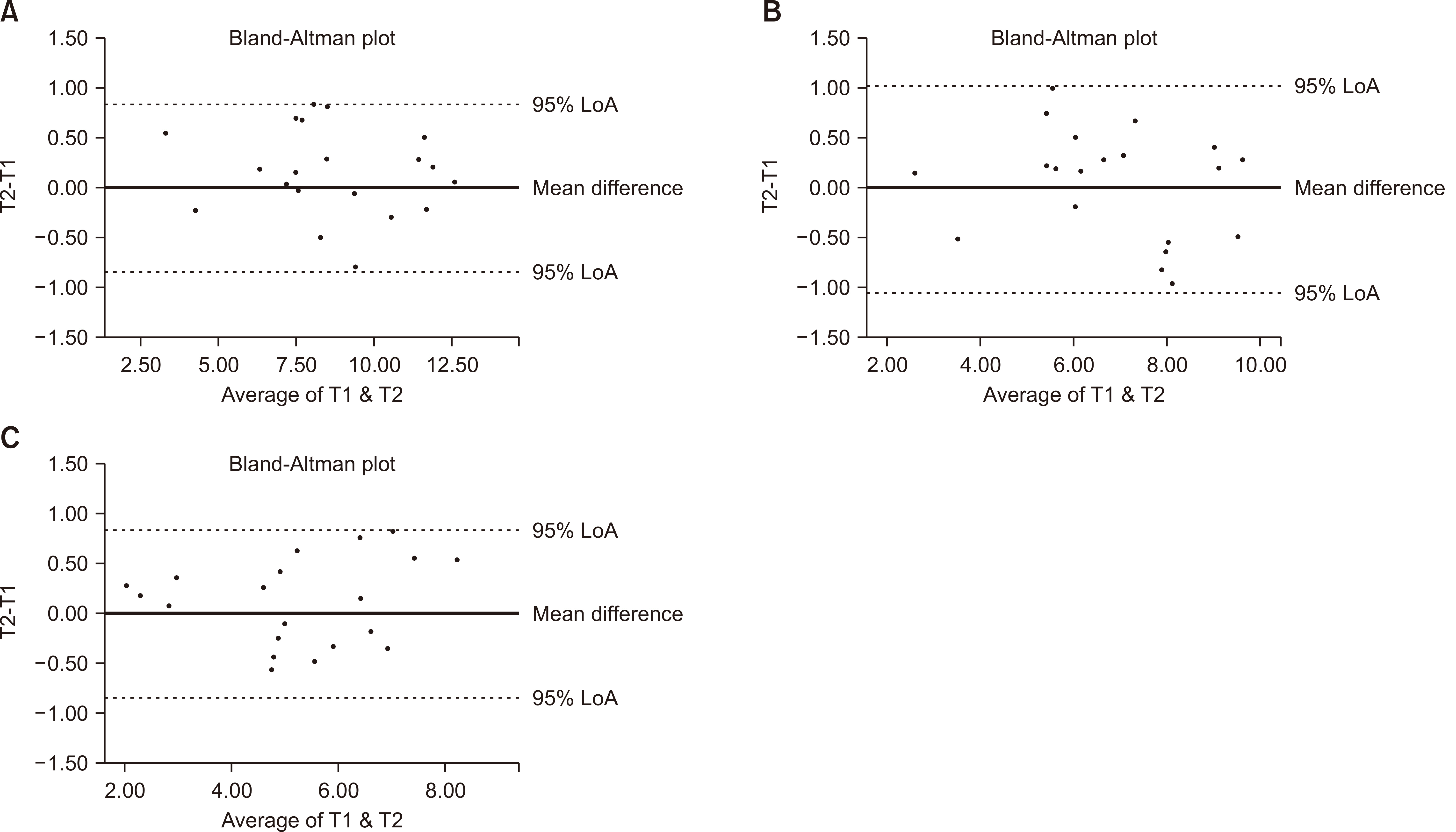
Figure 3 Intraexaminer agreements in mandibular posterior anatomic limit distance measurements by using the Bland-Altman plots. A, Depth of 4 mm, D4mm. B, Depth of 6 mm, D6mm. C, Depth of 8 mm from the cementoenamel junction, D8mm. “95% limit of agreement (LoA)” represents the mean difference ± 1.96 standard deviations. T1, the first measurement; T2, the second measurement, 2 weeks after T1.

Figure 4 Comparison of mandibular posterior anatomic limit distances between the groups. A, Depth of 4 mm, D4mm. B, Depth of 6 mm, D6mm. C, Depth of 8 mm from the cementoenamel junction, D8mm. Significant differencese are indicated by asterisk (**, ***).
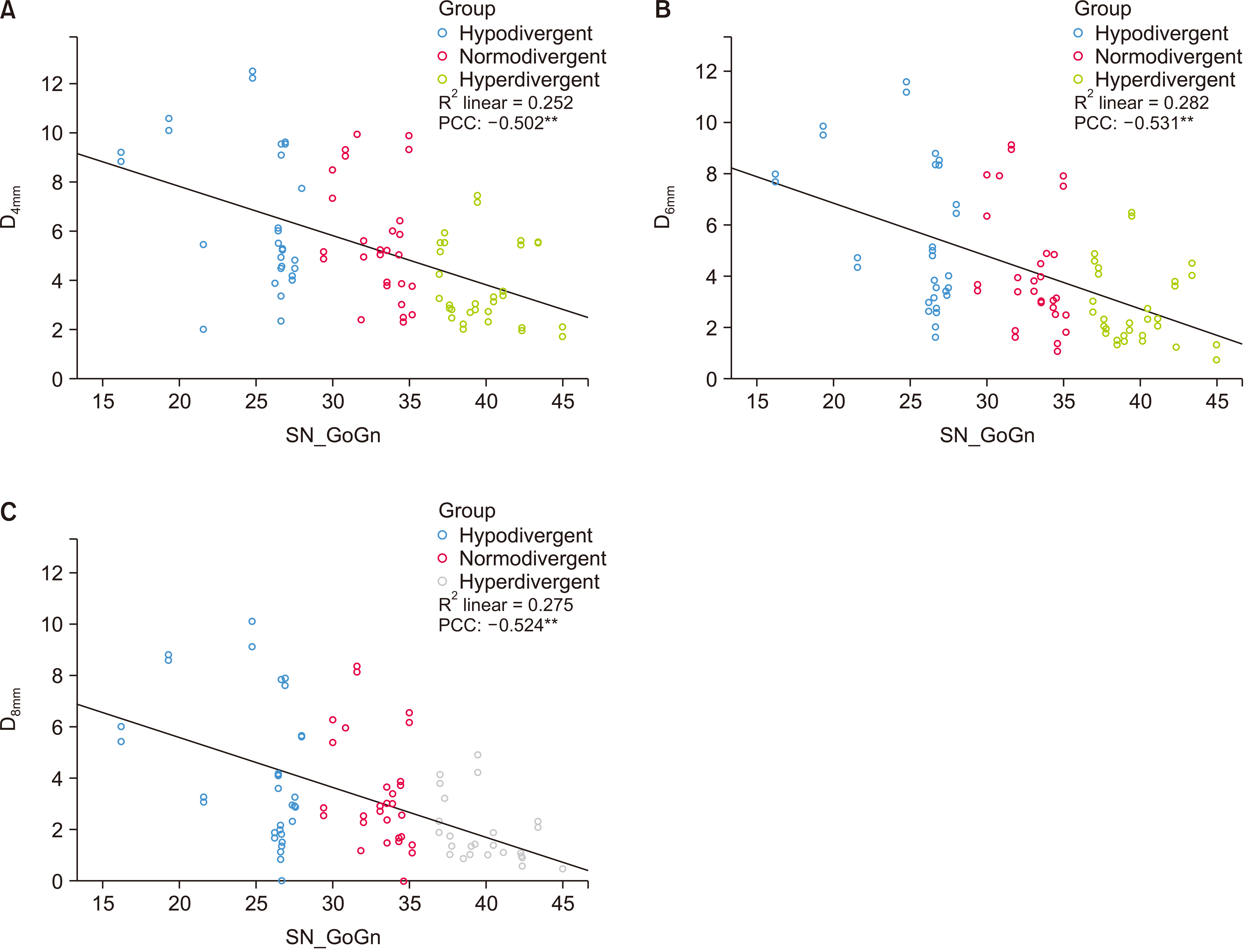
Figure 5 Scatter plots of depths according to the mandibular plane angles. A, Depth of 4 mm, D4mm. B, Depth of 6 mm, D6mm. C, Depth of 8 mm from the cementoenamel junction, D8mm. PCC, Pearson correlation coefficient. **Correlation is significant at the 0.01 level.
Reference
-
1. Yamada K, Kuroda S, Deguchi T, Takano-Yamamoto T, Yamashiro T. 2009; Distal movement of maxillary molars using miniscrew anchorage in the buccal interradicular region. Angle Orthod. 79:78–84. DOI: 10.2319/020408-68.1. PMID: 19123698.
Article2. Oh YH, Park HS, Kwon TG. 2011; Treatment effects of microimplant-aided sliding mechanics on distal retraction of posterior teeth. Am J Orthod Dentofacial Orthop. 139:470–81. DOI: 10.1016/j.ajodo.2009.05.037. PMID: 21457858.
Article3. Kim SJ, Choi TH, Baik HS, Park YC, Lee KJ. 2014; Mandibular posterior anatomic limit for molar distalization. Am J Orthod Dentofacial Orthop. 146:190–7. DOI: 10.1016/j.ajodo.2014.04.021. PMID: 25085302.
Article4. Edwards JG. 1976; A study of the anterior portion of the palate as it relates to orthodontic therapy. Am J Orthod. 69:249–73. DOI: 10.1016/0002-9416(76)90075-0. PMID: 1062165.
Article5. Handelman CS. 1996; The anterior alveolus: its importance in limiting orthodontic treatment and its influence on the occurrence of iatrogenic sequelae. Angle Orthod. 66:95–109. discussion 109–10. DOI: 10.1043/0003-3219(1996)066<0095:TAAIII>2.3.CO;2. PMID: 8712499.6. Nakada T, Motoyoshi M, Horinuki E, Shimizu N. 2016; Cone-beam computed tomography evaluation of the association of cortical plate proximity and apical root resorption after orthodontic treatment. J Oral Sci. 58:231–6. DOI: 10.2334/josnusd.15-0566. PMID: 27349544.
Article7. Ganss C, Hochban W, Kielbassa AM, Umstadt HE. 1993; Prognosis of third molar eruption. Oral Surg Oral Med Oral Pathol. 76:688–93. DOI: 10.1016/0030-4220(93)90035-3. PMID: 8284071.
Article8. Begtrup A, Grønastøð HÁ, Christensen IJ, Kjær I. 2013; Predicting lower third molar eruption on panoramic radiographs after cephalometric comparison of profile and panoramic radiographs. Eur J Orthod. 35:460–6. DOI: 10.1093/ejo/cjs012. PMID: 22474212.
Article9. Kim TW, Artun J, Behbehani F, Artese F. 2003; Prevalence of third molar impaction in orthodontic patients treated nonextraction and with extraction of 4 premolars. Am J Orthod Dentofacial Orthop. 123:138–45. DOI: 10.1067/mod.2003.13. PMID: 12594419.
Article10. Karad A. 2015. Clinical orthodontics: current concepts, goals and mechanics. 2nd ed. Elsevier;Gurgaon: p. 251.11. Hardy D, Cubas Y, Orellana M. 2012; Prevalence of angle class III malocclusion: a systematic review and meta-analysis. Open J Epidemiol. 2:75–82. DOI: 10.4236/ojepi.2012.24012.
Article12. Proffit WR, Fields HW, Sarver DM. 2013. Contemporary orthodontics. Mosby;St. Louis: p. 684p. 699–701.13. Choi YT, Kim YJ, Yang KS, Lee DY. 2018; Bone availability for mandibular molar distalization in adults with mandibular prognathism. Angle Orthod. 88:52–7. DOI: 10.2319/040617-237.1. PMID: 28949768.
Article14. Horner KA, Behrents RG, Kim KB, Buschang PH. 2012; Cortical bone and ridge thickness of hyperdivergent and hypodivergent adults. Am J Orthod Dentofacial Orthop. 142:170–8. DOI: 10.1016/j.ajodo.2012.03.021. PMID: 22858325.
Article15. Masumoto T, Hayashi I, Kawamura A, Tanaka K, Kasai K. 2001; Relationships among facial type, buccolingual molar inclination, and cortical bone thickness of the mandible. Eur J Orthod. 23:15–23. DOI: 10.1093/ejo/23.1.15. PMID: 11296507.
Article16. Ge J, Zheng JW, Yang C, Qian WT. 2016; Variations in the buccal-lingual alveolar bone thickness of impacted mandibular third molar: our classification and treatment perspectives. Sci Rep. 6:16375. DOI: 10.1038/srep16375. PMID: 26759181. PMCID: PMC4725356.
Article17. Wainwright WM. 1973; Faciolingual tooth movement: its influence on the root and cortical plate. Am J Orthod. 64:278–302. DOI: 10.1016/0002-9416(73)90021-3. PMID: 4199008.
Article18. Lemos Rinaldi MR, Azeredo F, Martinelli de Lima E, Deon Rizzatto SM, Sameshima G, Macedo de Menezes L. 2018; Cone-beam computed tomography evaluation of bone plate and root length after maxillary expansion using tooth-borne and tooth-tissue-borne banded expanders. Am J Orthod Dentofacial Orthop. 154:504–16. DOI: 10.1016/j.ajodo.2017.12.018. PMID: 30268261.
Article19. Ingervall B, Helkimo E. 1978; Masticatory muscle force and facial morphology in man. Arch Oral Biol. 23:203–6. DOI: 10.1016/0003-9969(78)90217-0. PMID: 278554.
Article20. Kiliaridis S, Mejersjö C, Thilander B. 1989; Muscle function and craniofacial morphology: a clinical study in patients with myotonic dystrophy. Eur J Orthod. 11:131–8. DOI: 10.1093/oxfordjournals.ejo.a035975. PMID: 2767145.
Article21. Beckmann SH, Kuitert RB, Prahl-Andersen B, Segner D, The RP, Tuinzing DB. 1998; Alveolar and skeletal dimensions associated with lower face height. Am J Orthod Dentofacial Orthop. 113:498–506. DOI: 10.1016/S0889-5406(98)70260-4. PMID: 9598607.
Article22. Veli I, Uysal T, Baysal A, Karadede I. 2014; Buccal cortical bone thickness at miniscrew placement sites in patients with different vertical skeletal patterns. J Orofac Orthop. 75:417–29. DOI: 10.1007/s00056-014-0235-7. PMID: 25344123.
Article23. Park HS. 2015. Orthodontic treatment using microimplant: clinical applications of microimplant anchorage. 3rd ed. DaehanNarae Publishing, Inc.;Seoul: p. 349–52.24. Vinay G, Mangala Gowri S R, Anbalagan J. 2013; Sex determination of human mandible using metrical parameters. J Clin Diagn Res. 7:2671–3. DOI: 10.7860/JCDR/2013/7621.3728. PMID: 24551607. PMCID: PMC3919368.25. Van der Weijden F, Dell'Acqua F, Slot DE. 2009; Alveolar bone dimensional changes of post-extraction sockets in humans: a systematic review. J Clin Periodontol. 36:1048–58. DOI: 10.1111/j.1600-051X.2009.01482.x. PMID: 19929956.
Article26. Verna C. 2016; Regional acceleratory phenomenon. Front Oral Biol. 18:28–35. DOI: 10.1159/000351897. PMID: 26599115.
Article27. Hansson S, Halldin A. 2012; Alveolar ridge resorption after tooth extraction: a consequence of a fundamental principle of bone physiology. J Dent Biomech. 3:1758736012456543. DOI: 10.1177/1758736012456543. PMID: 22924065. PMCID: PMC3425398.
Article28. de los Rios CM, Pustiglioni FE, Romito GA. 2002; Biometric study of the width, length and depth of the root trunk groove of human lower second molars. Pesqui Odontol Bras. 16:26–30. DOI: 10.1590/S1517-74912002000100005. PMID: 11938714.29. Poletti L, Silvera AA, Ghislanzoni LT. 2013; Dentoalveolar class III treatment using retromolar miniscrew anchorage. Prog Orthod. 14:7. DOI: 10.1186/2196-1042-14-7. PMID: 24325840. PMCID: PMC4384962.
Article30. Kook YA, Park JH, Bayome M, Kim S, Han E, Kim CH. 2016; Distalization of the mandibular dentition with a ramal plate for skeletal Class III malocclusion correction. Am J Orthod Dentofacial Orthop. 150:364–77. DOI: 10.1016/j.ajodo.2016.03.019. PMID: 27476370.
Article31. Yu J, Park JH, Bayome M, Kim S, Kook YA, Kim Y, et al. 2016; Treatment effects of mandibular total arch distalization using a ramal plate. Korean J Orthod. 46:212–9. DOI: 10.4041/kjod.2016.46.4.212. PMID: 27478798. PMCID: PMC4965592.
Article32. Torres MG, Campos PS, Segundo NP, Navarro M, Crusoé-Rebello I. 2012; Accuracy of linear measurements in cone beam computed tomography with different voxel sizes. Implant Dent. 21:150–5. DOI: 10.1097/ID.0b013e31824bf93c. PMID: 22382754.
Article
- Full Text Links
-
- Actions
-
Cited
- CITED
-
- Close
- Share
-
- Similar articles
-
- A study on the vertical dysplasia in the skeletal class iii malocclusion
- Posteroanterior cephalometric characteristics in skeletal Class III malocclusion
- A study on calcification of the second molars in skeletal Class III malocclusions
- A study on the effects of third molars on angle's Class III malocclusion
- The relationship between posterior dental compensation and skeletal discrepancy in class III malocclusion
