Rescue Maneuver of Migrated Coil Using the ERIC Device after Previous Attempts with Conventional Stentrievers
- Schüller-Arteaga M
 1
1 - Galván-Fernández J1
- Jiménez-Arribas P2
- Nogales-Martin L3
- Rodríguez-Arias C2
- Martínez-Galdámez M1
- Affiliations
-
- 1Interventional Neuroradiology/Endovascular Neurosurgery, Hospital Clínico Universitario de Valladolid, Valladolid, Spain
- 2Department of Neurosurgery, Hospital Clínico Universitario de Valladolid, Valladolid, Spain
- 3Department of Intensive Care, Hospital Clínico Universitario de Valladolid, Valladolid, Spain
- KMID: 2517741
- DOI: http://doi.org/10.5469/neuroint.2021.00066
Abstract
- Coil prolapse or migration is a rare but potentially serious complication that may occur during aneurysm embolization, with no standard management currently described. Here we describe our experience with the Embolus Retriever with Interlinked Cages (ERIC) device® (Microvention, Aliso Viejo, CA, USA) for the retrieval of prolapsed or migrated coils in a case series and Flow-Model analysis. First, a retrospective review was performed using our institution database for patients in which coil prolapse or migration occurred during aneurysm embolization, and data was collected and analyzed. Second, an in vitro Flow-Model analysis was performed comparing the ERIC device® with other stent retrievers for coil retrieval. In 2 cases, the ERIC device® successfully retrieved the displaced coil from intracranial circulation in 1 pass, after failure with other devices. In the Flow-Model, again the ERIC device® achieved success for retrieving a detached coil, whereas 2 other different stent retrievers failed to capture the coil after 2 attempts. The ERIC device® appears to be a safe and effective tool for retrieving a prolapsed or migrated coil from the intracranial circulation.
Keyword
Figure
-
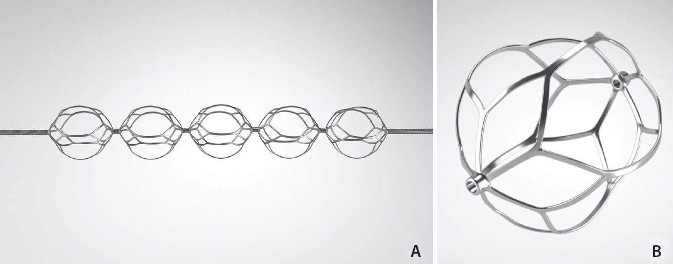
Fig. 1. Embolus Retriever with Interlinked Cages (ERIC) device design. (A) Construction of the device with interlinked spherical cages fixed on a pusher wire. The design of the device results in limited contact points with the vessel wall which can potentially reduce vascular trauma. (B) Magnified view of a single cage. Image from ERIC device® (Microvention, Aliso Viejo, CA, USA).

Fig. 2. (A) The aneurysm morphology can be seen prior to embolization (small arrow). (B) Partially migrated coil (small arrow) can be seen prior to first attempt with Embotrap® (Johnson & Johnson, New Brunswick, NJ, USA). Stryker® SL-10 (Kalamazoo, MI, USA) is navigated past the migrated coil (small arrowhead). (C) Proximal M3 segment coil migration (small arrow) can be seen in this posteroanterior (PA) view, with the ERIC device deployed (small arrowhead), with the ACoA aneurysm already coiled (big arrow). (D) Normal PA angiographic run demonstrating absence of the migrated coil and no aneurysm remnant. ACoA, anterior communicating artery; ERIC, Embolus Retriever with Interlinked Cages.
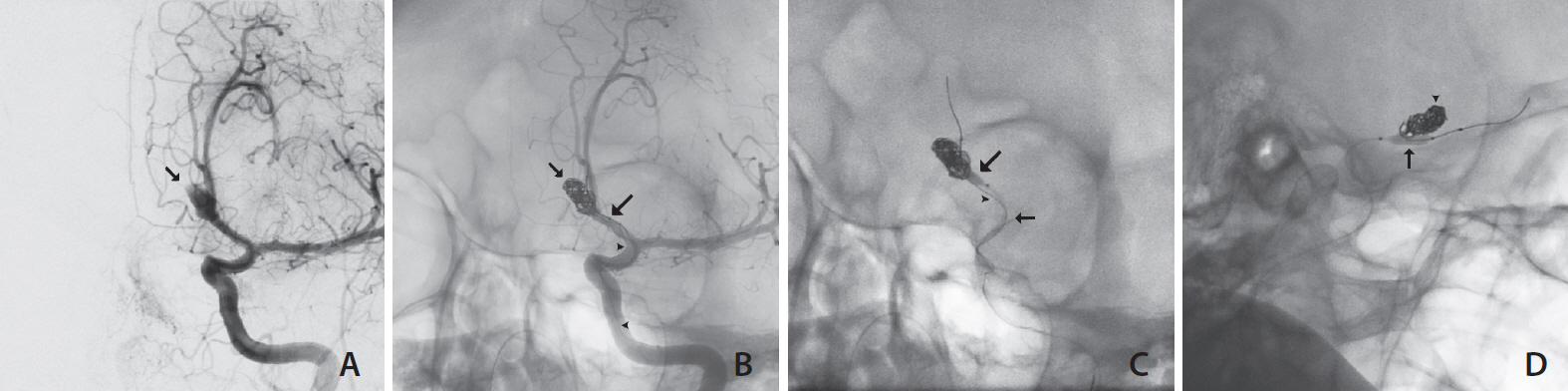
Fig. 3. (A) Ruptured ACoA aneurysm prior to embolization (arrow). (B) Partially coiled aneurysm (small arrow), with a proximally migrated coil (small arrowheads) and a balloon microcatheter (big arrow) covering the aneurysm neck. (C) ERIC device deployed (small arrow) during retrieval maneuver while inflating the balloon (big arrow) to maintain the rest of the coils inside the aneurysm. (D) Post-retrieval image with coil packing integrity maintained (small arrowhead) and the balloon still partially inflated (big arrow). ACoA, anterior communicating artery; ERIC, Embolus Retriever with Interlinked Cages.
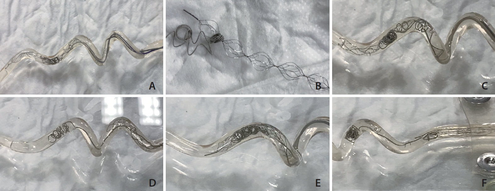
Fig. 4. (A) ERIC opened inside the flow model at the level of the migrated coil before the retrieval attempt. (B) Coil was retrieved from the flow model and found around one of the interlinked cages of the device. (C) Closed cell SR (Trevo; Stryker, Kalamazoo, MI, USA) positioned around the detached coil. (D) Coil remained in place after retrieval, unaffected after the closed cell SR retrieval attempt. (E) EmboTrap positioned in contact with the detached coil. (F) Attempt made with the articulated stent resulted in partial displacement of the coil (coil interaction), but not complete retrieval. ERIC, Embolus Retriever with Interlinked Cages; SR, stent retrievers.
Reference
-
1. Molyneux A, Kerr R, Stratton I, Sandercock P, Clarke M, Shrimpton J, International Subarachnoid Aneurysm Trial (ISAT) Collaborative Group, et al. International Subarachnoid Aneurysm Trial (ISAT) of neurosurgical clipping versus endovascular coiling in 2143 patients with ruptured intracranial aneurysms: a randomised trial. Lancet. 2002; 360:1267–1274.
Article2. Bradac GB, Bergui M, Stura G, Fontanella M, Daniele D, Gozzoli L, et al. Periprocedural morbidity and mortality by endovascular treatment of cerebral aneurysms with GDC: a retrospective 12-year experience of a single center. Neurosurg Rev. 2007; 30:117–125. discussion 125-126.
Article3. van Rooij WJ, Sluzewski M, Beute GN, Nijssen PC. Procedural complications of coiling of ruptured intracranial aneurysms: incidence and risk factors in a consecutive series of 681 patients. AJNR Am J Neuroradiol. 2006; 27:1498–1501.4. Borggrefe J, Behme D, Mpotsaris A, Weber W. Complications associated with cerebral aneurysm morphology in balloon-assisted coil embolization of ruptured and unruptured aneurysms-a single-center analysis of 116 consecutive cases. World Neurosurg. 2016; 91:483–489.
Article5. Leslie-Mazwi TM, Heddier M, Nordmeyer H, Stauder M, Velasco A, Mosimann PJ, et al. Stent retriever use for retrieval of displaced microcoils: a consecutive case series. AJNR Am J Neuroradiol. 2013; 34:1996–1999.
Article6. Vora N, Thomas A, Germanwala A, Jovin T, Horowitz M. Retrieval of a displaced detachable coil and intracranial stent with an L5 Merci Retriever during endovascular embolization of an intracranial aneurysm. J Neuroimaging. 2008; 18:81–84.
Article7. Lee CY. Use of wire as a snare for endovascular retrieval of displaced or stretched coils: rescue from a technical complication. Neuroradiology. 2011; 53:31–35.
Article8. Ding D, Liu KC. Management strategies for intraprocedural coil migration during endovascular treatment of intracranial aneurysms. J Neurointerv Surg. 2014; 6:428–431.9. Henkes H, Lowens S, Preiss H, Reinartz J, Miloslavski E, Kühne D. A new device for endovascular coil retrieval from intracranial vessels: alligator retrieval device. AJNR Am J Neuroradiol. 2006; 27:327–329.10. Zhou KZ, Maingard J, Kok HK, Wang J, Barras CD, O’hare A, et al. Endovascular retrieval of dislodged neurovascular devices with a stentriever: case series and technical review. World Neurosurg. 2019; 123:e661–e669.
Article11. Kabbani MR, Smith A, Leider M. Endovascular coil retrieval using a TrevoProVue stentriever. J Neurointerv Surg. 2015; 7:e19.
Article12. Nas OF, Kacar E, Kaya A, Erdogan C, Hakyemez B. Retrieval of a dislocated coil and stent-assisted coiling by Solitaire® stent during endovascular treatment of an intracranial aneurysm. Diagn Interv Imaging. 2016; 97:381–384.13. Amuluru K, Al-Mufti F, Romero CE. Endovascular retrieval of migrated coil within the distal middle cerebral artery using stentriever device. World Neurosurg. 2018; 117:382–385.
Article14. Nikoubashman O, Pjontek R, Brockmann MA, Tolba R, Wiesmann M. Retrieval of migrated coils with stent retrievers: an animal study. AJNR Am J Neuroradiol. 2015; 36:1162–1166.
Article15. Simgen A, Tomori T, Mühl-Benninghaus R, Bomberg H, Yilmaz U, Körner H, et al. Retrieval of migrated volume coils using different clot retrievers in a porcine model. Clin Neuroradiol. 2018; 28:593–600.16. Ducroux C, Renaud N, Bourcier R, Marnat G, Sibon I, Gory B, ETIS investigators, et al. Embolus Retriever with Interlinked Cages (ERIC) versus conventional stent retrievers for thrombectomy: a propensity score-based analysis. J Neurointerv Surg. 2021; 13:255–260.
Article17. Edwards NJ, Jones WH, Sanzgiri A, Corona J, Dannenbaum M, Chen PR. Antiplatelet therapy for the prevention of peri-coiling thromboembolism in high-risk patients with ruptured intracranial aneurysms. J Neurosurg. 2017; 127:1326–1332.
Article
- Full Text Links
-
- Actions
-
Cited
- CITED
-
- Close
- Share
-
- Similar articles
-
- Migrated coil and damaged stent removal during coil embolization, using an additional, retrievable stent: A case report
- Rescue Therapy of Inadvertent Coil Migration for Endovascular Treatment of Type II Endoleak
- Endovascular Rescue Method for Undesirably Stretched Coil
- Management of a Complicated Cerebral Aneurysm with Distal Migration of a Detachable Coil: A Case Report
- Retrieval of Distally Migrated Coils with Detachable Intracranial Stent during Coil Embolization of Cerebral Aneurysm
