Rescue endovascular treatment for rapid regrowth of aneurysm remnant on middle cerebral artery trunk after unsuccessful surgical clipping in patients with a ruptured cerebral aneurysm: A report of two cases
- Affiliations
-
- 1Department of Neurosurgery, Kangwon National University Hospital, Chuncheon, Korea
- 2Department of Neurosurgery, Kangwon National University College of Medicine, Chuncheon, Korea
- KMID: 2517024
- DOI: http://doi.org/10.7461/jcen.2020.E2020.08.004
Abstract
- We report two rare cases treated with coiling after rapid regrowth (within a month) of an aneurysm remnant on the middle cerebral artery (MCA) trunk after incomplete surgical clipping. The first case, a 47-year-old man with subarachonoid hemorrhage (SAH) (Hunt-Hess grade II, Fisher grade III) underwent clipping of a ruptured saccular aneurysm with a wide neck on the right early frontal branch arising from the MCA trunk. Incomplete clipping with a 1 mm sized remnant neck was performed to avoid sacrificing the lenticulostriate artery. In a follow-up cerebral angiogram on postoperative day 30, a rapid regrowth of the aneurysm remnant was observed, and on that day, complete obliteration was obtained by rescue endovascular treatment. The second case, a 48-year-old healthy woman with SAH (Hunt-Hess grade II, Fisher grade III) underwent clipping of an anteroposteriorly projecting bilobulated aneurysm on the left M1. Incomplete clipping with a minimal remnant neck was performed. In follow-up digital subtraction angiogram on postoperative day 30, a rapid regrowth of an aneurysm remnant involving only a part of the initial aneurysm near the neck was observed, and on that day, complete obliteration was obtained by rescue coiling. These patients were both discharged without any neurological deficits.
Keyword
Figure
-

Fig. 1. A 47-year-old man patient (Hunt- Hess grade II) with subarachnoid hemorrhage (Fisher grade III). Initial three-dimensional reconstruction images in brain computed tomographic angiogram (CTA) revealed a 3.9 mm sized saccular aneurysm (white arrow) with wide neck and it`s neck was incorporated with early frontal branch on right early frontal cortical branch of middle cerebral artery trunk (A). Immediate postoperative CTA demonstrated about 1 mm sized aneurysm remnant (AR, white arrow) behind a clipped aneurysm (white circle) (B: posterior view, C: magnified image in small box). In follow-up digital subtraction angiography (DSA) at postoperative day 30, a rapid regrowth of AR (white arrow) with two blebs (laterally projecting and depth: 7.39 mm×width: 2.58 mm×neck: 3.01 mm) was showed (D: magnified 180˚ reverse image in small box). A prompt endovascular treatment was done and complete obliteration (white arrow) of AR was done (E: magnified DSA working image in small box).
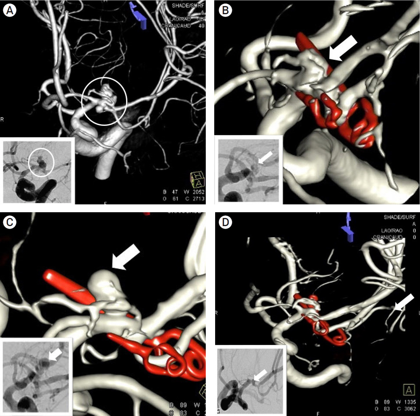
Fig. 2. A 48-year-old woman patient (Hunt- Hess grade II) with subarachnoid hemorrhage (Fisher grade III). Three-dimensional reconstruction image in initial digital subtraction angiography (DSA) showed a ruptured 8 mm sized aneurysm (white circle) with anteroposteriorly projecting bilobar figure and it`s neck was incorporated with lateral lenticulostriate artery (LSA) on left middle cerebral artery trunk (A: magnified DSA image in small box). Immediate postoperative three-dimensional -DSA demonstrated 0.6 mm x 3.2 mm sized remnant aneurysm (AR, white arrow) was located between LSA and a clipped posterior portion aneurysm (AR, white arrow) (B: magnified DSA image in small box). In follow-up three-dimensional -DSA at postoperative day 30, a rapid regrowth of AR (white arrow) with one bleb, superiorly projecting and depth: 7.10 mm×width: 3.48 mm×neck: 3.21 mm was showed (C: magnified DSA image in small box). A prompt endovascular treatment was done and complete obliteration (white arrow) of AR was done (D: magnified DSA working image in small box). At 3 months follow-up, DSA studies demonstrated complete obliteration of aneurysm.
Reference
-
1. Burkhardt JK, Chua MHJ, Weiss M, Do AS-MS, Winkler EA, Lawton MT. Risk of aneurysm residual regrowth, recurrence, and de novo aneurysm formation after microsurgical clip occlusion based on follow-up with catheter angiography. World Neurosurg. 2017; Oct. 106:74–84.
Article2. Cekirge HS, Islak C, Firat MM, Kocer N, Saatci I. Endovascular coil embolization of residual or recurrent aneurysms after surgical clipping. Acta Radiol. 2000; Mar. 41(2):111–5.
Article3. Dashti R, Rinne J, Hernesniemi J, Niemelä M, Kivipelto L, Lehecka M, et al. Microneurosurgical management of proximal middle cerebral artery aneurysms. Surg Neurol. 2007; Jan. 67(1):6–14.
Article4. Della Pepa GM, Bianchi F, Scerrati A, Albanese A, Cotroneo E, Delitala A, et al. Secondary coiling after incomplete surgical clipping of cerebral aneurysms: a rescue strategy or a treatment option for complex cases? Institutional series and systematic review. Neurosurg Rev. 2019; Jun. 42(2):337–50.
Article5. El Beltagy M, Muroi C, Roth P, Fandino J, Imhof H-G, Yonekawa Y. Recurrent intracranial aneurysms after successful neck clipping. World Neurosurg. 2010; Oct-Nov. 74(4-5):472–7.
Article6. Jun HS, Ahn J, Song JH, Chang IB. Spontaneous regression of aneurysm remnant after incomplete surgical clipping in a patient with ruptured cerebral aneurysm. J Cerebrovasc Endovasc Neurosurg. 2016; Dec. 18(4):402–6.
Article7. Kivisaari RP, Porras M, Ohman J, Siironen J, Ishii K, Hernesniemi J. Routine cerebral angiography after surgery for saccular aneurysms: is it worth it? Neurosurgery. 2004; Nov. 55(5):1015–24.
Article8. Kobayashi S, Moroi J, Hikichi K, Yoshioka S, Saito H, Tanabe J, et al. Treatment of Recurrent Intracranial Aneurysms After Neck Clipping: Novel Classification Scheme and Management Strategies. Oper Neurosurg (Hagerstown). 2017; Dec. 13(6):670–8.
Article9. Lin T, Fox AJ, Drake CG. Regrowth of aneurysm sacs from residual neck following aneurysm clipping. J Neurosurg. 1989; Apr. 70(4):556–60.
Article10. Park JC, Shim JH, Lee DH, Ahn JS, Lee DG, Yang K, et al. Three-dimensional angiographic evaluation of middle cerebral artery trunk aneurysms: demonstration of the close relationship between the early frontal cortical branches and lateral lenticulostriate arteries. World Neurosurg. 2016; Jul. 91:383–9.
Article11. Ravindra VM, Karsy M, Schmidt RH, Taussky P, Park MS, Bollo RJ. Rapid de novo aneurysm formation after clipping of a ruptured middle cerebral artery aneurysm in an infant with an MYH11 mutation. J Neurosurg Pediatr. 2016; Oct. 18(4):463–70.
Article12. Spiotta AM, Hui F, Schuette A, Moskowitz SI. Patterns of aneurysm recurrence after microsurgical clip obliteration. Neurosurgery. 2013; Jan. 72(1):65–9. discussion 69.
Article13. Tsutsumi K, Ueki K, Morita A, Usui M, Kirino T. Risk of aneurysm recurrence in patients with clipped cerebral aneurysms: results of long-term follow-up angiography. Stroke. 2001; May. 32(5):1191–4.14. Zhou G, Zhu Y, Yin Y, Su M, Li M. Association of wall shear stress with intracranial aneurysm rupture: systematic review and meta-analysis. Sci Rep. 2017; Jul. 7(1):5331.
Article
- Full Text Links
-
- Actions
-
Cited
- CITED
-
- Close
- Share
-
- Similar articles
-
- Spontaneous Regression of Aneurysm Remnant after Incomplete Surgical Clipping in a Patient with Ruptured Cerebral Aneurysm
- Surgical Clipping of Intracranial Aneurysm Regrown after Endovascular Coiling
- Endovascular Coil Embolization After Clipping: Endovascular Treatment of Incompletely Clipped or Recurred Cerebral Aneurysms
- Surgical Experience of Recurrent Hemorrhage from the Regrowth Cerebral Aneurysm after Initial Neck Clipping: Case Report
- Modified Clipping on Broad Neck Aneurysm of Middle Cerebral Artery
