Anterior Chest Wall Involvement in Spondyloarthritis Patients as Detected by Magnetic Resonance Imaging: A Case Series and Literature Review
- Affiliations
-
- 1Department of Rheumatology, Hanyang University Hospital for Rheumatic Diseases, Seoul, Korea
- 2Department of Radiology, Hanyang University Seoul Hospital, Seoul, Korea
- KMID: 2516952
- DOI: http://doi.org/10.4078/jrd.2021.28.3.159
Abstract
- Magnetic resonance imaging (MRI) plays an important role in diagnosing and classifying axial spondyloarthritis (SpA) and is also useful for appropriate evaluation of disease status owing to its ability to detect inflammation early and reveal structural changes. However, dedicated MRI for the anterior chest wall (ACW) is not routinely considered despite relatively frequent presence of ACW lesions. To date, no study has investigated the imaging findings and clinical features of ACW involvement in Korean SpA patients. Thus, we aimed to show ACW involvement in SpA patients using ACW lesions found by MRI. We describe 20 cases of ACW involvement in which MRI-detected manubriosternal joint lesions. The lesion types included subchondral bone marrow edema, marginal or central bone erosions, subchondral fat infiltration or deposition, and ankylosis, with erosions being the most prevalent finding. We also provide the literature review results describing MRI findings of ACW lesions in SpA patients.
Figure
-
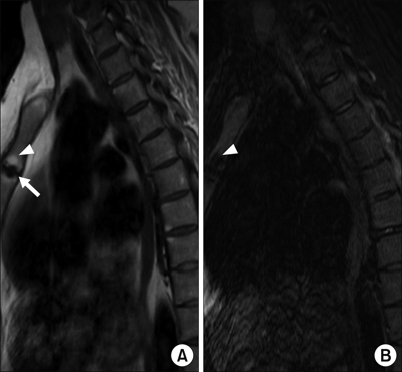
Figure 1 Sagittal MR images from Dixon sequence of a 40-year-old female AS patient. The patient underwent thoracic spine MRI because of ACW pain and worsening back pain during TNF inhibitor therapy. T1-weighted (A) and T2-weighted water-only (B) images of the ACW included in the field of view for thoracic spine MRI. The images show bony erosion (arrow) and periarticular fat deposition (arrowheads) at the MSJ. MRI: magnetic resonance imaging, AS: ankylosing spondylitis, TNF: tumor necrosis factor, ACW: anterior chest wall, MSJ: manubriosternal joint.
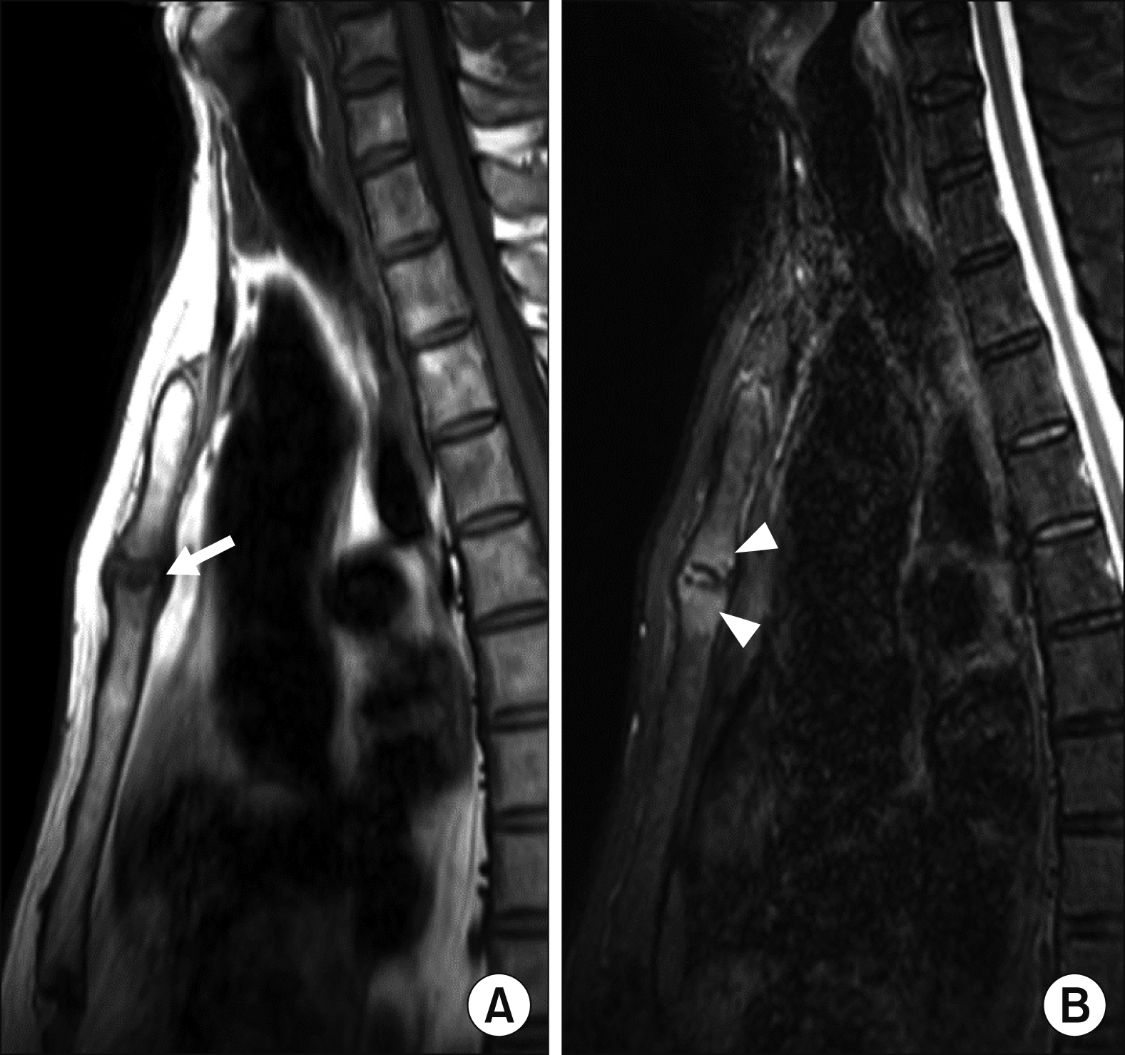
Figure 2 Sagittal MR images of a 53-year-old male patient with nr-axSpA, including T1-weighted (A) and T2-weighted STIR (B) images. Whole spine MRI was acquired following a clinical trial protocol in which the patient was enrolled. The patient did not experience ACW pain at the time of MRI. Bony erosion (arrow) and subchondral bone marrow edema (arrowheads) were observed at the MSJ. MRI: magnetic resonance imaging, nr-axSpA: non-radiographic axial spondyloarthritis, STIR: short tau inversion recovery, ACW: anterior chest wall, MSJ: manubriosternal joint.
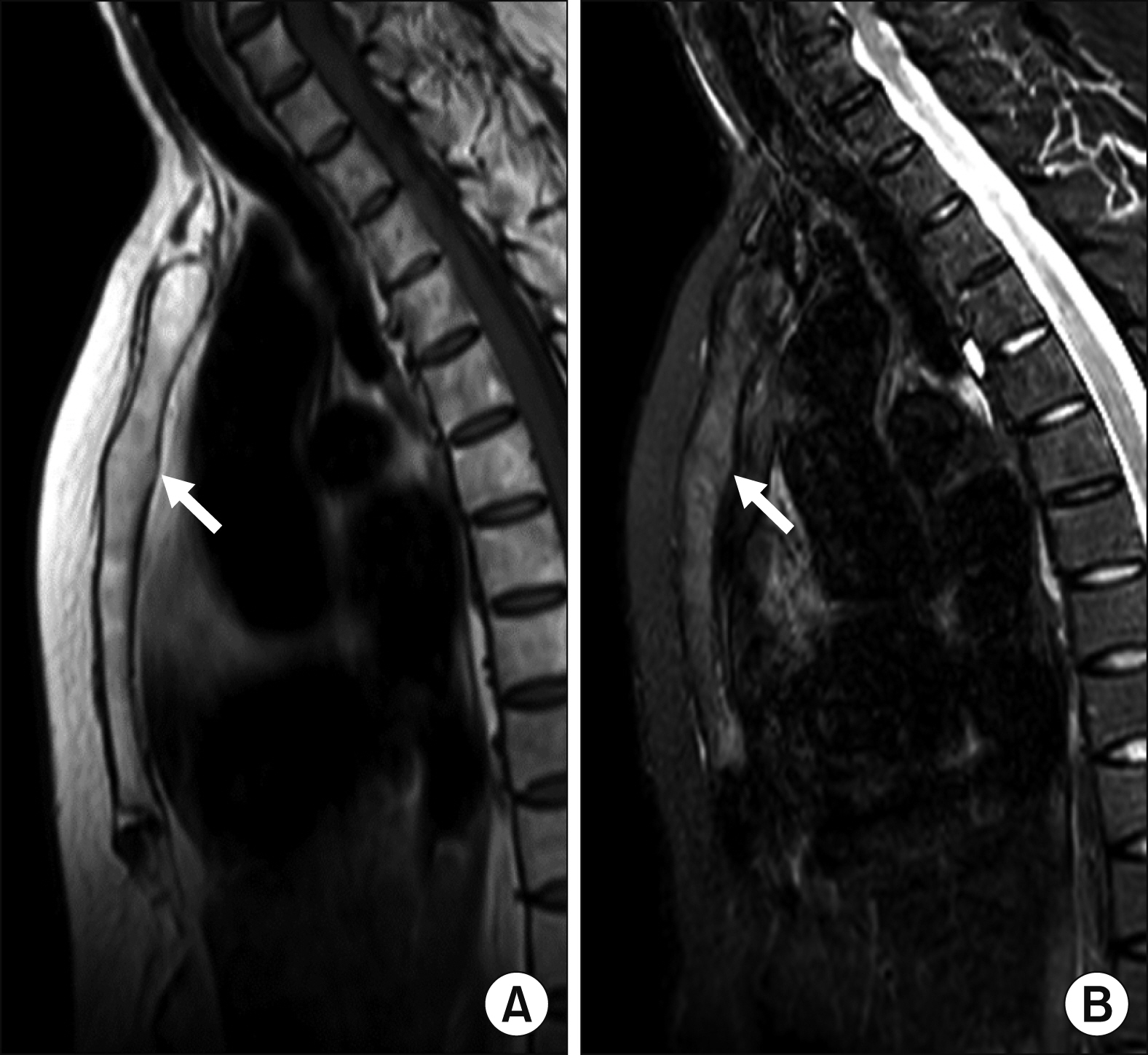
Figure 3 Sagittal T1-weighted (A) and T2-weighted STIR (B) images of a 52-year-old female AS patient, demonstrating ankylosis of the MSJ (arrows). The patient reported no ACW pain at the time of MRI. STIR: short tau inversion recovery, AS: ankylosing spondylotis, MSJ: manubriosternal joint, ACW: anterior chest wall, MRI: magnetic resonance imaging.
Reference
-
1. Kiltz U, Braun J. 2020; Assessments of functioning in patients with axial spondyloarthritis. J Rheum Dis. 27:22–9. DOI: 10.4078/jrd.2020.27.1.22.
Article2. Weber U, Lambert RG, Rufibach K, Maksymowych WP, Hodler J, Zejden A, et al. 2012; Anterior chest wall inflammation by whole-body magnetic resonance imaging in patients with spondyloarthritis: lack of association between clinical and imaging findings in a cross-sectional study. Arthritis Res Ther. 14:R3. DOI: 10.1186/ar3551. PMID: 22226453. PMCID: PMC3392789.
Article3. Elhai M, Paternotte S, Burki V, Durnez A, Fabreguet I, Koumakis E, et al. 2012; Clinical characteristics of anterior chest wall pain in spondyloarthritis: an analysis of 275 patients. Joint Bone Spine. 79:476–81. DOI: 10.1016/j.jbspin.2011.10.003. PMID: 22119315.
Article4. Wendling D, Prati C, Demattei C, Loeuille D, Richette P, Dougados M. 2013; Anterior chest wall pain in recent inflammatory back pain suggestive of spondyloarthritis. data from the DESIR cohort. J Rheumatol. 40:1148–52. DOI: 10.3899/jrheum.121460. PMID: 23678156.
Article5. Guglielmi G, Scalzo G, Cascavilla A, Salaffi F, Grassi W. 2008; Imaging of the seronegative anterior chest wall (ACW) syndromes. Clin Rheumatol. 27:815–21. DOI: 10.1007/s10067-008-0905-1. PMID: 18500440.
Article6. Jurik AG, Zejden A, Lambert RG, Rufibach K, Hodler J, Maksymowych WP, et al. 2013; Pitfalls in MR morphology of the sterno-costo-clavicular region using whole-body MRI. Clin Radiol. 68:785–91. DOI: 10.1016/j.crad.2013.02.007. PMID: 23561226.
Article7. Park EH, Yoon CH, Kang EH, Baek HJ. 2020; Utility of magnetic resonance imaging and positron emission tomography in rheumatic diseases. J Rheum Dis. 27:136–51. DOI: 10.4078/jrd.2020.27.3.136.
Article8. Mandl P, Navarro-Compán V, Terslev L, Aegerter P, van der Heijde D, D'Agostino MA, et al. European League Against Rheumatism (EULAR). 2015; EULAR recommendations for the use of imaging in the diagnosis and management of spondyloarthritis in clinical practice. Ann Rheum Dis. 74:1327–39. DOI: 10.1136/annrheumdis-2014-206971. PMID: 25837448.
Article9. Fournié B, Boutes A, Dromer C, Sixou L, Le Guennec P, Granel J, et al. 1997; Prospective study of anterior chest wall involvement in ankylosing spondylitis and psoriatic arthritis. Rev Rhum Engl Ed. 64:22–5. PMID: 9051856.10. Guglielmi G, Cascavilla A, Scalzo G, Salaffi F, Grassi W. 2009; Imaging of sternocostoclavicular joint in spondyloarthropaties and other rheumatic conditions. Clin Exp Rheumatol. 27:402–8. PMID: 19604431.11. Ramonda R, Lorenzin M, Lo Nigro A, Vio S, Zucchetta P, Frallonardo P, et al. 2012; Anterior chest wall involvement in early stages of spondyloarthritis: advanced diagnostic tools. J Rheumatol. 39:1844–9. DOI: 10.3899/jrheum.120107. PMID: 22798267.
Article12. Verhoeven F, Guillot X, Godfrin-Valnet M, Prati C, Wendling D. 2015; Ultrasonographic evaluation of the anterior chest wall in spondyloarthritis: a prospective and controlled study. J Rheumatol. 42:87–92. DOI: 10.3899/jrheum.140409. PMID: 25362653.
Article13. Verhoeven F, Sondag M, Chouk M, Prati C, Wendling D. 2020; Ultrasonographic involvement of the anterior chest wall in spondyloarthritis: factors associated with 5-years structural progression. A prospective study in 58 patients. Joint Bone Spine. 87:321–5. DOI: 10.1016/j.jbspin.2020.02.008. PMID: 32147567.
Article14. Krabbe S, Eshed I, Sørensen IJ, Jensen B, Møller JM, Balding L, et al. 2020; Whole-body magnetic resonance imaging inflammation in peripheral joints and entheses in axial spondyloarthritis: distribution and changes during adalimumab treatment. J Rheumatol. 47:50–8. DOI: 10.3899/jrheum.181159. PMID: 30936290.
Article15. Althoff CE, Sieper J, Song IH, Weiß A, Diekhoff T, Haibel H, et al. 2016; Comparison of clinical examination versus whole-body magnetic resonance imaging of enthesitis in patients with early axial spondyloarthritis during 3 years of continuous etanercept treatment. J Rheumatol. 43:618–24. DOI: 10.3899/jrheum.150659. PMID: 26834218.
Article
- Full Text Links
-
- Actions
-
Cited
- CITED
-
- Close
- Share
-
- Similar articles
-
- Clinical Manifestation and Diagnosis of Ankylosing Spondylitis
- MRI Features of Axial Spondyloarthritis and Differential Diagnosis: Focusing on the Spine and Sacroiliac Joint
- A Case of Muscular Sarcoidosis diagnosed by Gallium-67 Scintigraphy and Magnetic Resonance Imaging
- Reconstruction of Anterior Chest Wall Defect Induced by Sterile Costochondritis: A Case Report of Non-radiographic Axial Spondyloarthritis
- Piriformis syndrome as an overlooked cause of pain in a patient with axial spondyloarthritis: a case report
