A rare variation of the glossopharyngeal nerve
- Affiliations
-
- 1Department of Anatomy, Lake Erie College of Osteopathic Medicine, Erie, PA, USA
- KMID: 2516913
- DOI: http://doi.org/10.5115/acb.21.005
Abstract
- The glossopharyngeal nerve (CN IX) provides innervation to the parotid gland, carotid body/sinus, mucosa of the middle ear, tongue and oropharynx and the stylopharyngeus muscle. The vagus nerve provides innervation to the remaining skeletal muscle of the pharynx. CN IX contributes to the pharyngeal plexus and normally provides innervation to the mucosa of the oropharynx. Herein, we describe a previously undescribed variation of CN IX. CN IX was observed to enter the pharyngeal wall but instead of forming terminal branches in the tonsillar fossa, CN IX descended along the posterior wall between the mucosa and pharyngeal constrictors to the esophagus. This unusual branch of CN IX gave rise to numerous branches along the pharynx but did not intermingle with laryngeal branches from the vagus nerve. From this dissection, we developed innervation maps of the pharynx and propose a central miswiring mechanism for this unusual variation.
Keyword
Figure
-
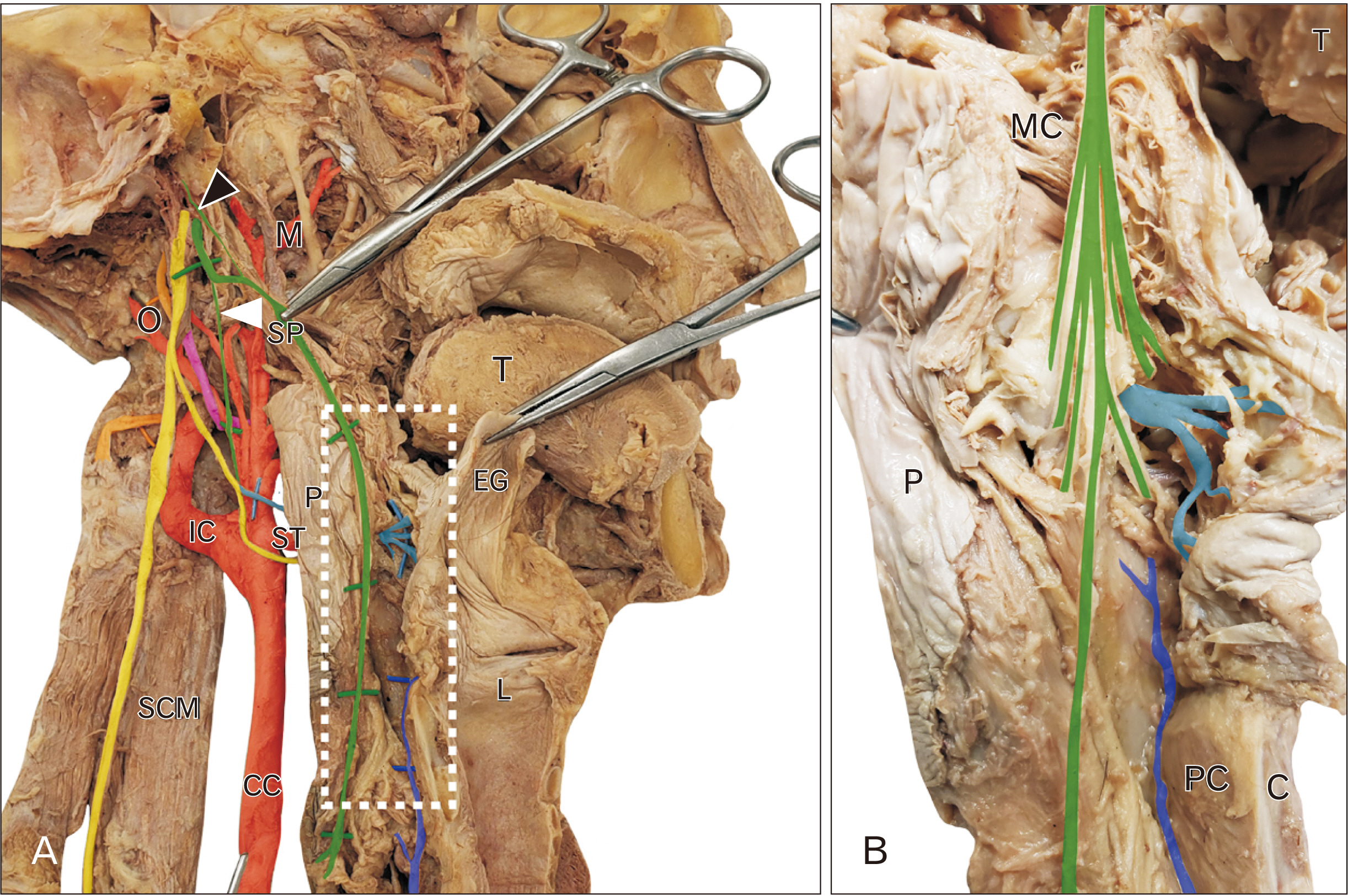
Fig. 1 Dissection of CN IX. (A) is the dissection of CN IX (green) and X (yellow). CN IX emerges from the skull and gives off a tympanic nerve (black arrowhead) and sinus nerve (solid arrowhead). CN IX sits on the stylopharyngeus muscle (SP) and enters the pharyngeal wall (P) deep to the middle constrictor muscle (MC). CN IX continues to descend along the posterior wall of the pharynx between the mucosa and muscle layers. The region indicated by the white rectangle is enlarged in (B) and demonstrates the numerous branches arising from CN IX in this specimen, distinct from the internal laryngeal nerve (cyan) and recurrent laryngeal nerve (blue). C, cricoid cartilage; CC, common carotid; EG, epiglottis; IC, internal carotid artery; L, larynx; L, lingual artery; M, maxillary artery; MC, middle constrictor; O, occipital artery; P, pharynx; PC, posterior cricoarytenoid; SCM, sternocleidomastoid; SP, stylopharyngeus; ST, superior thyroid artery; T, tongue.
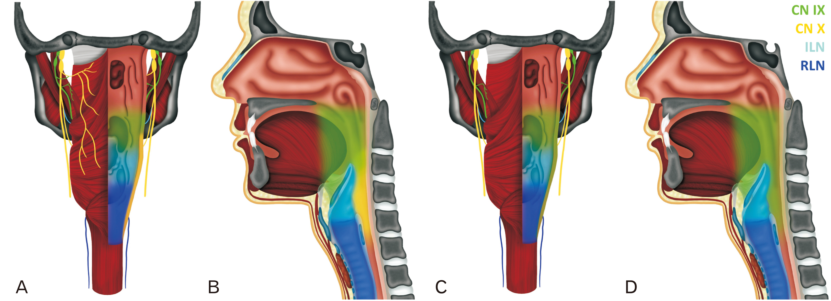
Fig. 2 Distribution of CN IX. Posterior (A) and midsagittal (B) show the normal distribution of CN IX (green) and X (yellow). Pharyngeal and tonsillar branches from CN IX supply the oropharynx and pharyngeal branches from CN X supply the hypopharynx. The ILN (cyan) and RLN (blue) provide innervation to the larynx. In the dissected specimen (C, D), CN IX appears to cover the entire posterior pharyngeal wall. ILN, internal laryngeal nerve; RLN, recurrent laryngeal nerve.
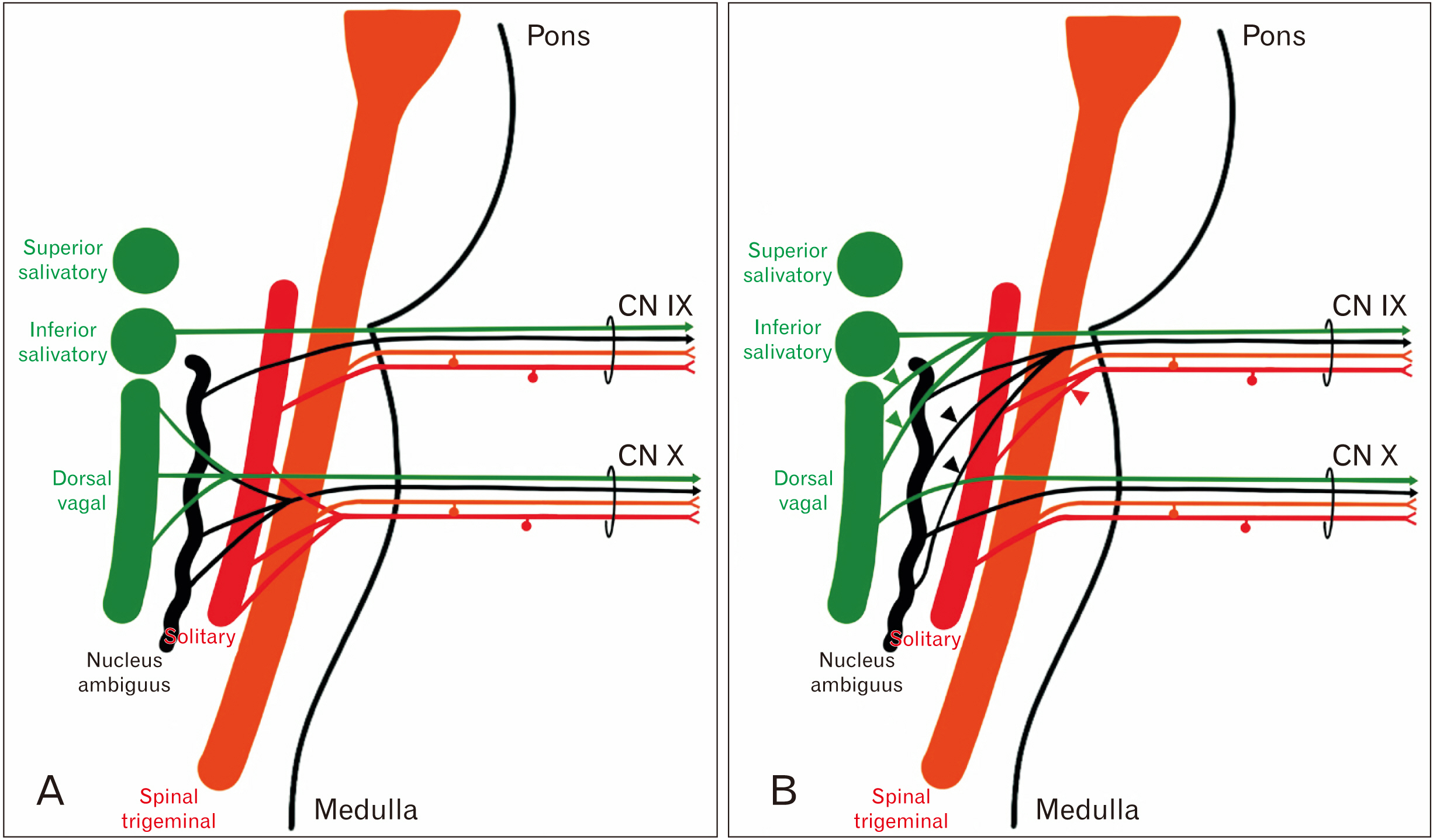
Fig. 3 Formation of CN IX and X. (A) A wiring diagram showing the brainstem nuclei associated with CN IX and X. (B) The proposed wiring pattern for CN IX in this subject. Since CN IX innervates the entire posterior pharyngeal wall, it takes a more extensive contribution from the ISN, NA and caudal SN. ISN, inferior salivatory nucleus; NA, nucleus ambiguus; SN, solitary nucleus.
Reference
-
References
1. Crosby EC, Humphrey T, Lauer EW. 1962. Correlative anatomy of the nervous system. Macmillan Co;New York:2. Standring S. 2016. Gray's anatomy: the anatomical basis of clinical practice. 41st ed. Elsevier;Philadelphia:3. Bergman RA, Thompson SA, Afifi AK. 1988. Compendium of human anatomic variation: text, atlas, and world literature. Urban & Schwarzenberg;Baltimore:4. Quain J, Schaefer EA, Thane GD. 1897. Quain's Elements of anatomy. 10th ed. Longmans, Green;London:5. Tubbs RS, Shoja MM, Loukas M. 2016. Bergman's comprehensive encyclopedia of human anatomic variation. Wiley Blackwell;Hoboken: DOI: 10.1002/9781118430309.6. Exner ID. 1884. Innervation des Kehlkopfes. Sitzungsber. d. Akad. d. Wissensch;Wien:7. Singh PM, Kaur M, Trikha A. 2013; An uncommonly common: glossopharyngeal neuralgia. Ann Indian Acad Neurol. 16:1–8. DOI: 10.4103/0972-2327.107662. PMID: 23661955. PMCID: PMC3644765.
Article
- Full Text Links
-
- Actions
-
Cited
- CITED
-
- Close
- Share
-
- Similar articles
-
- The Effect of Percutaneous Glossopharyngeal Nerve Block for Glossopharyngeal Neuralgia: A case report
- Glossopharyngeal Nerve Block for Idiopathic Glossopharyngeal Neuralgia: A case report
- A Case of Transient Glossopharyngeal and Hypoglossal Nerve Palsy after Laryngomicrosurgery
- Glossopharyngeal Neuralgia - A case report
- A Case of Herpes Zoster Oticus Involving Glossopharyngeal Nerve without Facial Nerve Palsy
