Application of three-dimensional printing for intraoperative guidance during liver resection of a hepatocellular carcinoma with sophisticated location
- Affiliations
-
- 1Department of Surgery, Samsung Medical Center, Sungkyunkwan University School of Medicine, Seoul, Korea
- KMID: 2516249
- DOI: http://doi.org/10.14701/ahbps.2021.25.2.265
Abstract
- While 3D printing is adapted usefully in certain field of surgery, its application in liver surgery was limited. Here, we introduce our experience for using 3D printing for intraoperative guidance during liver resection in a case for HCC with an intrahepatic metastasis at a sophisticated location. A 50 years old male patient was diagnosed 4.7 cm-sized hepatocellular carcinoma located on segment 3 with and an intrahepatic metastasis located on segment 8 which was between right anterior portal vein, middle hepatic vein and right hepatic vein. Since radiofrequency ablation appeared to be inappropriate, surgical resection was planned. However, the patient had a cirrhotic liver and left liver was estimated to be 47% according to volume measurement. Therefore, we planned a two-step procedure by performing left hemihepatectomy preserving the middle hepatic vein and additionally removing the intrahepatic metastasis by tumorectomy. For better guidance, we made a 3D printed model tailored for using it as a guidance during operation, and the accuracy of 3D-printed model helped the surgical team perform a safe operation. The patient underwent adjuvant proton beam therapy on the site of tumorectomy and did not experience recurrence.
Keyword
Figure
-

Fig. 1 Preoperative 3D modeling of the liver. (A) A 4.7 cm-sized hepatocellular carcinoma (arrowhead) was located on segment 3 and intrahepatic metastasis (arrow) located between the right anterior portal vein, middle hepatic vein and right hepatic vein. Preoperative magnetic resonance imaging shows (B) the main mass on segment 3 and (C) segment 8 right above right anterior portal vein.
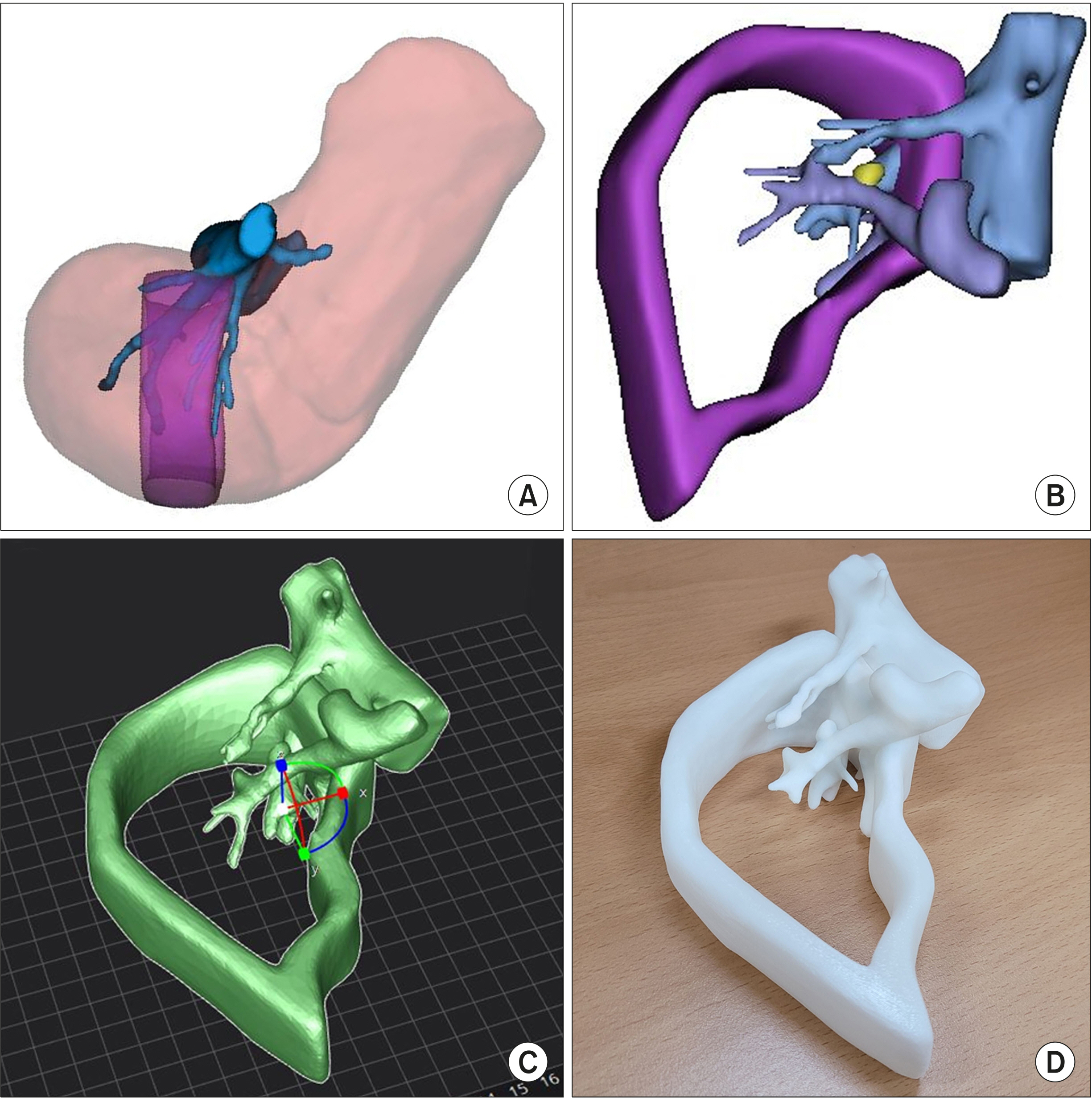
Fig. 2 Three-dimensional print-ing of the model. (A) The re-gion of interest was set between the middle hepatic vein and 3 cm right side to the midline. (B) The liver parenchyma was removed to allow visualization of the inner vascular structure and intrahepatic metastasis. (C) The 3D model was edited (D) and printed using a 3D printer.

Fig. 3 Intraoperative finding during surgery. After left hemi-hepatectomy, (A) the 3D printed model was compared directly side by side as a guide. (B) After dissecting the liver parenchyma between the right anterior glisso-nean pedicle and middle hepa-tic vein, intrahepatic metastasis (arrow) was identified. (C) The 3D model was compared to the actual field with the intrahepa-tic metastasis exposed. (D) The intrahepatic metastasis was re-moved and (E) the main mass was removed with the left hemi-liver with a 3 cm margin.
Reference
-
1. Ercolani G, Grazi GL, Ravaioli M, Del Gaudio M, Gardini A, Cescon M, et al. 2003; Liver resection for hepatocellular carcinoma on cirrhosis: univariate and multivariate analysis of risk factors for intrahepatic recurrence. Ann Surg. 237:536–543. DOI: 10.1097/01.SLA.0000059988.22416.F2. PMID: 12677151. PMCID: PMC1514472.2. Fong Y, Sun RL, Jarnagin W, Blumgart LH. 1999; An analysis of 412 cases of hepatocellular carcinoma at a Western center. Ann Surg. 229:790–799. discussion 799–800. DOI: 10.1097/00000658-199906000-00005. PMID: 10363892. PMCID: PMC1420825.
Article3. Rustgi VK. 1987; Epidemiology of hepatocellular carcinoma. Gastroenterol Clin North Am. 16:545–551. DOI: 10.1016/S0889-8553(21)00328-9.
Article4. Grazi GL, Ercolani G, Pierangeli F, Del Gaudio M, Cescon M, Cavallari A, et al. 2001; Improved results of liver resection for hepatocellular carcinoma on cirrhosis give the procedure added value. Ann Surg. 234:71–78. DOI: 10.1097/00000658-200107000-00011. PMID: 11420485. PMCID: PMC1421950.
Article5. Regimbeau JM, Kianmanesh R, Farges O, Dondero F, Sauvanet A, Belghiti J. 2002; Extent of liver resection influences the outcome in patients with cirrhosis and small hepatocellular carcinoma. Surgery. 131:311–317. DOI: 10.1067/msy.2002.121892. PMID: 11894036.
Article6. Rhu J, Kim SJ, Choi GS, Kim JM, Joh JW, Kwon CHD. 2018; Laparoscopic versus open right posterior sectionectomy for hepatocellular carcinoma in a high-volume center: a propensity score matched analysis. World J Surg. 42:2930–2937. DOI: 10.1007/s00268-018-4531-z. PMID: 29426971.
Article7. Louvrier A, Marty P, Barrabé A, Euvrard E, Chatelain B, Weber E, et al. 2017; How useful is 3D printing in maxillofacial surgery? J Stomatol Oral Maxillofac Surg. 118:206–212. DOI: 10.1016/j.jormas.2017.07.002. PMID: 28732777.
Article8. Witowski JS, Coles-Black J, Zuzak TZ, Pędziwiatr M, Chuen J, Major P, et al. 2017; 3D printing in liver surgery: a systematic review. Telemed J E Health. 23:943–947. DOI: 10.1089/tmj.2017.0049. PMID: 28530492.
Article9. Kuroda S, Kobayashi T, Ohdan H. 2017; 3D printing model of the intrahepatic vessels for navigation during anatomical resection of hepatocellular carcinoma. Int J Surg Case Rep. 41:219–222. DOI: 10.1016/j.ijscr.2017.10.015. PMID: 29096348. PMCID: PMC5683889.
Article
- Full Text Links
-
- Actions
-
Cited
- CITED
-
- Close
- Share
-
- Similar articles
-
- Virtual Surgical Planning and Three-Dimensional Printing for Reconstruction of Mandibular Defect by Fibula Free Flap: A Case Report
- Comparative analysis of intraoperative radiofrequency ablation versus non-anatomical hepatic resection for small hepatocellular carcinoma: short-term result
- Application of Three-Dimensional Printing in the Fracture Management
- Clinical Application of Three-Dimensional Printing Technology in Craniofacial Plastic Surgery
- A Review of Current Clinical Applications of Three-Dimensional Printing in Spine Surgery
