Nonsurgical maxillary expansion in a 60-year-old patient with gingival recession and crowding
- Affiliations
-
- 1Department of Orthodontics, Institute of Craniofacial Deformity, Yonsei University College of Dentistry, Seoul, Korea
- 2Postgraduate Orthodontic Program, Arizona School of Dentistry & Oral Health, A. T. Still University, Mesa, AZ, USA
- KMID: 2515824
- DOI: http://doi.org/10.4041/kjod.2021.51.3.217
Abstract
- Maxillary transverse deficiency often manifests as a posterior crossbite or edgeto-edge bite and anterior crowding. However, arbitrary arch expansion in mature patients has been considered to be challenging due to the possible periodontal adverse effects such as alveolar bone dehiscence and gingival recession. To overcome these limitations, nonsurgical maxillary expansion of the basal bone has been demonstrated in young adults. However, the age range for successful orthopedic expansion has remained a topic of debate, possibly due to the underlying individual variations in suture maturity. This case report illustrates nonsurgical, miniscrew-assisted rapid palatal expansion (MARPE) in a 60-yearold patient with maxillary transverse deficiency accompanied by anterior and posterior crossbites, crowding, and gingival recession. The use of MARPE allowed relief of crowding and correction of the crossbite without causing significant periodontal adverse effects.
Keyword
Figure
-
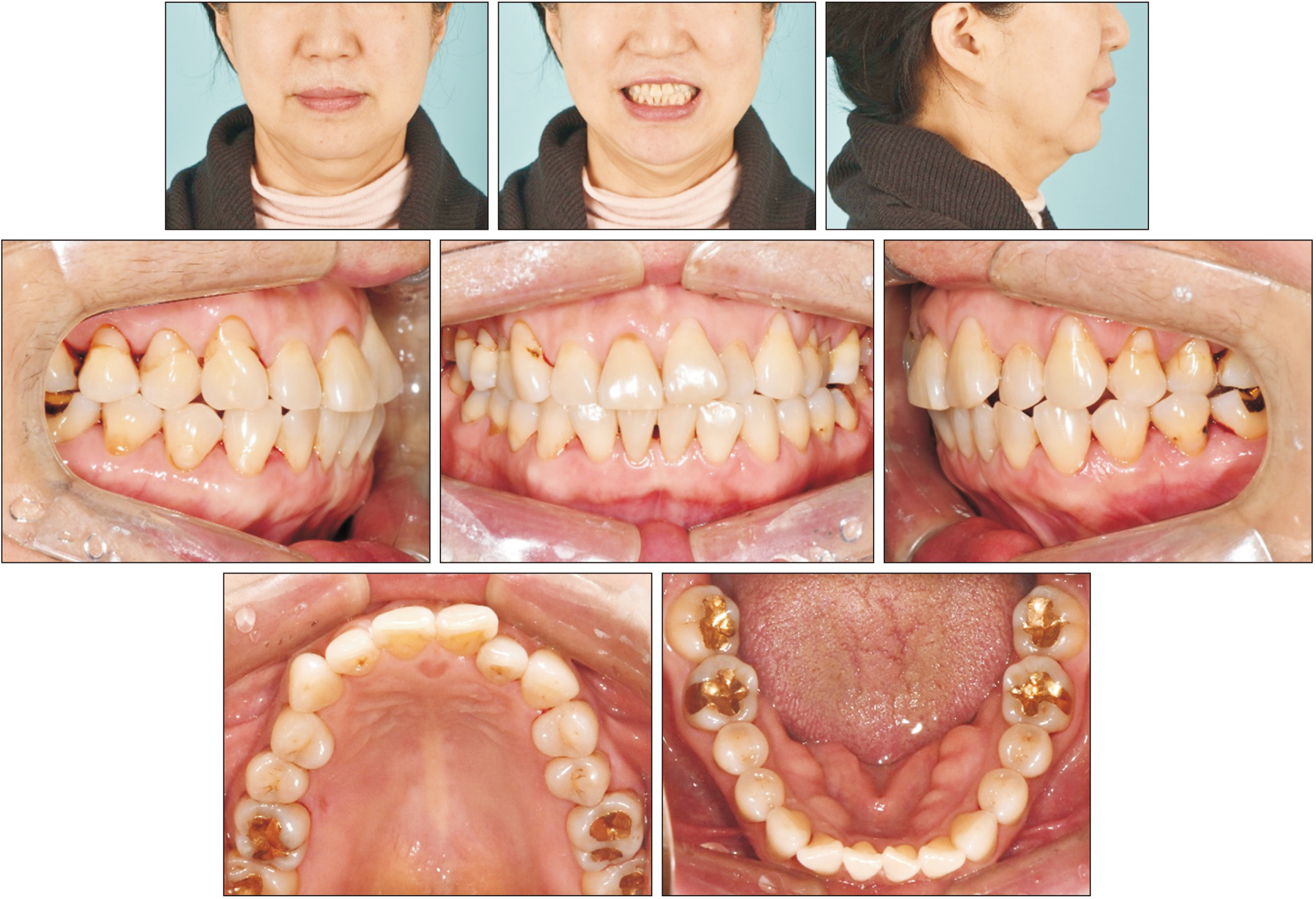
Figure 1 Pretreatment facial and intraoral photographs.

Figure 2 Pretreatment radiographs and cephalometric tracing.
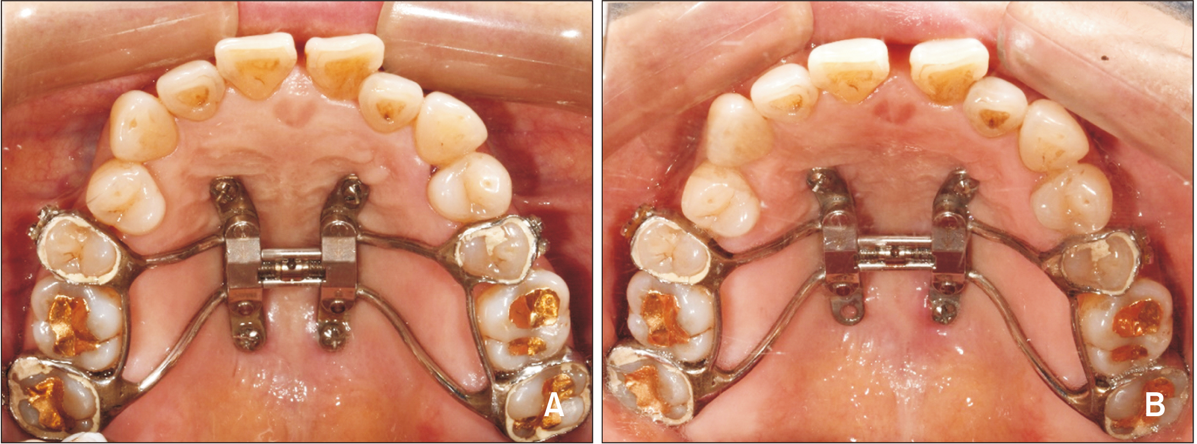
Figure 3 Treatment progress using the miniscrew-assisted rapid palatal expansion appliance. A, Opening of diastema after 4 weeks of activation (0.2 mm/day). B, Post-expansion.

Figure 4 Periapical radiographs. A, Pre-expansion. B, Post-expansion. C, At the end of active treatment.

Figure 5 Final facial and intraoral photographs.

Figure 6 Cephalometric superimposition.
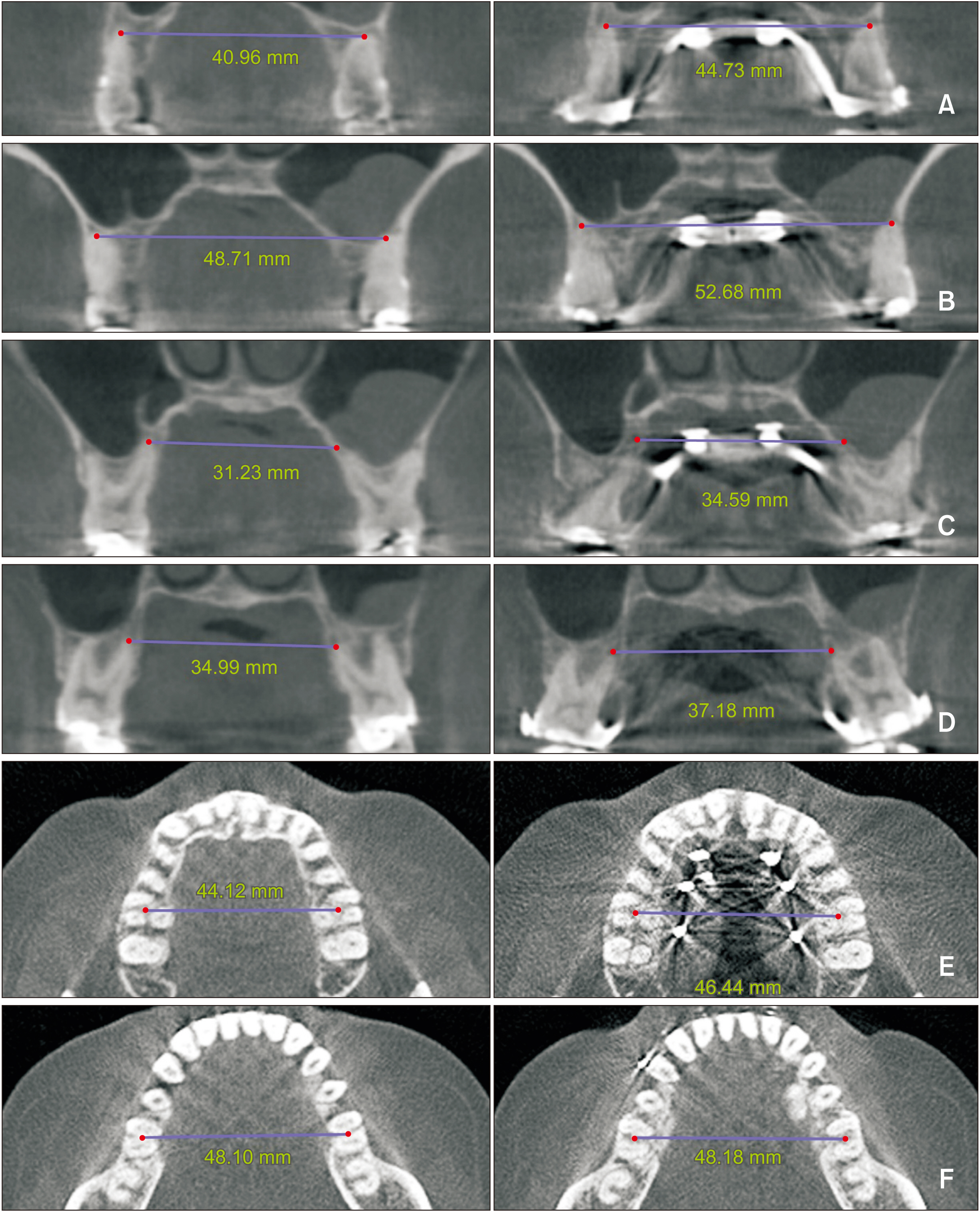
Figure 7 Interapex width before and after palatal expansion measured between the maxillary first premolars (A); second premolars (B); first molars (C); second molars (D); and Yonsei Transverse Index measured before and after palatal expansion in the maxilla (E) and mandible (F).

Figure 8 Comparison of cone-beam computed tomography (CBCT) images before and after palatal expansion. A, Superimposition of pre- and post-expansion CBCT scans. B, Axial palatal section before expansion. C, Axial view showing suture opening after expansion.
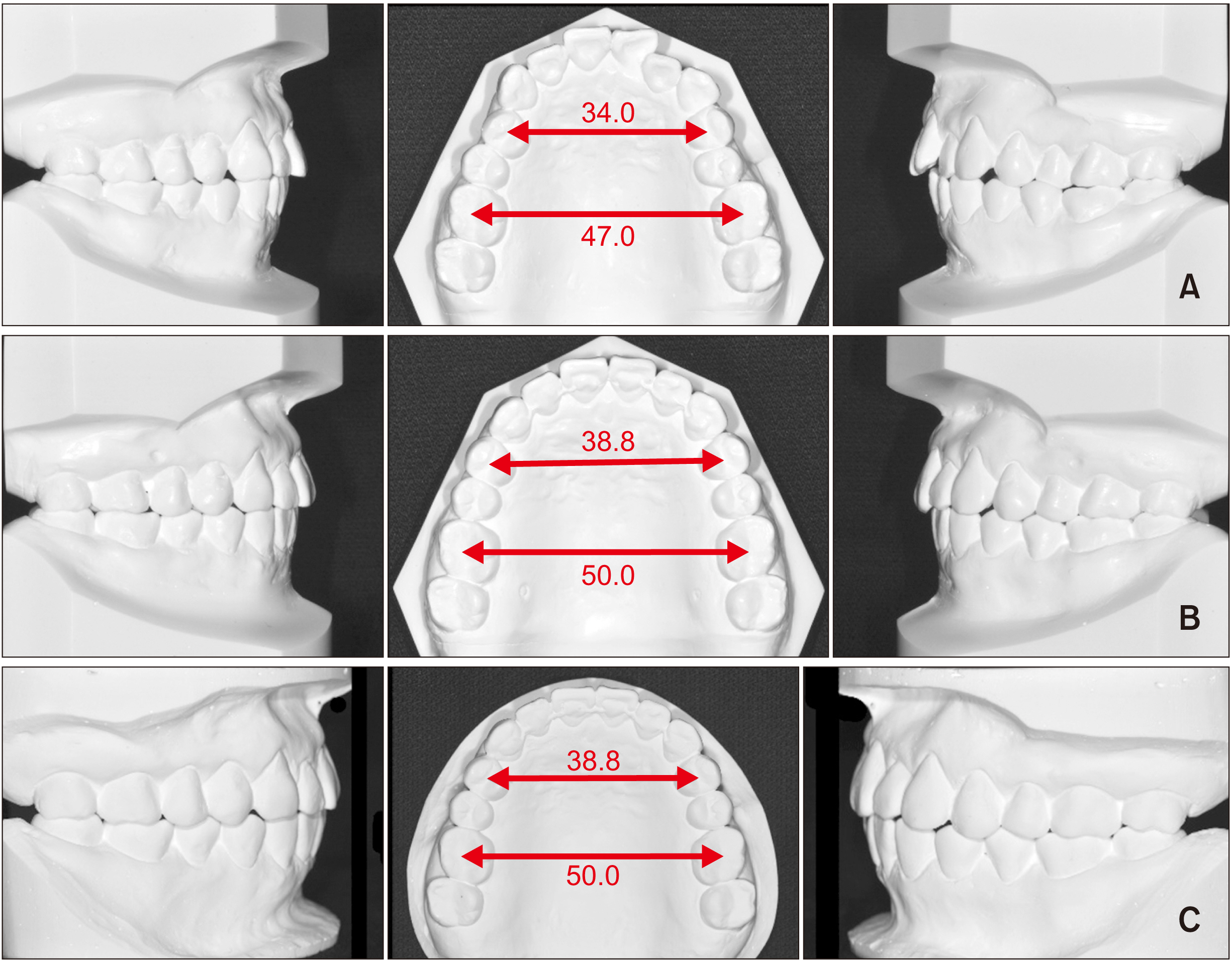
Figure 9 Comparison of pretreatment (A), posttreatment (B), and follow-up (C) dental casts.
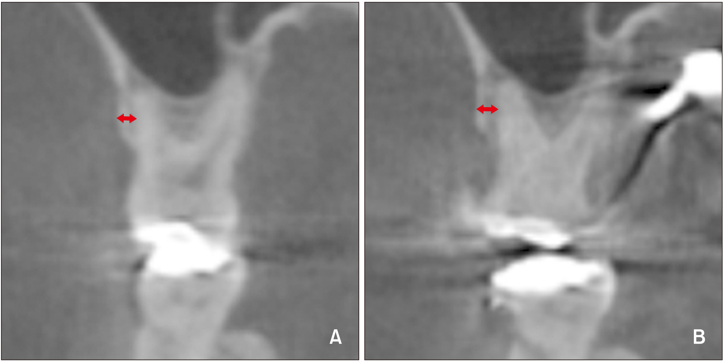
Figure 10 Buccal bone plate before (A) and after (B) palatal expansion.
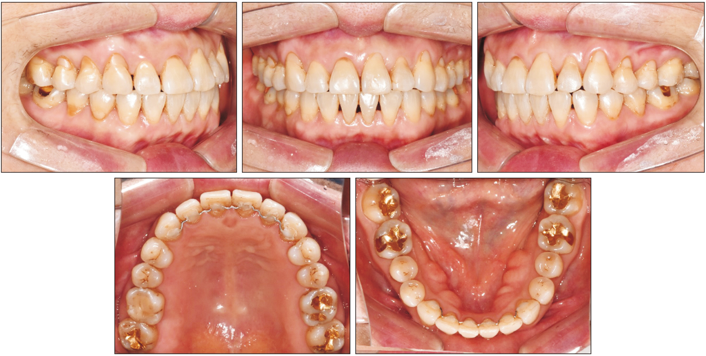
Figure 11 Intraoral photographs taken at the 6-month follow-up.
Reference
-
1. Atik E, Taner T. 2017; Stability comparison of two different dentoalveolar expansion treatment protocols. Dental Press J Orthod. 22:75–82. DOI: 10.1590/2177-6709.22.5.075-082.oar. PMID: 29160347. PMCID: PMC5730139.
Article2. Papapanou PN, Wennström JL, Gröndahl K. 1988; Periodontal status in relation to age and tooth type. A cross-sectional radiographic study. J Clin Periodontol. 15:469–78. DOI: 10.1111/j.1600-051X.1988.tb01602.x. PMID: 3263400.
Article3. Van der Velden U. 1984; Effect of age on the periodontium. J Clin Periodontol. 11:281–94. DOI: 10.1111/j.1600-051X.1984.tb01325.x. PMID: 6371061.
Article4. Angell EH. White JD, McQuillen JH, Ziegler GJ, White JW, Kirk EC, Anthony LP, editors. 1860. Treatment of irregularities of the permanent or adult teeth. The dental cosmos. S.S. White Dental Manufacturing Company;Philadelphia: p. 540–4. p. 99–600.5. Haas AJ. 1965; The treatment of maxillary deficiency by opening the midpalatal suture. Angle Orthod. 35:200–17. DOI: 10.1043/0003-3219(1965)035<0200:TTOMDB>2.0.CO;2. PMID: 14331020.6. Choi SH, Shi KK, Cha JY, Park YC, Lee KJ. 2016; Nonsurgical miniscrew-assisted rapid maxillary expansion results in acceptable stability in young adults. Angle Orthod. 86:713–20. DOI: 10.2319/101415-689.1. PMID: 26938955.
Article7. Lee KJ, Park YC, Park JY, Hwang WS. 2010; Miniscrew-assisted nonsurgical palatal expansion before orthognathic surgery for a patient with severe mandibular prognathism. Am J Orthod Dentofacial Orthop. 137:830–9. DOI: 10.1016/j.ajodo.2007.10.065. PMID: 20685540.
Article8. Persson M, Thilander B. 1977; Palatal suture closure in man from 15 to 35 years of age. Am J Orthod. 72:42–52. DOI: 10.1016/0002-9416(77)90123-3. PMID: 267435.
Article9. Melsen B. 1975; Palatal growth studied on human autopsy material. A histologic microradiographic study. Am J Orthod. 68:42–54. DOI: 10.1016/0002-9416(75)90158-X. PMID: 1056143.10. Shetty V, Caridad JM, Caputo AA, Chaconas SJ. 1994; Biomechanical rationale for surgical-orthodontic expansion of the adult maxilla. J Oral Maxillofac Surg. 52:742–9. discussion 750–1. DOI: 10.1016/0278-2391(94)90492-8. PMID: 8006740.
Article11. Asscherickx K, Govaerts E, Aerts J, Vande Vannet B. 2016; Maxillary changes with bone-borne surgically assisted rapid palatal expansion: a prospective study. Am J Orthod Dentofacial Orthop. 149:374–83. DOI: 10.1016/j.ajodo.2015.08.018. PMID: 26926025.
Article12. Kokich VG. 1976; Age changes in the human frontozygomatic suture from 20 to 95 years. Am J Orthod. 69:411–30. DOI: 10.1016/0002-9416(76)90209-8. PMID: 1062937.
Article13. Uysal T, Memili B, Usumez S, Sari Z. 2005; Dental and alveolar arch widths in normal occlusion, class II division 1 and class II division 2. Angle Orthod. 75:941–7. DOI: 10.1043/0003-3219(2005)75[941:DAAAWI]2.0.CO;2. PMID: 16448235.14. Koo YJ, Choi SH, Keum BT, Yu HS, Hwang CJ, Melsen B, et al. 2017; Maxillomandibular arch width differences at estimated centers of resistance: comparison between normal occlusion and skeletal Class III malocclusion. Korean J Orthod. 47:167–75. DOI: 10.4041/kjod.2017.47.3.167. PMID: 28523243. PMCID: PMC5432438.
Article15. Jang W, Shin C, Hwang S, Kim KH, Jackson T, Nguyen T, et al. 2020; Nonsurgical treatment of an adult with a skeletal Class III malocclusion combined with a functional anterior shift, severely overclosed vertical dimension, and a reverse smile. Am J Orthod Dentofacial Orthop. 157:561–70. DOI: 10.1016/j.ajodo.2019.01.029. PMID: 32241363.
Article16. Choi NC, Park YC, Lee HA, Lee KJ. 2007; Treatment of Class II protrusion with severe crowding using indirect miniscrew anchorage. Angle Orthod. 77:1109–18. DOI: 10.2319/112106-476.1. PMID: 18004931.
Article17. Bechtold TE, Kim JW, Choi TH, Park YC, Lee KJ. 2013; Distalization pattern of the maxillary arch depending on the number of orthodontic miniscrews. Angle Orthod. 83:266–73. DOI: 10.2319/032212-123.1. PMID: 22970751.
Article18. Kraus CD, Campbell PM, Spears R, Taylor RW, Buschang PH. 2014; Bony adaptation after expansion with light-to-moderate continuous forces. Am J Orthod Dentofacial Orthop. 145:655–66. DOI: 10.1016/j.ajodo.2014.01.017. PMID: 24785930.
Article19. Seong EH, Choi SH, Kim HJ, Yu HS, Park YC, Lee KJ. 2018; Evaluation of the effects of miniscrew incorporation in palatal expanders for young adults using finite element analysis. Korean J Orthod. 48:81–9. DOI: 10.4041/kjod.2018.48.2.81. PMID: 29564217. PMCID: PMC5854885.
Article20. Wehrbein H, Merz BR, Diedrich P. 1999; Palatal bone support for orthodontic implant anchorage--a clinical and radiological study. Eur J Orthod. 21:65–70. DOI: 10.1093/ejo/21.1.65. PMID: 10191579.21. Kang S, Lee SJ, Ahn SJ, Heo MS, Kim TW. 2007; Bone thickness of the palate for orthodontic mini-implant anchorage in adults. Am J Orthod Dentofacial Orthop. 131(4 Suppl):S74–81. DOI: 10.1016/j.ajodo.2005.09.029. PMID: 17448390.
Article22. McNamara JA. 2000; Maxillary transverse deficiency. Am J Orthod Dentofacial Orthop. 117:567–70. DOI: 10.1016/S0889-5406(00)70202-2. PMID: 18249297.23. Nascimento MM, Dilbone DA, Pereira PN, Duarte WR, Geraldeli S, Delgado AJ. 2016; Abfraction lesions: etiology, diagnosis, and treatment options. Clin Cosmet Investig Dent. 8:79–87. DOI: 10.2147/CCIDE.S63465. PMID: 27217799. PMCID: PMC4861607.
Article24. Moon W. Park JH, editor. 2020. Maxillary expansion in skeletally mature patients with TADs. Temporary anchorage devices in clinical orthodontics. John Wiley & Sons, Inc.;Hoboken: DOI: 10.1002/9781119513636.ch24.
Article25. Proffit WR, Fields HW, Sarver DM. 2014. Contemporary orthodontics. Elsevier Health Sciences;St. Louis:26. Cohen MM Jr. 1993; Sutural biology and the correlates of craniosynostosis. Am J Med Genet. 47:581–616. DOI: 10.1002/ajmg.1320470507. PMID: 8266985.
Article27. Opperman LA, Sweeney TM, Redmon J, Persing JA, Ogle RC. 1993; Tissue interactions with underlying dura mater inhibit osseous obliteration of developing cranial sutures. Dev Dyn. 198:312–22. DOI: 10.1002/aja.1001980408. PMID: 8130378.
Article28. Wehrbein H, Yildizhan F. 2001; The mid-palatal suture in young adults. A radiological-histological investigation. Eur J Orthod. 23:105–14. DOI: 10.1093/ejo/23.2.105. PMID: 11398548.
Article29. Castello JR, Olaso AS, Chao JJ, McCarthy JG, Molina F. 2000; Craniofacial shortening by contraction osteogenesis: an experimental model. Plast Reconstr Surg. 105:617–25. discussion 626–7. DOI: 10.1097/00006534-200002000-00021. PMID: 10697169.
Article30. Garib DG, Henriques JF, Janson G, de Freitas MR, Fernandes AY. 2006; Periodontal effects of rapid maxillary expansion with tooth-tissue-borne and tooth-borne expanders: a computed tomography evaluation. Am J Orthod Dentofacial Orthop. 129:749–58. DOI: 10.1016/j.ajodo.2006.02.021. PMID: 16769493.
Article31. Reitan K. 1959; Tissue rearrangement during retention of orthodontically rotated teeth. Angle Orthod. 29:105–13.32. Kassab MM, Cohen RE. 2003; The etiology and prevalence of gingival recession. J Am Dent Assoc. 134:220–5. DOI: 10.14219/jada.archive.2003.0137. PMID: 12636127.
Article
- Full Text Links
-
- Actions
-
Cited
- CITED
-
- Close
- Share
-
- Similar articles
-
- Crowding with no posterior crossbite treatment byrapid palatal expansion
- Orthodontic treatment for maxillary anterior pathologic tooth migration by periodontitis using clear aligner
- Clinical study of gingival recession and dentine hypersensitivity
- Periodontal regenerative treatment with connective tissue grafts in deep intrabony defect and gingival recession in the maxillary canine: a case report with 3-year follow-up
- Long-term stability after multidisciplinary treatment involving maxillary distraction osteogenesis, and sagittal split ramus osteotomy for unilateral cleft lip and palate with severe occlusal collapse and gingival recession: A case report
