Korean Circ J.
2021 Apr;51(4):379-381. 10.4070/kcj.2020.0529.
Ortner's Syndrome Discovered by a Routine Echocardiographic Examination: a Huge Aneurysmal Dilatation of the Aortic Arch as a Cause of Hoarseness
- Affiliations
-
- 1Division of Cardiology, Department of Internal Medicine, Chungnam National University Hospital, Chungnam National University College of Medicine, Daejeon, Korea
- KMID: 2514644
- DOI: http://doi.org/10.4070/kcj.2020.0529
Figure
-

Figure 1 Chest X-ray shows an abnormally round shadow above the aortic knob (A, arrows) and rightward deviation of the trachea. Echocardiographic examination reveals about 55×42 mm sized oval-shaped mass lesion was noted on the aortic arch filled with an echogenic lesion suggesting thrombus (B, arrow heads). Illustration shows aneurysmal dilatation on the aortic arch with compression of the left recurrent laryngeal nerve (C).Asc Ao = ascending aorta; LSA = left subclavian artery; Dsc Ao = descending aorta.
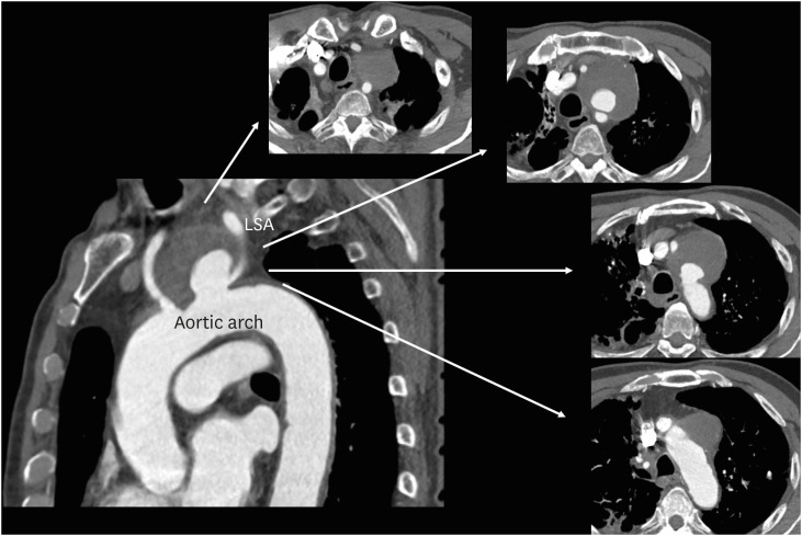
Figure 2 Contrast-enahced computerized tomography demonstrates about 65×60 mm sized large saccular aneurysm on the aortic arch filled with mural thrombus near the origin of left subclavian artery.LSA = left subclavian artery.
Reference
-
1. Kheok SW, Salkade PR, Bangaragiri A, Koh NS, Chen RC. Cardiovascular hoarseness (Ortner's syndrome): a pictorial review. Curr Probl Diagn Radiol. 2020; S0363-0188(20)30190-0.2. Rommens KL, Estrera AL. Contemporary management of aortic arch aneurysm. Semin Thorac Cardiovasc Surg. 2019; 31:697–702. PMID: 30980932.3. Choi BK, Lee HC, Lee HW, et al. Successful treatment of a ruptured aortic arch aneurysm using a hybrid procedure. Korean Circ J. 2011; 41:469–473. PMID: 21949532.
- Full Text Links
-
- Actions
-
Cited
- CITED
-
- Close
- Share
-
- Similar articles
-
- A case of thoracic aortic aneurysm presenting as hoarseness of voice: ortner's syndrome
- Reversible Ortner’s Syndrome as a Presenting Feature of Thyrotoxicosis in an Adolescent: A Rare Case Report
- Ortner Syndrome due to Concomitant Mitral Stenosis and Bronchiectasis
- Comparison of the Mid-term Changes at the Remnant Distal Aorta after Aortic Arch Replacement or Ascending Aortic Replacement for Treating Type A Aortic Dissection
- Total Arch Replacement for Chronic Aortic Aneurysmal Dissection Patient with Aberrant Subclavian Artery
