Korean Circ J.
2021 Apr;51(4):376-378. 10.4070/kcj.2020.0516.
A Case of Aneurysm Occurring at the Dissection Site after Intervention with Drug-Coated Balloon
- Affiliations
-
- 1Department of Cardiology, Ulsan Medical Center, Ulsan, Korea
- 2Department of Cardiology, Dong-A University Hospital, Busan, Korea
- 3Department of Cardiology, Ulsan University Hospital, University of Ulsan College of Medicine, Ulsan, Korea
- 4Department of Cardiology, East Lancashire Hospitals NHS Trust, Blackburn, Lancashire, UK
- KMID: 2514643
- DOI: http://doi.org/10.4070/kcj.2020.0516
Figure
-
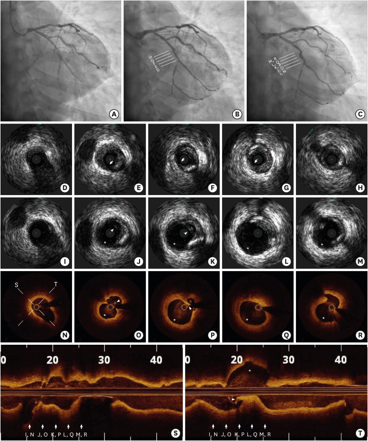
Figure 1 (A) Pre-procedure; (B) Post-DCB treatment; (C) Follow-up angiographic images. In the IVUS image immediately after DCB treatment, plaque can be seen separated from the subintimal space by dissection (arrows). In the follow-up IVUS and OCT, the aneurysm is shown (asterisks) at the site of a plaque dissection (arrows) and cavities with dissection flaps in the plaque that were not observed immediately after DCB is observed (arrowheads). Longitudinal OCT views (S and T) demonstrate the extent and direction of the aneurysm and ruptured plaque.DCB = drug-coated balloon; IVUS = intravascular ultrasound; OCT = optical coherence tomography.
Reference
-
1. Bal ET, Thijs Plokker HW, van den Berg EM, et al. Predictability and prognosis of PTCA-induced coronary artery aneurysms. Cathet Cardiovasc Diagn. 1991; 22:85–88. PMID: 2009568.2. Vassanelli C, Turri M, Morando G, Menegatti G, Zardini P. Coronary arterial aneurysms after percutaneous transluminal coronary angioplasty--a not uncommon finding at elective follow-up angiography. Int J Cardiol. 1989; 22:151–156. PMID: 2521614.
- Full Text Links
-
- Actions
-
Cited
- CITED
-
- Close
- Share
-
- Similar articles
-
- No-Reflow Phenomenon During Treatment of Coronary In-Stent Restenosis With a Paclitaxel-Coated Balloon Catheter
- Impact of Dissection after Drug-Coated Balloon Treatment of De Novo Coronary Lesions: Angiographic and Clinical Outcomes
- Acute Type B Aortic Dissection in a Patient with Previous Endovascular Abdominal Aortic Aneurysm Repair
- A Case of Coarctation of the Aorta Treated with Balloon Angioplasty
- Treatment of Stent Dislodgement Complicated by Coronary Artery Dissection using Parallel Wire Technique and Small Balloon
