Prediction of Post-resection Prognosis Using the ADV Score for Huge Hepatocellular Carcinomas ≥13 cm
- Affiliations
-
- 1Department of Surgery, Asan Medical Center, University of Ulsan College of Medicine, Seoul, Korea
- KMID: 2514234
- DOI: http://doi.org/10.17998/jlc.21.1.45
Abstract
- Background/Aims
Multiplication of α-fetoprotein, des-γ-carboxy prothrombin, and tumor volume (ADV score) is a surrogate marker for post-resection prognosis of hepatocellular carcinoma (HCC). This study aimed to validate the predictive power of the ADV score-based prognostic prediction model for patients with solitary huge HCC.
Methods
Of 3,018 patients, 100 patients who underwent hepatic resection for solitary HCC ≥13 cm between 2008 and 2012 were selected.
Results
The median tumor diameter and tumor volume were 15.0 cm and 886 mL, respectively. Tumor recurrence and overall survival (OS) rates were 70.7% and 66.0% at one year and 84.9% and 34.0% at five years, respectively. Microvascular invasion (MVI) was the only independent risk factor for disease-free survival (DFS) and OS. DFS and OS, stratified by ADV score with 1-log intervals, showed significant prognostic contrasts (P=0.007 and P=0.017, respectively). DFS and OS, stratified by ADV score with a cut-off of 8-log, showed significant prognostic contrasts (P=0.014 and P=0.042, respectively). The combination of MVI and ADV score with a cut-off of 8-log also showed significant prognostic contrasts in DFS (P<0.001) and OS (P=0.001) considering the number of risk factors. Prognostic contrast was enhanced after combining the MVI and ADV score.
Conclusions
The prognostic prediction model with the ADV score could reliably predict the risk of tumor recurrence and long-term patient survival outcomes in patients with solitary huge HCC ≥13 cm. The results of this study suggest that our prognostic prediction models can be used to guide surgical treatment and post-resection follow-up for patients with huge HCCs.
Figure
-

Figure 1 Comparison of preoperative computed tomography (CT) scan, resected right liver specimen, and CT scan taken 3 months after hepatic resection in patients who underwent right hepatectomy. The maximal tumor diameters were 13 cm (A), 17 cm (B), and 20 cm (C).
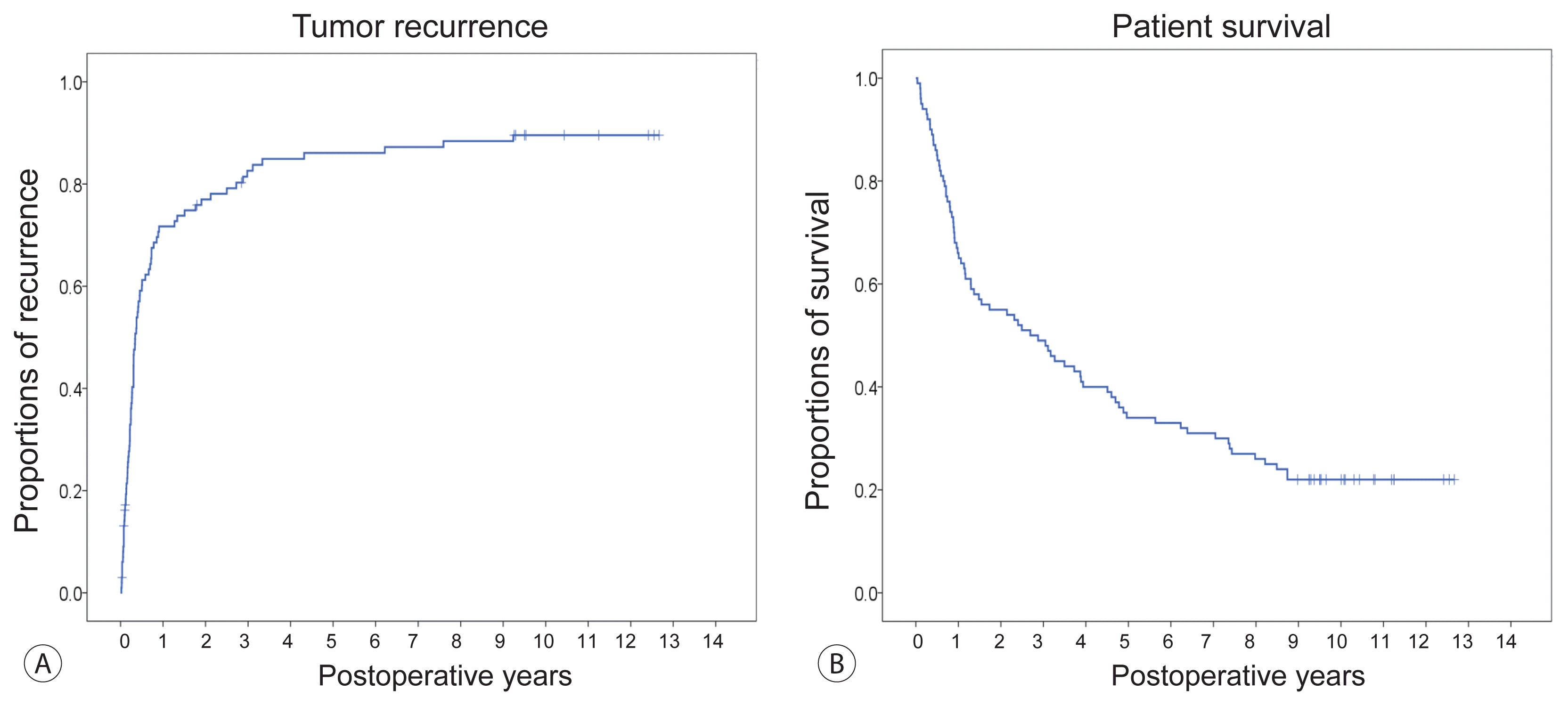
Figure 2 Kaplan-Meier estimation of post-resection tumor recurrence (A) and overall patient survival (B).
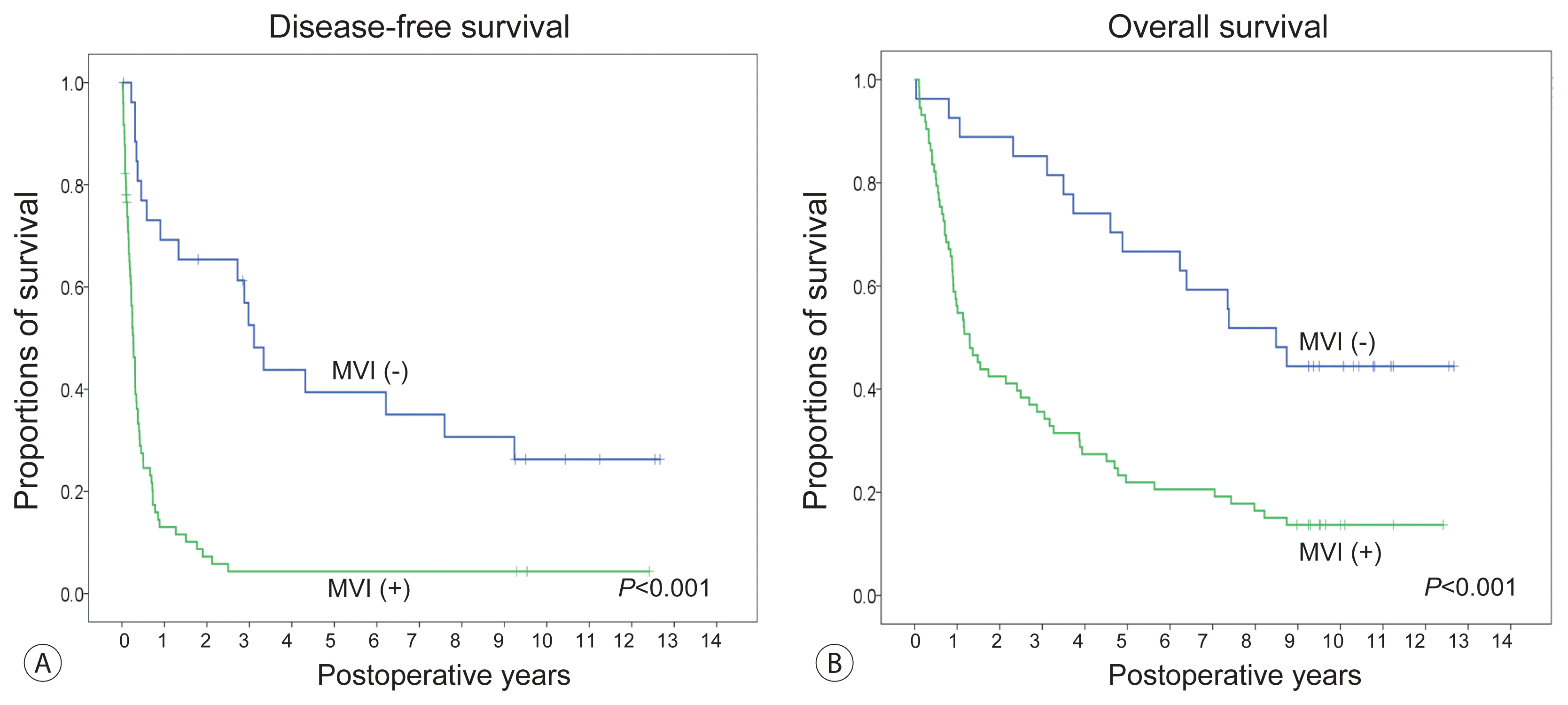
Figure 3 Comparison of the disease-free survival (A) and overall survival (B) curves according to the status of microvascular invasion (MVI).
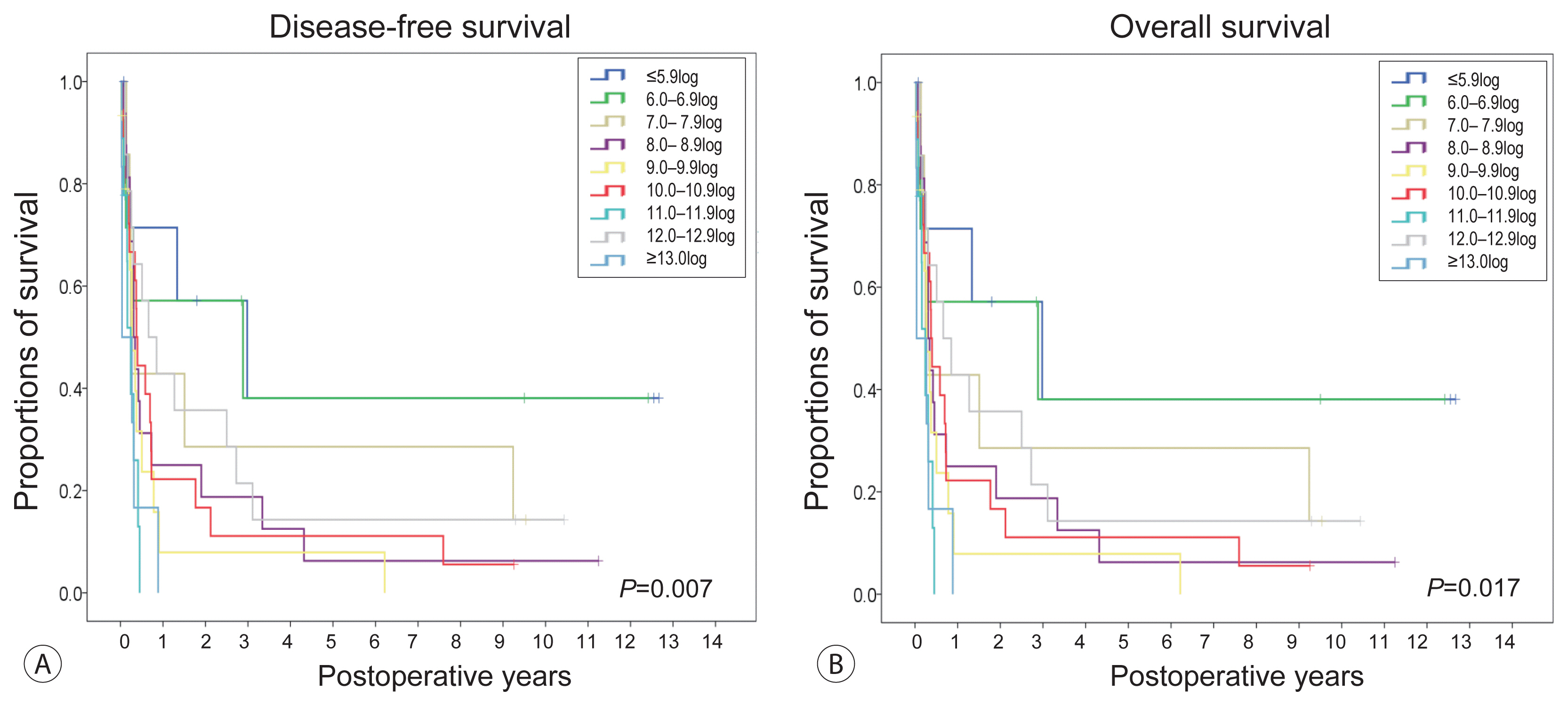
Figure 4 Comparison of the disease-free survival (A) and overall survival (B) curves according to the α-fetoprotein, des-γ-carboxy prothrombin, and tumor volume (ADV) score of 1log intervals.

Figure 5 Comparison of the disease-free survival (A) and overall survival (B) curves according to the α-fetoprotein, des-γ-carboxy prothrombin, and tumor volume (ADV) score with a cut-off of 8.0log.

Figure 6 Comparison of the disease-free survival (A) and overall survival (B) curves according to the number of risk factors (presence of microvascular invasion and α-fetoprotein, des-γ-carboxy prothrombin, and tumor volume [ADV] score ≥8.0log).
Reference
-
1. Forner A, Llovet JM, Bruix J. Hepatocellular carcinoma. Lancet. 2012; 379:1245–1255.2. Huang YH, Wu JC, Chen SC, Chen CH, Chiang JH, Huo TI, et al. Survival benefit of transcatheter arterial chemoembolization in patients with hepatocellular carcinoma larger than 10 cm in diameter. Aliment Pharmacol Ther. 2006; 23:129–135.3. Hwang S, Lee YJ, Kim KH, Ahn CS, Moon DB, Ha TY, et al. Long-term outcome after resection of huge hepatocellular carcinoma ≥ 10 cm: single-institution experience with 471 patients. World J Surg. 2015; 39:2519–2528.4. Yamashita Y, Taketomi A, Shirabe K, Aishima S, Tsuijita E, Morita K, et al. Outcomes of hepatic resection for huge hepatocellular carcinoma (≥10 cm in diameter). J Surg Oncol. 2011; 104:292–298.5. Zhou YM, Li B, Xu DH, Yang JM. Safety and efficacy of partial hepatectomy for huge (≥10 cm) hepatocellular carcinoma: a systematic review. Med Sci Monit. 2011; 17:RA76–RA83.6. Tsoulfas G, Mekras A, Agorastou P, Kiskinis D. Surgical treatment for large hepatocellular carcinoma: does size matter? ANZ J Surg. 2012; 82:510–517.7. Hwang S, Song GW, Lee YJ, Kim KH, Ahn CS, Moon DB, et al. Multiplication of tumor volume by two tumor markers is a post-resection prognostic predictor for solitary hepatocellular carcinoma. J Gastrointest Surg. 2016; 20:1807–1820.8. Jung DH, Hwang S, Lee YJ, Kim KH, Song GW, Ahn CS, et al. Small hepatocellular carcinoma with low tumor marker expression benefits more from anatomical resection than tumors with aggressive biology. Ann Surg. 2019; 269:511–519.9. Hwang S, Moon DB, Kim KH, Ahn CS, Song GW, Jung DH, et al. Prognostic accuracy of the ADV score following resection of hepatocellular carcinoma with portal vein tumor thrombosis. J Gastrointest Surg. 2020; Sep. 19. DOI: 10.1007/s11605-020-04800-6. [Epub ahead of print].10. Cho HD, Hwang S, Lee YJ, Park KM, Kim KH, Kim JC, et al. Changes in the types of liver diseases requiring hepatic resection: a single-institution experience of 9016 cases over a 10-year period. Korean J Hepatobiliary Pancreat Surg. 2016; 20:49–52.11. Hwang S, Lee YJ, Kim KH, Ahn CS, Moon DB, Ha TY, et al. The impact of tumor size on long-term survival outcomes after resection of solitary hepatocellular carcinoma: single-institution experience with 2558 patients. J Gastrointest Surg. 2015; 19:1281–1290.12. Hwang S, Ha TY, Song GW, Jung DH, Ahn CS, Moon DB, et al. Quantified risk assessment for major hepatectomy via the indocyanine green clearance rate and liver volumetry combined with standard liver volume. J Gastrointest Surg. 2015; 19:1305–1314.13. The Korean Association for the Study of the Liver. KASL clinical practice guidelines: management of chronic hepatitis B. Clin Mol Hepatol. 2016; 22:18–75.14. Pawlik TM, Delman KA, Vauthey JN, Nagorney DM, Ng IO, Ikai I, et al. Tumor size predicts vascular invasion and histologic grade: Implications for selection of surgical treatment for hepatocellular carcinoma. Liver Transpl. 2005; 11:1086–1092.15. Yang LY, Fang F, Ou DP, Wu W, Zeng ZJ, Wu F. Solitary large hepatocellular carcinoma: a specific subtype of hepatocellular carcinoma with good outcome after hepatic resection. Ann Surg. 2009; 249:118–123.16. Zhou YM, Sui CJ, Zhang XF, Li B, Yang JM. Anterior approach combined with infrahepatic inferior vena cava clamping right hepatic resection for large hepatocellular carcinoma: a prospective randomized controlled trial. Medicine (Baltimore). 2016; 95:e4159.17. Wang XB, Yu QM, Yu PF, Zhang YL, Yang LT, Zhang ZW, et al. Surgical treatment of huge hepatocarcinoma with invasion or severe adhesion of diaphragm using the technique of orthotopic liver resection. Hepatogastroenterology. 2015; 62:153–156.18. Huang JS, Dai WD, Miao XY, Zhong DW, Xiong SZ, Hu JX. Null-margin bisegmentectomy VII–VIII for hepatocellular carcinoma in cirrhotic patients. Hepatogastroenterology. 2012; 59:1706–1709.19. Shin HN, Hwang S, Kim KH, Ahn CS, Moon DB, Ha TY, et al. Role of the 1-month protocol transarterial chemoinfusion in detecting intrahepatic metastasis after resection of large hepatocellular carcinoma greater than 10 cm. Korean J Hepatobiliary Pancreat Surg. 2013; 17:157–161.20. Eguchi S, Kanematsu T, Arii S, Okazaki M, Okita K, Omata M, et al. Comparison of the outcomes between an anatomical subsegmentectomy and a non-anatomical minor hepatectomy for single hepatocellular carcinomas based on a Japanese nationwide survey. Surgery. 2008; 143:469–475.21. Kitamura K, Hatano E, Higashi T, Seo S, Nakamoto Y, Yamanaka K, et al. Preoperative FDG-PET predicts recurrence patterns in hepatocellular carcinoma. Ann Surg Oncol. 2012; 19:156–162.22. Ishii T, Hatano E, Yasuchika K, Taura K, Seo S, Uemoto S. High risk of lung metastasis after resection of hepatocellular carcinoma more than 7 cm in diameter. Surg Today. 2014; 44:1900–1905.23. Park MJ, Kim YK, Lee MW, Lee WJ, Kim YS, Kim SH, et al. Small hepatocellular carcinomas: improved sensitivity by combining gadoxetic acid-enhanced and diffusion-weighted MR imaging patterns. Radiology. 2012; 264:761–770.24. Sano K, Ichikawa T, Motosugi U, Sou H, Muhi AM, Matsuda M, et al. Imaging study of early hepatocellular carcinoma: usefulness of gadoxetic acid-enhanced MR imaging. Radiology. 2011; 261:834–844.25. Hwang S, Joh JW, Wang HJ, Kim DG, Kim KS, Suh KS, et al. Prognostic prediction models for resection of large hepatocellular carcinoma: a Korean multicenter study. World J Surg. 2018; 42:2579–2591.26. Lencioni R, Llovet JM. Modified RECIST (mRECIST) assessment for hepatocellular carcinoma. Semin Liver Dis. 2010; 30:52–60.27. Botea F, Marconi M, Lutman F, Balzarini L, Roncalli M, Montorsi M, et al. Radiological estimation of size in colorectal liver metastases: is it reliable? Comparison with post-resectional measurements. Updates Surg. 2010; 62:21–26.28. Galizia MS, Töre HG, Chalian H, Yaghmai V. Evaluation of hepatocellular carcinoma size using two-dimensional and volumetric analysis: effect on liver transplantation eligibility. Acad Radiol. 2011; 18:1555–1560.29. Kim HD, Shim JH, Kim GA, Shin YM, Yu E, Lee SG, et al. Optimal methods for measuring eligibility for liver transplant in hepatocellular carcinoma patients undergoing transarterial chemoembolization. J Hepatol. 2015; 62:1076–1784.30. Hwang S, Song GW, Ahn CS, Kim KH, Moon DB, Ha TY, et al. Quantitative prognostic prediction using ADV score for hepatocellular carcinoma following living donor liver transplantation. J Gastrointest Surg. 2021; Feb. 2. DOI: 10.1007/s11605-021-04939-w. [Epub ahead of print].31. Lau WY, Lai EC, Lau SH. The current role of neoadjuvant/adjuvant/chemoprevention therapy in partial hepatectomy for hepatocellular carcinoma: a systematic review. Hepatobiliary Pancreat Dis Int. 2009; 8:124–133.32. Li KW, Li X, Wen TF, Lu WS. The effect of postoperative TACE on prognosis of HCC: an update. Hepatogastroenterology. 2013; 60:248–251.
- Full Text Links
-
- Actions
-
Cited
- CITED
-
- Close
- Share
-
- Similar articles
-
- Validation of the OncoHepa test, a multigene expression profile test, and the tumor marker-volume score to predict postresection outcome in small solitary hepatocellular carcinomas
- Impact of tumor size on hepatectomy outcomes in hepatocellular carcinoma: a nationwide propensity score matching analysis
- Salvage living-donor liver transplantation and ADV score
- Outcome of Hepatectomy for Huge Hepatocellular Carcinoma
- A Clinical Study of Surgically Resected Primary Liver Cancer
