Xanthogranulomatous Cholecystitis Suspected as Metastatic Gallbladder Cancer
- Affiliations
-
- 1Division of Gastroenterology, Department of Internal Medicine, International St. Mary’s Hospital, Catholic Kwandong University College of Medicine, Incheon, Korea
- 2Department of Pathology, International St. Mary’s Hospital, Catholic Kwandong University College of Medicine, Incheon, Korea
- 3Department of Internal Medicine and Liver Research Institute, Seoul National University College of Medicine, Seoul, Korea
- 4Division of Gastroenterology, Department of Internal Medicine, Seoul National University Hospital, Seoul, Korea
- KMID: 2514091
- DOI: http://doi.org/10.4166/kjg.2021.026
Figure
-
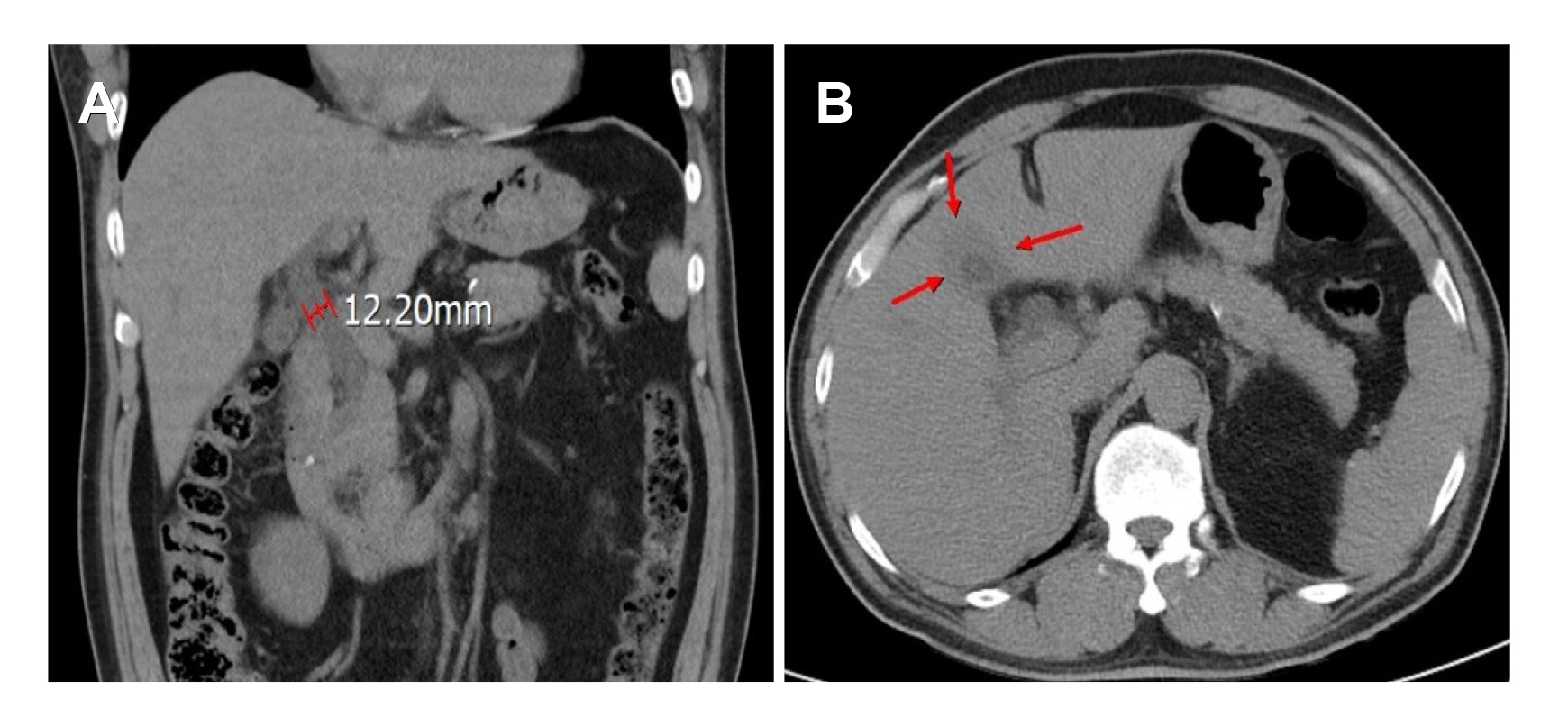
Fig. 1 Abdomen computed tomography findings. (A) Impacted distal common bile duct stone with proportional biliary dilatation. (B) Collapsed gallbladder, suspicious of chronic cholecystitis (arrows).
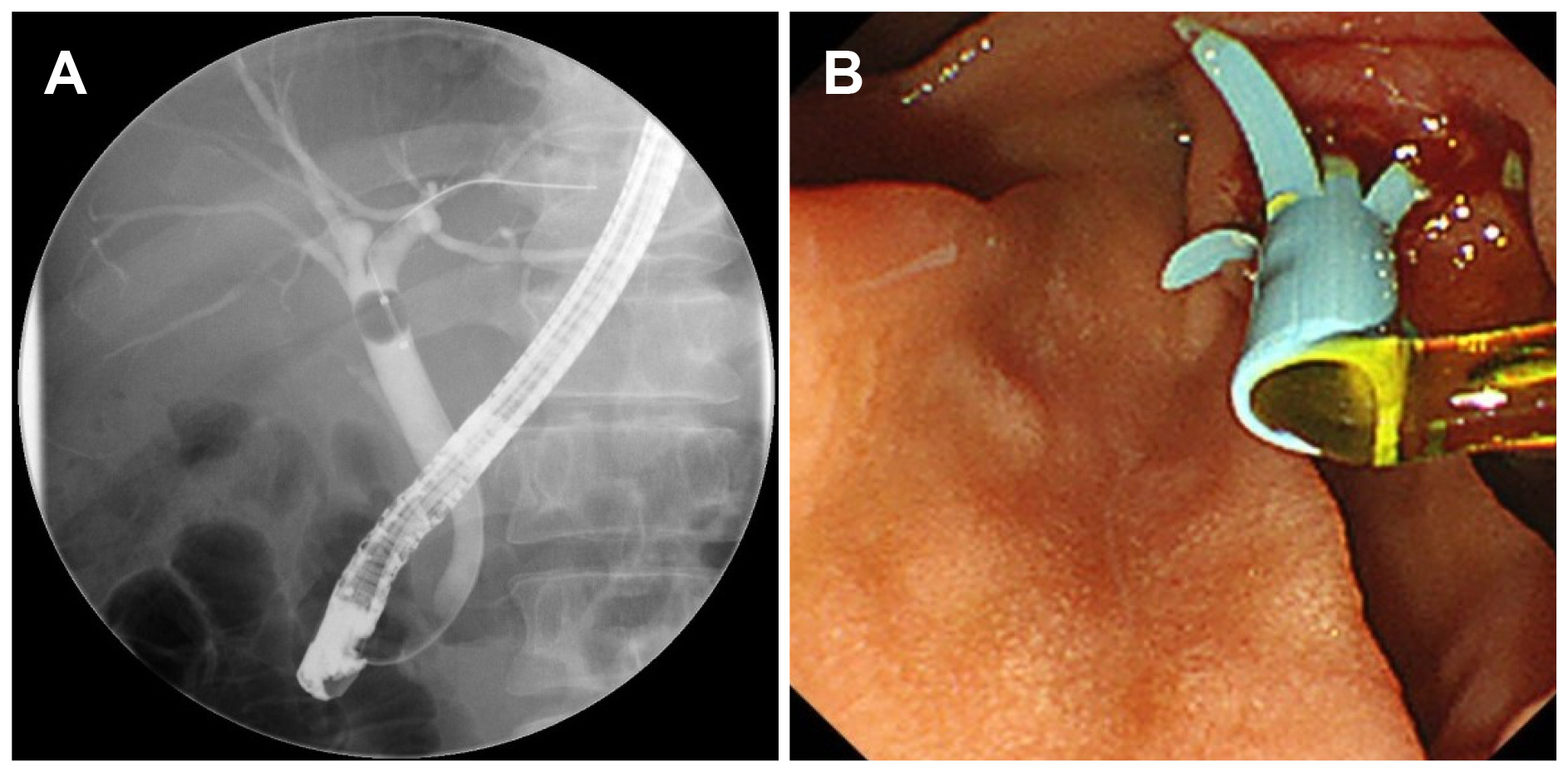
Fig. 2 (A) Endoscopic retrograde cholangiopancreatography showing mild common bile duct dilatation without a definite filling defect. (B) Stone extraction using balloon catheter and biliary stent insertion were performed.
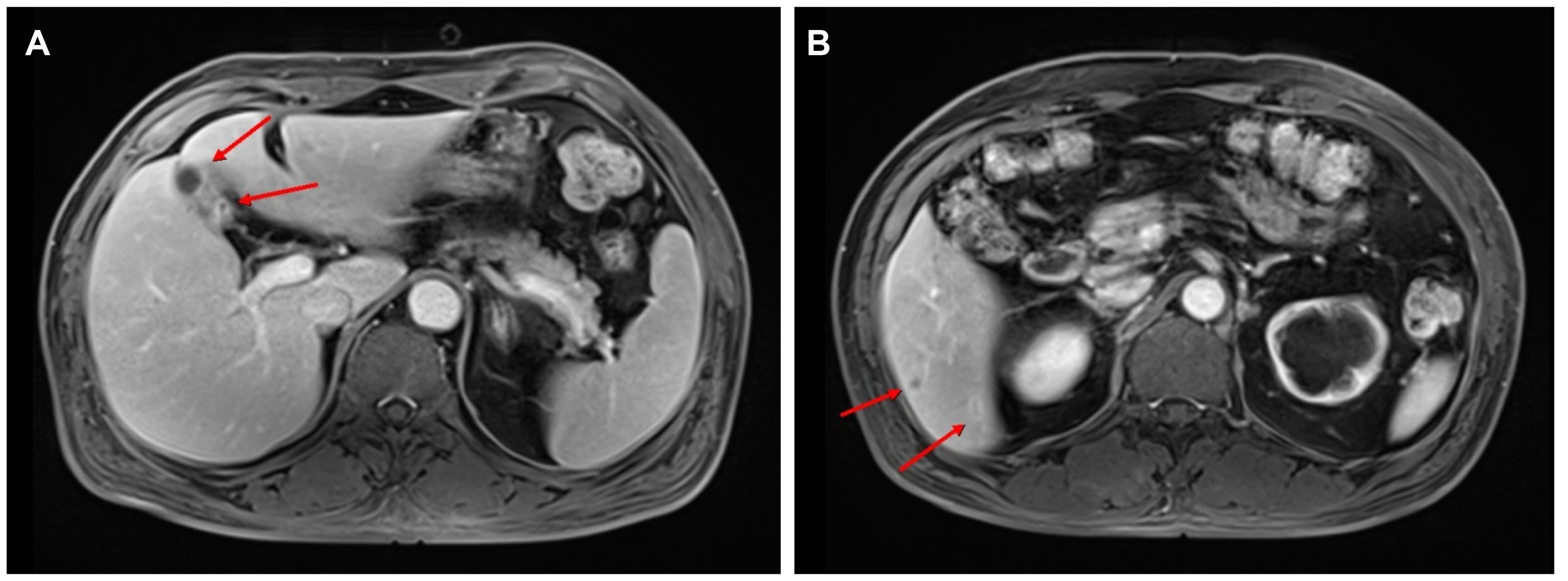
Fig. 3 Magnetic resonance imaging findings. (A) Irregular enhanced wall thickening of the gallbladder with diffusion restriction and direct invasion of the adjacent liver parenchyma (arrows), suspicious of gallbladder cancer with direct invasion (T3 vs. T4Nx). (B) Multiple subcentimeter peripherally enhancing lesions in the liver (arrows).
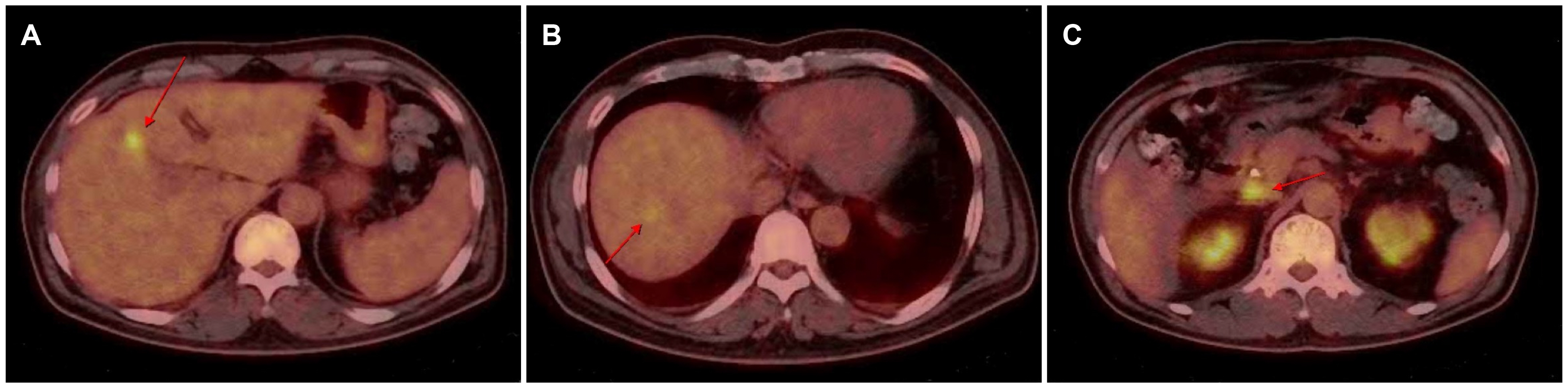
Fig. 4 Positron emission tomography-computed tomography shows (A) increased FDG uptake in the gallbladder (arrow), (B) multifocal mildly increased FDG uptake in the liver (arrow), and (C) focally increased FDG uptake in the portocaval space (arrow).

Fig. 5 Microscopically, (A) thickened gallbladder wall revealed a relatively preserved mucosal epithelium and partly disrupted muscular layers (left side), in which collections of lipid-laden macrophages were found admixed with mixed inflammatory cells and fibrosis (H&E, ×40). (B) Xanthogranuloma, a characteristic feature of xanthogranulomatous cholecystitis, was noted, which contained foamy histiocytes, multinucleated giant cells, and lymphoplasmacytic cells (H&E, ×200).
Reference
-
1. Ros PR, Goodman ZD. 1997; Xanthogranulomatous cholecystitis versus gallbladder carcinoma. Radiology. 203:10–12. DOI: 10.1148/radiology.203.1.9122374. PMID: 9122374.
Article2. Kang J, Lee SH, Lee JW, et al. 2015; A case of xanthogranulomatous cholecystitis decreased in size after steroid treatment and avoided extended resection. Korean J Pancreas Biliary Tract. 20:37–41. DOI: 10.15279/kpba.2015.20.1.37.
Article3. 전 훈배, 이 승규, 이 영주, et al. 1999; Xanthogranulomatous cholecystitis. Korean Journal of HBP Surgery. 3:203–209. DOI: 10.1007/s00464-020-07828-6,. PMID: 32720174.4. Goodman ZD, Ishak KG. 1981; Xanthogranulomatous cholecystitis. Am J Surg Pathol. 5:653–659. DOI: 10.1097/00000478-198110000-00007. PMID: 7337158.
Article5. Joo YE, Lee JJ, Chung IJ, et al. 1999; A case of xanthogranulomatous cholecystitis. Korean J Intern Med. 14:90–93. DOI: 10.3904/kjim.1999.14.2.90. PMID: 10461432. PMCID: PMC4531919.
Article6. Yang T, Zhang BH, Zhang J, Zhang YJ, Jiang XQ, Wu MC. 2007; Surgical treatment of xanthogranulomatous cholecystitis: experience in 33 cases. Hepatobiliary Pancreat Dis Int. 6:504–508. PMID: 17897914.7. Chang BJ, Kim SH, Park HY, et al. 2010; Distinguishing xanthogranulomatous cholecystitis from the wall-thickening type of early-stage gallbladder cancer. Gut Liver. 4:518–523. DOI: 10.5009/gnl.2010.4.4.518. PMID: 21253302. PMCID: PMC3021609.
Article8. Spinelli A, Schumacher G, Pascher A, et al. 2006; Extended surgical resection for xanthogranulomatous cholecystitis mimicking advanced gallbladder carcinoma: a case report and review of literature. World J Gastroenterol. 12:2293–2296. DOI: 10.3748/wjg.v12.i14.2293. PMID: 16610041. PMCID: PMC4087666.
Article9. Singh VP, Rajesh S, Bihari C, Desai SN, Pargewar SS, Arora A. 2016; Xanthogranulomatous cholecystitis: what every radiologist should know. World J Radiol. 8:183–191. DOI: 10.4329/wjr.v8.i2.183. PMID: 26981227. PMCID: PMC4770180.
Article10. Chun KA, Ha HK, Yu ES, et al. 1997; Xanthogranulomatous cholecystitis: CT features with emphasis on differentiation from gallbladder carcinoma. Radiology. 203:93–97. DOI: 10.1148/radiology.203.1.9122422. PMID: 9122422.
Article11. Kim PN, Ha HK, Kim YH, Lee MG, Kim MH, Auh YH. 1998; US findings of xanthogranulomatous cholecystitis. Clin Radiol. 53:290–292. DOI: 10.1016/S0009-9260(98)80129-3. PMID: 9585046.
Article12. Kim PN, Lee SH, Gong GY, et al. 1999; Xanthogranulomatous cholecystitis: radiologic findings with histologic correlation that focuses on intramural nodules. AJR Am J Roentgenol. 172:949–953. DOI: 10.2214/ajr.172.4.10587127. PMID: 10587127.
Article13. Nacif LS, Hessheimer AJ, Rodríguez Gómez S, Montironi C, Fondevila C. 2017; Infiltrative xanthogranulomatous cholecystitis mimicking aggressive gallbladder carcinoma: a diagnostic and therapeutic dilemma. World J Gastroenterol. 23:8671–8678. DOI: 10.3748/wjg.v23.i48.8671. PMID: 29358875. PMCID: PMC5752727.
Article14. Seo SH, Park JI, Kim JS, Kim KH, Choi CS, Choi YK. 2009; Xanthogranulomatous cholecystitis: a retrospective analysis of 36 cases. J Korean Surg Soc. 76:317–377. DOI: 10.4174/jkss.2009.76.6.371.
Article15. Kwon AH, Sakaida N. 2007; Simultaneous presence of xanthogranulomatous cholecystitis and gallbladder cancer. J Gastroenterol. 42:703–704. DOI: 10.1007/s00535-007-2072-6. PMID: 17701136.
Article
- Full Text Links
-
- Actions
-
Cited
- CITED
-
- Close
- Share
-
- Similar articles
-
- Xanthogranulomatous cholecystitis
- A Case of Xanthogranulomatous Cholecystitis
- A Case of Xanthogranulomatous Cholecystitis Decreased in Size after Steroid Treatment and Avoided Extended Resection
- Xanthogranulomatous Cholecystitis Mimicking Gallbladder Cancer
- A Case of Xanthogranulomatous Cholecystitis Associated with Mirizzi Syndrome
