Footsteps of the Innovations in Rhinology
- Affiliations
-
- 1Department of Otorhinolaryngology-Head and Neck Surgery, Guro Hospital, Korea University College of Medicine, Seoul, Korea
- KMID: 2513839
- DOI: http://doi.org/10.18787/jr.2020.00332
Abstract
- Rhinology is the study of nose, paranasal sinus, and nasopharynx. The nose is the most prominent structure on the human face and has been a subject of study since ancient human civilization. The history of rhinology has reflected the sociocultural aspects of the times, and rhinology has achieved remarkable growth with innovative discoveries by numerous pioneers. The focus of surgical procedures of the paranasal sinus shifted from mucosal stripping to functional endoscopic surgery with advancement of technology. Furthermore, the field of rhinology is gradually expanding due to cutting-edge technologies such as image-guided surgery, three-dimensional endoscopy, and robotic surgery. Additional clinical experiences and technological developments are expected to further advance rhinology.
Figure
-
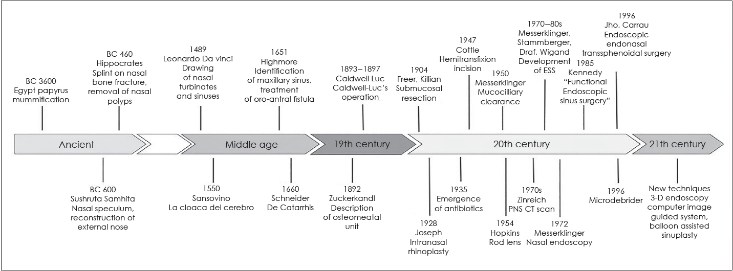
Fig. 1. Timelines of major events of rhinology. PNS CT: paranasal computed tomography, 3-D: 3-dimensional.
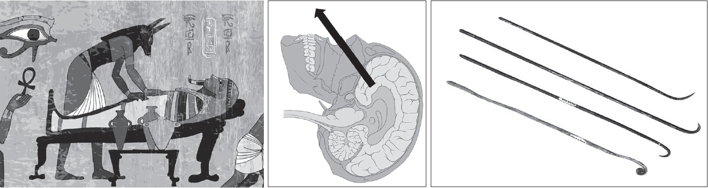
Fig. 2. Removal of brain via transnasal apporach using hook during mummification in ancient Egypt mural. Purchased image from Shutterstock.com.
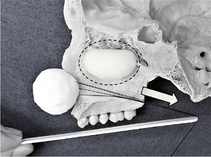
Fig. 3. Hippocrates’ removal technique of nasal polyposis (dotted circle) with a stringed sponge in the direction of arrow.
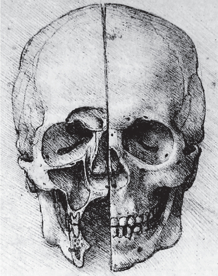
Fig. 4. Leonardo da Vinci’s drawing of human skull. The sketch describes a coronal section of frontal sinus, maxillary sinus, and inferior turbinate. Purchased image from Shutterstock.com.
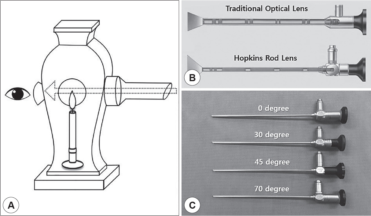
Fig. 5. Evolution of endoscopy. A: The first modern endoscopy invented by Phillip Bozzini in 1804. The “light conductor” was consisted of an optical part with the illumination device and a mechanical part which had to be modified to fit to the respective body opening. B: Rod lens system established by Korl Storz and Harold Hopkins. It provides better light transmission resulting in advanced resolution and contrast, and wider viewing angles compared to traditional optical system. C: The modern nasal endoscopy provide available in various angles from 0 to 30, 45, and 70 degree.

Fig. 6. Steroid-releasing sinus implants indicated for ethmoid sinus and frontal recess. A: PROPEL® is an cylindrical-shaped, expandable and biodegradable polymer (polyactide-co-glycolic acid) coated with steroid (mometasone fuorate). 23 mm in length of PROPEL® , 16 mm of PROPEL® Mini. B: PROPEL® Contour is an hourglass-shaped implant, 8 mm in length. Adapted from https://www.intersectent.com.
Reference
-
References
1. Whitaker IS, Karoo RO, Spyrou G, Fenton OM. The birth of plastic surgery: the story of nasal reconstruction from the Edwin Smith Papyrus to the twenty-first century. Plas Reconstr Surg. 2007; 120:327–36.2. Lascaratos JG, Trompoukis CC, Segas JV, Assimakopoulos DA. From the roots of rhinology: the reconstruction of nasal injuries by Hippocrates. Annals of Otology, Rhinology & Laryngology. 2003; 112:159–62.3. Breasted JH. The Edwin Smith Surgical Papyrus: published in facsimile and hieroglyphic transliteration with translation and commentary in two volumes. Chicago: University of Chicago Press;1930.4. Wright J. A history of laryngology and rhinology. 2nd ed. Philadelphia: Lea & Febiger;1914.5. Stevenson RS, Guthrie D. A History of Otolaryngology. Edinburgh: E. & S. Livingstone;1949.6. Cingi C, Onerci M, Leopold D. History of Rhinology. All Around the Nose. 1st ed. Springer;2020.7. Bhishagratna KL. An English translation of The Sushruta Samhita: based on original Sanskrit text. Calcutta: 1911.8. Voltolini FR. Die Krankheiten der Nase und des Nasenrachenraums. Breslau: 1888.9. Mudry A. An octopus in the nostrils. European Annals of Otorhinolaryngology, Head and Neck Diseases. 2020; 137:211–2.10. Frenkiel S, Wright ED. The specialty of rhinology, part 1: a historical glimpse. The Journal of Otolaryngology. 2001; 30:26–31.11. Feldmann H. The maxillary sinus and its illness in the history of rhinology. Images from the history of otorhinolaryngology, highlighted by instruments from the collection of the German Medical History Museum in Ingolstadt. Laryngo-Rhino-Otol. 1998; 77:587–95.12. Pirsig W. Early depictions of the nasal turbinates in the 15th century. Rhinology. 2002; 40:104–6.13. Yakoot AA. The journey of the evolution of rhinology. The Egyptian Journal of Otolaryngology. 2013; 29:136–42.14. Nicholl C. Leonardo da Vinci: the flights of the Mind. 1st ed. New York: Penguin;2005.15. Toledo-Pereyra LH. De humani corporis fabrica surgical revolution. Journal of Investigative Surgery. 2008; 21:232–6.16. Mavrodi A, Paraskevas G. Evolution of the paranasal sinuses’ anatomy through the ages. Anat Cell Biol. 2013; 46:235–8.17. Helidonis ES. The history of otolaryngology from ancient to modern times. American Journal of Otolaryngology. 1993; 14:382–93.18. Grand W. The anatomy of the brain, by Thomas Willis. Neurosurgery. 1999; 45:1234–7.19. Finger S, Boller F, Tyler KL. History of neurology. 1st ed. Amsterdam: Elsevier;2009.20. Stammberger H. History of rhinology: anatomy of the paranasal sinuses. Rhinology. 1989; 27:197–210.21. Tange RA. Some historical aspects of the surgical treatment of the infected maxillary sinus. Rhinology. 1991; 29:155–62.22. Deschamps JL. Dissertation sur les maladies des fosses nasales. Paris: 1804.23. Pirsig W. History of rhinology: nasal specula around the turn of the 19th-20th century. Rhinology. 1990; 28:113–22.24. Roe JO. The deformity termed “pug nose” and its correction, by a simple operation. Plastic and Reconstructive Surgery. 1970; 45:78–81.25. Kaluskar S. Evolution of rhinology. Indian J Otolaryngol Head Neck Surg. 2008; 60:101–5.26. Shoja MM, Tubbs RS, Loukas M, Shokouhi G, Oakes WJ. Emil Zuckerkandl (1849-1910): anatomist and pathologist. Ann Anat. 2008; 190:33–6.27. Freer OT. The correction of deflections of the nasal septum with a minimum of traumatism. Journal of the American Medical Association. 1902; 38:636–42.28. Killian G. Die submukose Fensterresektion der Nasenscheidewand. Arch. Laryngol. 1904; 16:203.29. King ED, Ashley FL. The correction of the internally and externally deviated nose. Plastic and Reconstructive Surgery. 1952; 10:116–20.30. Subramaniam V, Basheer M, Hosagadde RS. Evolution of correction of the deviated nasal septum-A historical overview. Archives of Medicine and Health Sciences. 2018; 6:293.31. Stammberger H, Posawetz W. Functional endoscopic sinus surgery. Eur Arch Otorhinolaryngol. 1990; 247:63–76.32. Lanza DC, Kennedy D, Zinreich S. Nasal endoscopy and its surgical applications. Essential otolaryngology: head and neck surgery. 5th ed. New York: Medical Examination Publishing;1991.33. Na’ara S, Kaptzan B, Gil Z, Ostrovsky D. Endoscopic Septoplasty Versus Traditional Septoplasty for Treating Deviated Nasal Septum: A Prospective, Randomized Controlled Trial. Ear, Nose & Throat Journal. 2020.34. Chung BJ, Batra PS, Citardi MJ, Lanza DC. Endoscopic septoplasty: revisitation of the technique, indications, and outcomes. American Journal of Rhinology. 2007; 21:307–11.35. Maltz M. New instrument: the sinuscope. Laryngoscope. 1925; 35:805–11.36. Jacobs JB. 100 years of frontal sinus surgery. Laryngoscope. 1997; 107:1–36.37. Messerklinger W. Endoscopy of the nose. 1st ed. Baltimore: Lippincott Williams and Wilkins;1978.38. Reynolds W, Brandow E. Recent advances in microsurgery of the maxillary antrum. Acta Oto-Laryngologica. 1975; 80:161–6.39. Mann W, Beck C. Inferior meatal antrostomy in chronic maxillary sinusitis. Arch Otorhinolaryngol. 1978; 221:289–95.40. Kennedy DW, Zinreich SJ, Rosenbaum AE, Johns ME. Functional endoscopic sinus surgery: theory and diagnostic evaluation. Archives of Otolaryngology. 1985; 111:576–82.41. Honda Y, Takahashi R. The Evolution of Endonasal Sinus Surgery in Japan. American Journal of Rhinology. 1991; 5:29–31.42. Takahashi R. Endonasal operation of chronic ethmoiditis. A Collection of Ear, Nose, and Throat Studied. 1971; 372–89.43. Ashikawa R, Ohkushi H, Ohmae T, Matsuda T. Clinical effects of endonasal sinusectomy with reconstruction of the nasal cavity (Takahashi’s method). Rhinology. 1981; 19:93–100.44. Moriyama H, Yanagi K, Ohtori N, Asai K, Fukami M. Healing process of sinus mucosa after endoscopic sinus surgery. Am J Rhinol. 1996; 10:61–6.45. Kane K. The early history and development of functional endoscopic sinus surgery. The Journal of Laryngology & Otology. 2020; 134:8–13.46. Jankowski R, Auque J, Simon C. How i do it: Head and neck and plastic surgery: Endoscopic pituitary tumor surgery. Laryngoscope. 1992; 102:198–202.47. Carrau RL, Jho HD, Ko Y. Transnasal-transsphenoidal endoscopic surgery of the pituitary gland. Laryngoscope. 1996; 106:914–8.48. Kennedy DW, Goodstein ML, Miller NR, Zinreich SJ. Endoscopic transnasal orbital decompression. Arch Otolaryngol Head Neck Surg. 1990; 116:275–82.49. Bleier BS, Castelnuovo P, Battaglia P, Turri-Zanoni M, Dallan I, Metson R, et al. Endoscopic endonasal orbital cavernous hemangioma resection: global experience in techniques and outcomes. International forum of allergy & rhinology: Wiley Online Library. 2016; 6:156–61.50. Anon JB, Klimek L, Mosges R, Zinreich SJ. Computer-assisted endoscopic sinus surgery. An international review. Otolaryngologic Clinics of North America. 1997; 30:389–401.51. Huang BY, Senior BA, Castillo M. Current trends in sinonasal imaging. Neuroimag Clin N Am. 2015; 25:507–25.52. Tabrizi LB, Mahvash M. Augmented reality-guided neurosurgery: accuracy and intraoperative application of an image projection technique. Journal of Neurosurgery. 2015; 123:206–11.53. Winne C, Khan M, Stopp F, Jank E, Keeve E. Overlay visualization in endoscopic ENT surgery. International Journal of Computer Assisted Radiology and Surgery. 2011; 6:401–6.54. Citardi MJ, Agbetoba A, Bigcas JL, Luong A. Augmented reality for endoscopic sinus surgery with surgical navigation: a cadaver study. International forum of allergy & rhinology: Wiley Online Library. 2016; 6:523–8.55. Hanna EY, Holsinger C, DeMonte F, Kupferman M, Surgery N. Robotic endoscopic surgery of the skull base: a novel surgical approach. Arch Otolaryngol Head Neck Surg. 2007; 133:1209–14.56. Singh A, Saraiya R. Three-dimensional endoscopy in sinus surgery. Current Opinion in Otolaryngology & Head and Neck Surgery. 2013; 21:3–10.57. Ogino-Nishimura E, Nakagawa T, Sakamoto T, Ito J. Efficacy of three-dimensional endoscopy in endonasal surgery. Auris Nasus Larynx. 2015; 42:203–7.58. Tomazic PV, Stammberger H, Braun H, Habermann W, Schmid C, Hammer GP, et al. Feasibility of balloon sinuplasty in patients with chronic rhinosinusitis: the Graz experience. Rhinology. 2013; 51:120–7.59. Bolger WE, Brown CL, Church CA, Goldberg AN, Karanfilov B, Kuhn FA, et al. Safety and outcomes of balloon catheter sinusotomy: a multicenter 24-week analysis in 115 patients. Otolaryngology-Head and Neck Surgery. 2007; 137:10–20.60. Kuhn FA, Church CA, Goldberg AN, Levine HL, Sillers MJ, Vaughan WC, et al. Balloon catheter sinusotomy: one-year follow-up-outcomes and role in functional endoscopic sinus surgery. Otolaryngology-Head and Neck Surgery. 2008; 139:S27–37.61. Massey CJ, Singh A. Advances in absorbable biomaterials and nasal packing. Otolaryngologic Clinics of North America. 2017; 50:545–63.62. Bednarski KA, Kuhn FA. Stents and drug-eluting stents. Otolaryngologic Clinics of North America. 2009; 42:857–66.63. Marple BF, Smith TL, Han JK, Gould AR, Jampel HD, Stambaugh JW, et al. Advance II: a prospective, randomized study assessing safety and efficacy of bioabsorbable steroid-releasing sinus implants. Otolaryngology-Head and Neck Surgery. 2012; 146:1004–11.64. Han JK, Marple BF, Smith TL, Murr AH, Lanier BJ, Stambaugh JW, et al. Effect of steroid-releasing sinus implants on postoperative medical and surgical interventions: an efficacy meta-analysis. International Forum of Allergy & Rhinology: Wiley Online Library. 2012; 2:271–9.65. Lee JT, Han JK. Sinus implants for chronic rhinosinusitis: technology evaluation. Expert Opinion on Drug Delivery. 2013; 10:1735–48.66. Low CM, Morris JM, Price DL, Matsumoto JS, Stokken JK, O’Brien EK, et al. Three-dimensional printing: current use in rhinology and endoscopic skull base surgery. American Journal of Rhinology & Allergy. 2019; 33:770–81.67. Sánchez-Gómez S, Herrero-Salado TF, Maza-Solano JM, RoperoRomero F, González-García J, Ambrosiani-Fernández J. Improved planning of endoscopic sinonasal surgery from 3-dimensional images with Osirix® and stereolithography. Acta Otorrinolaringologica (English Edition). 2015; 66:317–25.68. Grau S, Kellermann S, Faust M, Perrech M, Beutner D, Drzezga A, et al. Repair of cerebrospinal fluid leakage using a transfrontal, radial adipofascial flap: an individual approach supported by threedimensional printing for surgical planning. World neurosurgery. 2018; 110:315–8.69. Shinomiya A, Shindo A, Kawanishi M, Miyake K, Nakamura T, Matsubara S, et al. Usefulness of the 3D virtual visualization surgical planning simulation and 3D model for endoscopic endonasal transsphenoidal surgery of pituitary adenoma: technical report and review of literature. Interdisciplinary Neurosurgery. 2018; 13:13–9.70. Altunay ZO, Bly JA, Edwards PK, Holmes DR, Hamilton GS, O’Brien EK, et al. Three-dimensional printing of large nasal septal perforations for optimal prosthetic closure. American Journal of Rhinology & Allergy. 2016; 30:287–93.71. Xue R, Lai Q, Sun S, Lai L, Tang X, Ci J, et al. Application of three-dimensional printing technology for improved orbital-maxillary-zygomatic reconstruction. Journal of Craniofacial Surgery. 2019; 30:127–31.72. Tack P, Victor J, Gemmel P, Annemans L. 3D-printing techniques in a medical setting: a systematic literature review. Biomedical engineering online. 2016; 15:115–35.
- Full Text Links
-
- Actions
-
Cited
- CITED
-
- Close
- Share
-
- Similar articles
-
- Recent innovations in renal replacement technology and potential applications to transplantation and dialysis patients: a review of current methods
- HIV Service Delivery Innovations in the Philippines during the COVID-19 Pandemic
- Innovations: a dawning of a new age
- Innovations in Clinical Research Design and Conduct in Psychiatry: Shifting to Pragmatic Approaches
- Waiting for innovations in periodontal disease diagnosis
