Relationship between rotational disc displacement of the temporomandibular joint and the dentoskeletal morphology
- Affiliations
-
- 1Department of Orthodontics, Dankook University Jukjeon Dental Hospital, Yongin, Korea
- 2Department of Oral and Maxillofacial Radiology, College of Dentistry, Dankook University, Cheonan, Korea
- 3Department of Orthodontics, College of Dentistry, Dankook University, Cheonan, Korea
- 4Department of Orthodontics, Seoul National University Dental Hospital, Seoul, Korea
- 5Dental Research Institute and Department of Orthodontics, School of Dentistry, Seoul National University, Seoul, Korea
- KMID: 2513579
- DOI: http://doi.org/10.4041/kjod.2021.51.2.105
Abstract
Objective
The purpose of this study was to evaluate the relationship between rotational disk displacement (DD) of the temporomandibular joint (TMJ) and the dentoskeletal morphology.
Methods
Women aged > 17 years were included in this study. Each subject had a primary complaint of malocclusion and underwent routine cephalometric examinations. They were divided into five groups according to the findings on sagittal and coronal magnetic resonance images of their TMJs: bilateral normal disk position, bilateral anterior DD with reduction (ADDR), bilateral rotational DD with reduction (RDDR), bilateral anterior DD without reduction (ADDNR), and bilateral rotational DD without reduction (RDDNR). Twenty-three cephalometric variables were analyzed, and the Kruskal–Wallis test was used to evaluate differences in the dentoskeletal morphology among the five groups.
Results
Patients with TMJ DD exhibited a hyperdivergent pattern with a retrognathic mandible, unlike those with a normal disk position. These specific skeletal characteristics were more severe in patients exhibiting DD without reduction than in those with reduction, regardless of the presence of rotational DD. Rotational DD significantly influenced horizontal and vertical skeletal patterns only in the stage of DD with reduction, and the mandible exhibited a more backward position and rotation in patients with RDDR than in those with ADDR. However, there were no significant dentoskeletal differences between ADDNR and RDDNR.
Conclusions
The results of this study suggest that rotational DD of TMJ plays an important role in the dentoskeletal morphology, particularly in patients showing DD with reduction.
Figure
-

Figure 1 Coronal images of the temporomandibular joint showing the disk (arrow) interposed between the condyle (Co) and articular eminence (Em) in the closed mouth position. A, Normal disk position. B, Medial disk displacement. C, Lateral disk displacement.
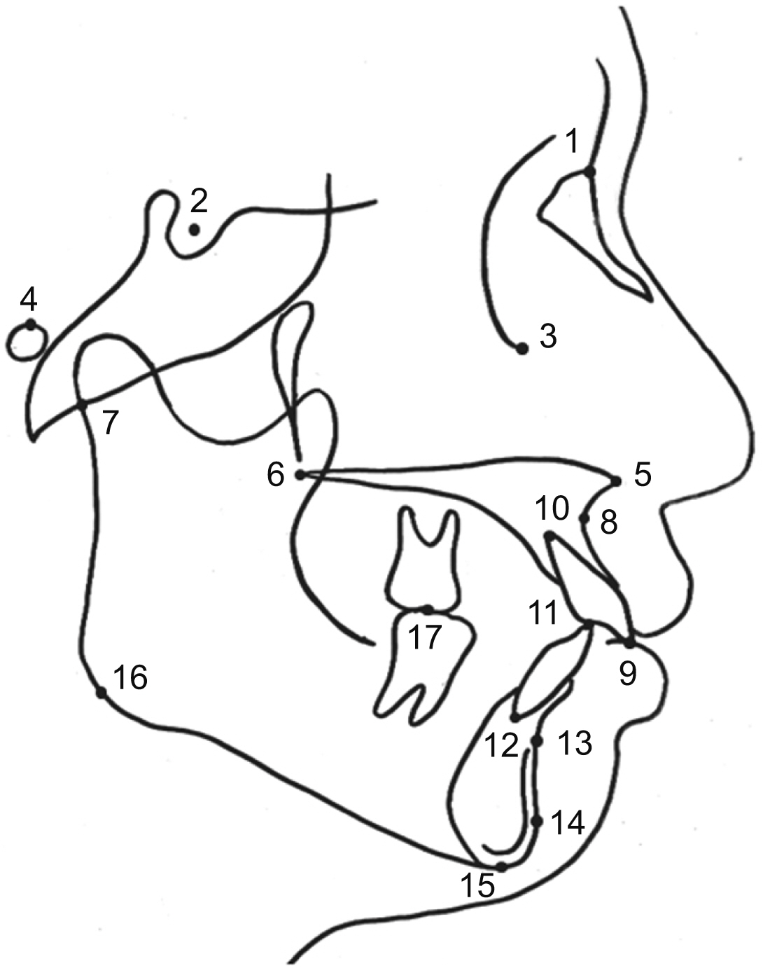
Figure 2 Landmarks used for measurements in the study. 1, nasion; 2, sella; 3, orbitale; 4, porion; 5, anterior nasal spine; 6, posterior nasal spine; 7, articulare; 8, point A; 9, incisal end of maxillary incisor; 10, apex of maxillary incisor; 11, incisal end of mandibular incisor; 12, apex of mandibular incisor; 13, point B; 14, pogonion; 15, menton; 16, gonion; and 17, articulation of maxillary and mandibular molars.
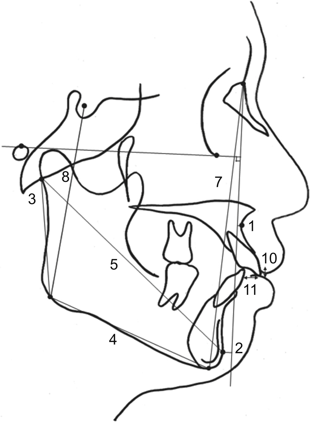
Figure 3 Linear measurements obtained in the study. 1, N-perpendicular to point A; 2, N-perpendicular to pogonion; 3, ramus height; 4, mandibular body length; 5, total mandibular length; 6, mandibular body length ratio (mandibular body length/total mandibular length); 7, anterior facial height; 8, posterior facial height; 9, facial height ratio (posterior facial height/anterior facial height); 10, overbite; and 11, overjet.

Figure 4 Angular measurements obtained in the study. 1, gonial angle; 2, maxillomandibular plane angle; 3, Frankfort horizontal plane to mandibular plane angle (FMA); 4, sella-nasion-A point angle (SNA); 5, sella-nasion-B point angle (SNB); 6, A point-nasion-B point angle (ANB); 7, ramus inclination; 8, interincisal angle; 9, mandibular incisor to Frankfort horizontal plane angle (FMIA); 10, mandibular incisor to mandibular plane angle (IMPA); 11, maxillary incisor to sella-nasion plane angle; and 12, maxillary incisor to Frankfort horizontal plane angle.
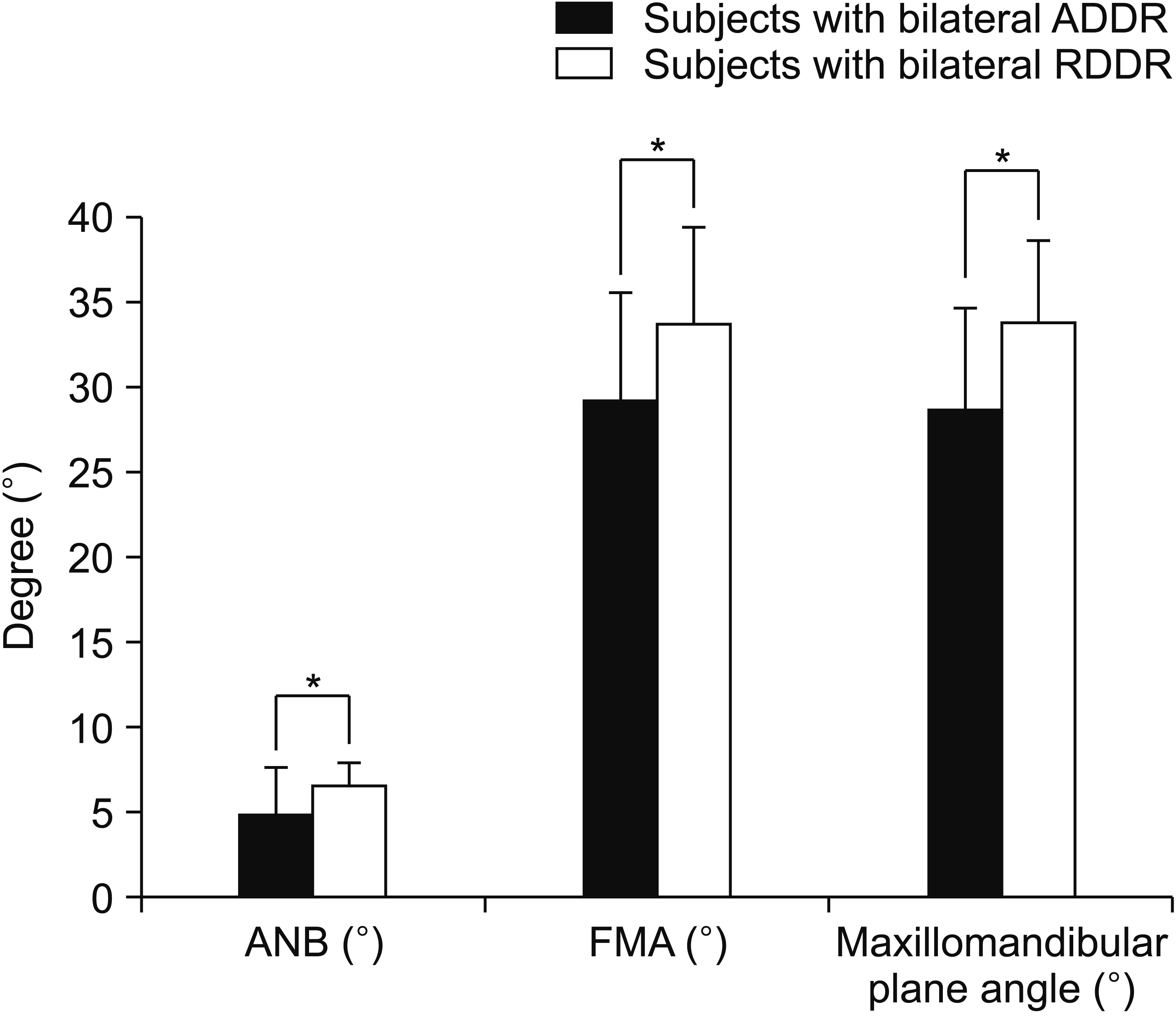
Figure 5 Dentoskeletal variables with significant differences between subjects with bilateral anterior disk displacement with reduction (ADDR) and those with bilateral rotational disk displacement with reduction (RDDR). ANB, A point-Nasion-B point angle; FMA, Frankfort horizontal plane to mandibular plane angle. Mann–Whitney U test; *p < 0.05.

Figure 6 Profilogram comparisons among five groups stratified according to the position of the temporomandibular joint disk on coronal and sagittal magnetic resonance images.
Reference
-
1. Ernberg M. 2017; The role of molecular pain biomarkers in temporomandibular joint internal derangement. J Oral Rehabil. 44:481–91. DOI: 10.1111/joor.12480. PMID: 28054366.
Article2. Larheim TA. 2005; Role of magnetic resonance imaging in the clinical diagnosis of the temporomandibular joint. Cells Tissues Organs. 180:6–21. DOI: 10.1159/000086194. PMID: 16088129.
Article3. Tasaki MM, Westesson PL, Isberg AM, Ren YF, Tallents RH. 1996; Classification and prevalence of temporomandibular joint disk displacement in patients and symptom-free volunteers. Am J Orthod Dentofacial Orthop. 109:249–62. DOI: 10.1016/S0889-5406(96)70148-8. PMID: 8607470.
Article4. Talaat WM, Adel OI, Al Bayatti S. 2018; Prevalence of temporomandibular disorders discovered incidentally during routine dental examination using the Research Diagnostic Criteria for Temporomandibular Disorders. Oral Surg Oral Med Oral Pathol Oral Radiol. 125:250–9. DOI: 10.1016/j.oooo.2017.11.012. PMID: 29274723.
Article5. Tamimi D, Jalali E, Hatcher D. 2018; Temporomandibular joint imaging. Radiol Clin North Am. 56:157–75. DOI: 10.1016/j.rcl.2017.08.011. PMID: 29157545.
Article6. Ahn SJ, Kim TW, Nahm DS. 2004; Cephalometric keys to internal derangement of temporomandibular joint in women with Class II malocclusions. Am J Orthod Dentofacial Orthop. 126:485–94. discussion 494–5. DOI: 10.1016/j.ajodo.2003.08.029. PMID: 15470352.
Article7. Nebbe B, Major PW, Prasad Ng. 1999; Female adolescent facial pattern associated with TMJ disk displacement and reduction in disk length: part I. Am J Orthod Dentofacial Orthop. 116:168–76. DOI: 10.1016/S0889-5406(99)70214-3. PMID: 10434090.
Article8. Khoury MB, Dolan E. 1986; Sideways dislocation of the temporomandibular joint meniscus: the edge sign. AJNR Am J Neuroradiol. 7:869–72. PMID: 3096106.9. Tasaki MM, Westesson PL. 1993; Temporomandibular joint: diagnostic accuracy with sagittal and coronal MR imaging. Radiology. 186:723–9. DOI: 10.1148/radiology.186.3.8430181. PMID: 8430181.
Article10. Liedberg J, Westesson PL, Kurita K. 1990; Sideways and rotational displacement of the temporomandibular joint disk: diagnosis by arthrography and correlation to cryosectional morphology. Oral Surg Oral Med Oral Pathol. 69:757–63. DOI: 10.1016/0030-4220(90)90362-V. PMID: 2356088.
Article11. Aoyama S, Kino K, Amagasa T, Sakamoto I, Omura K, Honda E, et al. 2002; Clinical and magnetic resonance imaging study of unilateral sideways disc displacements of the temporomandibular joint. J Med Dent Sci. 49:89–94. PMID: 12627814.12. Jeon DM, Jung WS, Mah SJ, Kim TW, Ahn SJ. 2014; The effects of TMJ symptoms on skeletal morphology in orthodontic patients with TMJ disc displacement. Acta Odontol Scand. 72:776–82. DOI: 10.3109/00016357.2014.906650. PMID: 24702009.
Article13. Ahn SJ, Lee SP, Nahm DS. 2005; Relationship between temporomandibular joint internal derangement and facial asymmetry in women. Am J Orthod Dentofacial Orthop. 128:583–91. DOI: 10.1016/j.ajodo.2004.06.038. PMID: 16286205.
Article14. Ahn SJ, Baek SH, Kim TW, Nahm DS. 2006; Discrimination of internal derangement of temporomandibular joint by lateral cephalometric analysis. Am J Orthod Dentofacial Orthop. 130:331–9. DOI: 10.1016/j.ajodo.2005.02.019. PMID: 16979491.
Article15. Dahlberg G. 1940. Statistical methods for medical and biological students. G. Allen & Unwin Ltd;London: p. 232.16. Katzberg RW, Westesson PL, Tallents RH, Anderson R, Kurita K, Manzione JV Jr, et al. 1988; Temporomandibular joint: MR assessment of rotational and sideways disk displacements. Radiology. 169:741–8. DOI: 10.1148/radiology.169.3.3186996. PMID: 3186996.
Article17. Foucart JM, Carpentier P, Pajoni D, Marguelles-Bonnet R, Pharaboz C. 1998; MR of 732 TMJs: anterior, rotational, partial and sideways disc displacements. Eur J Radiol. 28:86–94. DOI: 10.1016/S0720-048X(97)00102-2. PMID: 9717628.
Article18. Brand JW, Nielson KJ, Tallents RH, Nanda RS, Currier GF, Owen WL. 1995; Lateral cephalometric analysis of skeletal patterns in patients with and without internal derangement of the temporomandibular joint. Am J Orthod Dentofacial Orthop. 107:121–8. DOI: 10.1016/S0889-5406(95)70126-5. PMID: 7847269.
Article19. Schellhas KP, Pollei SR, Wilkes CH. 1993; Pediatric internal derangements of the temporomandibular joint: effect on facial development. Am J Orthod Dentofacial Orthop. 104:51–9. DOI: 10.1016/0889-5406(93)70027-L. PMID: 8179643.
Article20. Chang MS, Choi JH, Yang IH, An JS, Heo MS, Ahn SJ. 2018; Relationships between temporomandibular joint disk displacements and condylar volume. Oral Surg Oral Med Oral Pathol Oral Radiol. 125:192–8. DOI: 10.1016/j.oooo.2017.11.001. PMID: 29233525.21. Schellhas KP, Piper MA, Omlie MR. 1990; Facial skeleton remodeling due to temporomandibular joint degeneration: an imaging study of 100 patients. AJNR Am J Neuroradiol. 11:541–51. DOI: 10.2214/ajr.155.2.2115271. PMID: 2112321.
Article22. Milam SB. 2005; Pathogenesis of degenerative temporomandibular joint arthritides. Odontology. 93:7–15. DOI: 10.1007/s10266-005-0056-7. PMID: 16170470.
Article23. Legrell PE, Reibel J, Nylander K, Hörstedt P, Isberg A. 1999; Temporomandibular joint condyle changes after surgically induced non-reducing disk displacement in rabbits: a macroscopic and microscopic study. Acta Odontol Scand. 57:290–300. DOI: 10.1080/000163599428724. PMID: 10614908.24. Legrell PE, Isberg A. 1999; Mandibular length and midline asymmetry after experimentally induced temporomandibular joint disk displacement in rabbits. Am J Orthod Dentofacial Orthop. 115:247–53. DOI: 10.1016/S0889-5406(99)70325-2. PMID: 10066971.
Article25. Osborn JW. 1985; The disc of the human temporomandibular joint: design, function and failure. J Oral Rehabil. 12:279–93. DOI: 10.1111/j.1365-2842.1985.tb01283.x. PMID: 3862791.
Article26. Takahara N, Nakagawa S, Sumikura K, Kabasawa Y, Sakamoto I, Harada H. 2017; Association of temporomandibular joint pain according to magnetic resonance imaging findings in temporomandibular disorder patients. J Oral Maxillofac Surg. 75:1848–55. DOI: 10.1016/j.joms.2017.03.026. PMID: 28413149.
Article27. Torres MG, Crusoé-Rebello IM, Rosário M, Albuquerque MC, Campos PS. 2016; Morphometric features of the mandibular condyle and association with disk abnormalities. Oral Surg Oral Med Oral Pathol Oral Radiol. 121:566–72. DOI: 10.1016/j.oooo.2016.01.020. PMID: 27068314.
Article28. Nebbe B, Major PW. 2000; Prevalence of TMJ disc displacement in a pre-orthodontic adolescent sample. Angle Orthod. 70:454–63. DOI: 10.1043/0003-3219(2000)070<0454:POTDDI>2.0.CO;2. PMID: 11138649.29. Kwon HB, Kim H, Jung WS, Kim TW, Ahn SJ. 2013; Gender differences in dentofacial characteristics of adult patients with temporomandibular disc displacement. J Oral Maxillofac Surg. 71:1178–86. DOI: 10.1016/j.joms.2012.12.015. PMID: 23455416.
Article30. Ikeda R, Ikeda K. 2016; Directional characteristics of incipient temporomandibular joint disc displacements: a magnetic resonance imaging study. Am J Orthod Dentofacial Orthop. 149:39–45. DOI: 10.1016/j.ajodo.2015.06.021. PMID: 26718376.
Article
- Full Text Links
-
- Actions
-
Cited
- CITED
-
- Close
- Share
-
- Similar articles
-
- Relation between shape of the articular eminence and disc displacement in the temporomandibular joint
- Positional and morphologic changes of the temporomandibular joint disc using magnetic resonance imaging
- Correlation between internal derangement and osteoarthrosis in the temporomandibular joint using magnetic resonance imaging
- CHANGE OF JOINT SPACE ACCORDING TO ANTERIOR DISC DISPLACEMENT OF TMJ
- A Clinical Study Of Temporomandibular Joint Disorders By Using Arthrography
