Evaluation of a multi-stage convolutional neural network-based fully automated landmark identification system using cone-beam computed tomographysynthesized posteroanterior cephalometric images
- Affiliations
-
- 1Department of Orthodontics, Graduate School, Kyung Hee University, Seoul, Korea
- 2Department of Orthodontics, Peking University School of Stomatology, Beijing, China
- 3Department of Oral and Maxillofacial Radiology, Graduate School, Kyung Hee University, Seoul, Korea
- 4Division of Orthodontics, Department of Orofacial Science, University of California San Francisco, CA, USA
- KMID: 2513576
- DOI: http://doi.org/10.4041/kjod.2021.51.2.77
Abstract
Objective
To evaluate the accuracy of a multi-stage convolutional neural network (CNN) model-based automated identification system for posteroanterior (PA) cephalometric landmarks.
Methods
The multi-stage CNN model was implemented with a personal computer. A total of 430 PA-cephalograms synthesized from cone-beam computed tomography scans (CBCT-PA) were selected as samples. Twenty-three landmarks used for Tweemac analysis were manually identified on all CBCT-PA images by a single examiner. Intra-examiner reproducibility was confirmed by repeating the identification on 85 randomly selected images, which were subsequently set as test data, with a two-week interval before training. For initial learning stage of the multi-stage CNN model, the data from 345 of 430 CBCT-PA images were used, after which the multi-stage CNN model was tested with previous 85 images. The first manual identification on these 85 images was set as a truth ground. The mean radial error (MRE) and successful detection rate (SDR) were calculated to evaluate the errors in manual identification and artificial intelligence (AI) prediction.
Results
The AI showed an average MRE of 2.23 ± 2.02 mm with an SDR of 60.88% for errors of 2 mm or lower. However, in a comparison of the repetitive task, the AI predicted landmarks at the same position, while the MRE for the repeated manual identification was 1.31 ± 0.94 mm.
Conclusions
Automated identification for CBCT-synthesized PA cephalometric landmarks did not sufficiently achieve the clinically favorable error range of less than 2 mm. However, AI landmark identification on PA cephalograms showed better consistency than manual identification.
Keyword
Figure
-

Figure 1 Flow diagram showing the processing of the cone-beam computed tomography (CBCT)-synthesized posteroanterior (PA) cephalograms. The raw CBCT data were imported using Dolphin software 11.95 Premium (Dolphin Imaging & Management Solutions, Chatsworth, CA, USA). The head position was adjusted to reduce layered bilateral structures. The ‘Build X-ray’ button in the software was used to synthesize the CBCT-PA with orthogonal X-ray exposure while eliminating virtual magnification, which causes image distortion.

Figure 2 Schematic experimental design summary of the multi-stage convolutional neural network (CNN) model. Conv, convolution; FC, fully connected; AI, artificial intelligence.
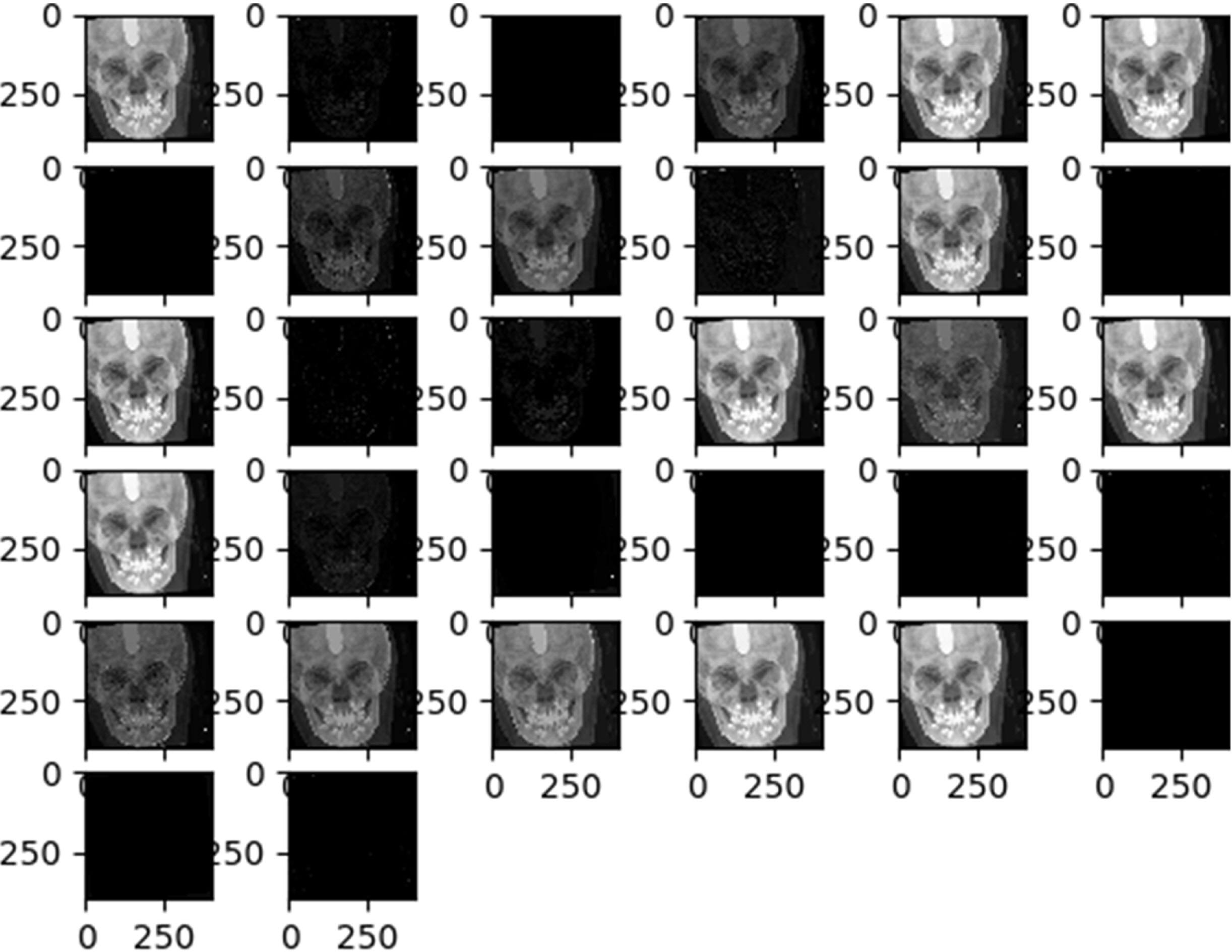
Figure 3 The visualized effect of each convolutional layer during the first stage training.
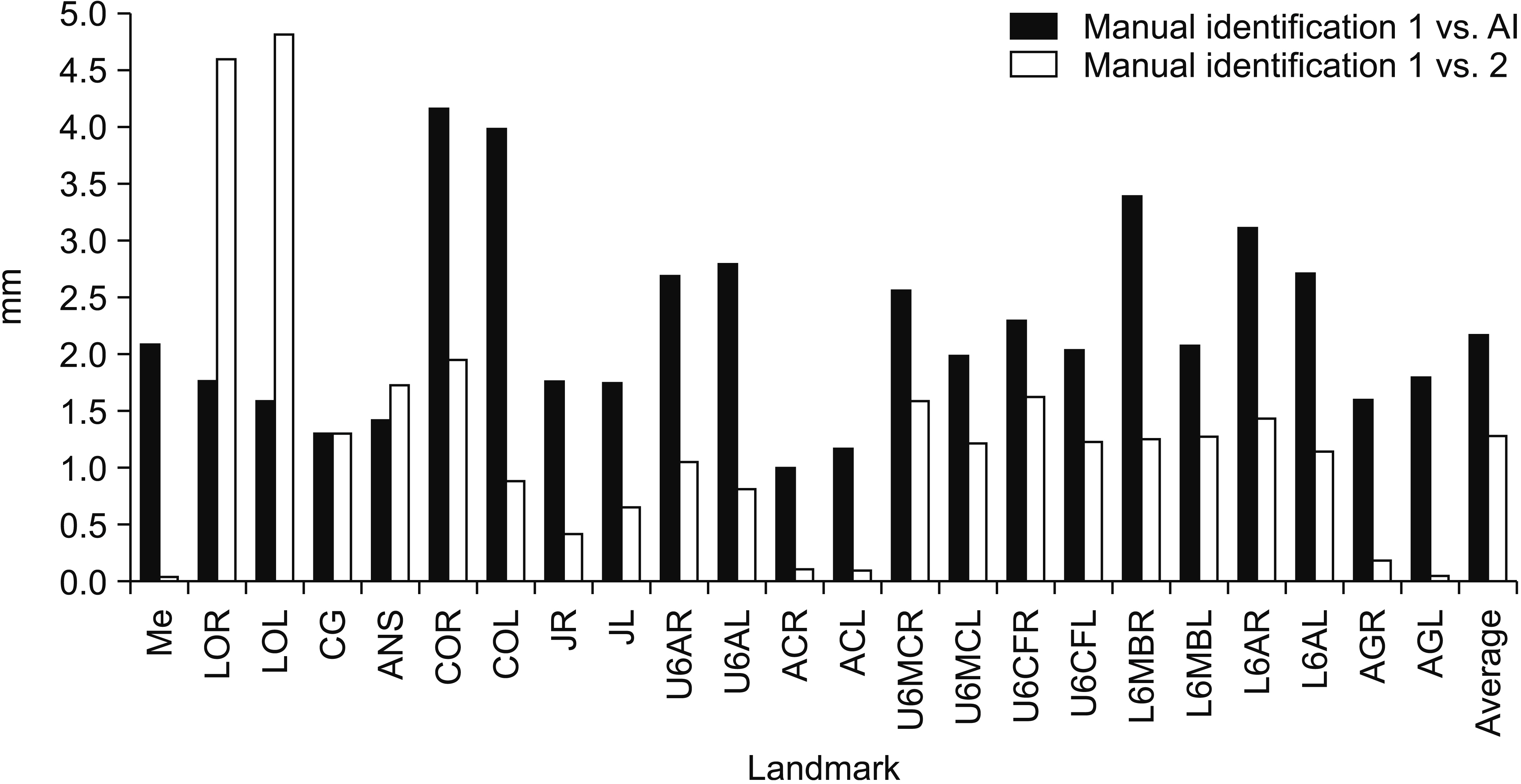
Figure 4 Mean radial error (MRE) for each landmark, and the average MRE between manual identification 1 and artificial intelligence (AI) (black) and manual identifications 1 and 2 (white). See Table 1 for definitions of the other landmarks.
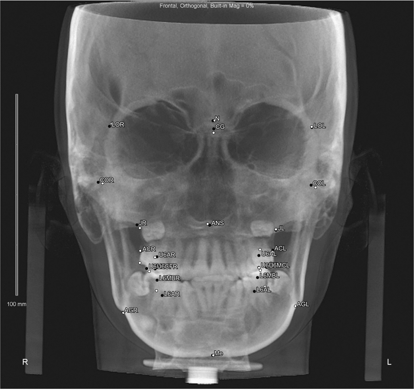
Figure 5 Accuracy of the convolutional neural network based on the automatic landmark identification system for cone-beam computed tomography using the synthesized posteroanterior cephalograms. The black dot represents the manually identified landmark and the white dot indicates the automatically identified landmark. N, nasion. See Table 1 for definitions of the other landmarks.
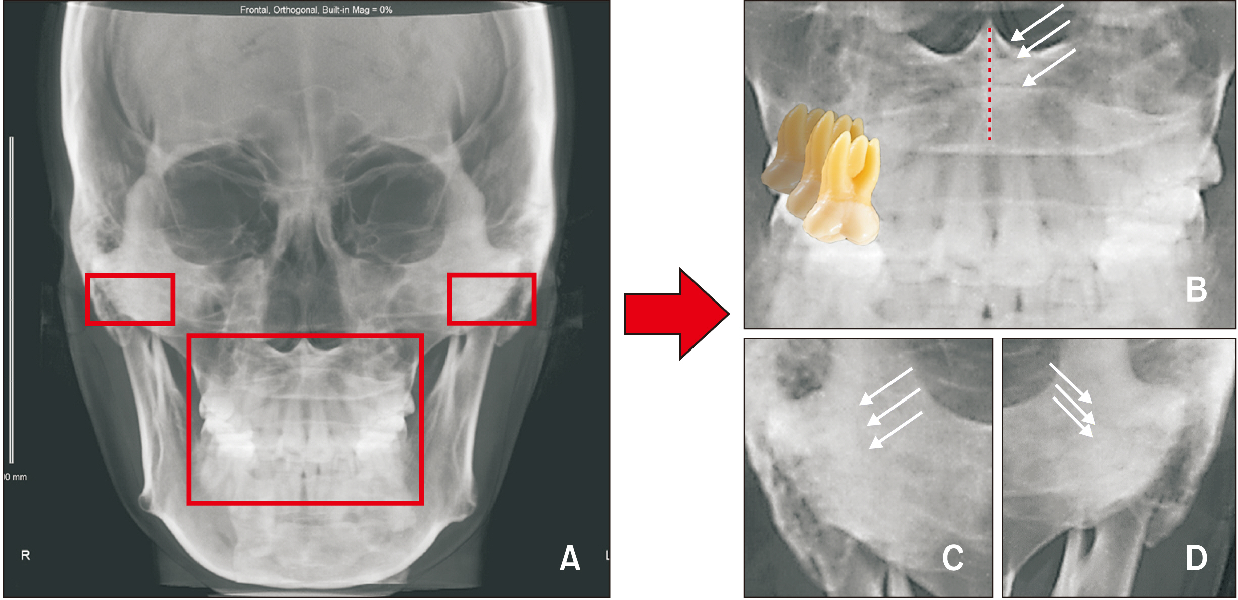
Figure 6 Many structures layered in vertical dimension. The condyle and dentoalveolar areas of the posteroanterior (PA) cephalograms interfere with the artificial intelligence image recognition ability. A, Cone-beam computed tomography-synthesized PA cephalogram. B, Vertical layered images at the intersection of the nasal septum and palatal area (white arrow) and vertical layered and horizontal superimposed images in the dental area. C, D, Vertical layered images in the condyle area.
Reference
-
1. Major PW, Johnson DE, Hesse KL, Glover KE. 1994; Landmark identification error in posterior anterior cephalometrics. Angle Orthod. 64:447–54. DOI: 10.1043/0003-3219(1994)064<0447:LIEIPA>2.0.CO;2. PMID: 7864466.2. Ulkur F, Ozdemir F, Germec-Cakan D, Kaspar EC. 2016; Landmark errors on posteroanterior cephalograms. Am J Orthod Dentofacial Orthop. 150:324–31. DOI: 10.1016/j.ajodo.2016.01.016. PMID: 27476366.
Article3. Na ER, Aljawad H, Lee KM, Hwang HS. 2019; A comparative study of the reproducibility of landmark identification on posteroanterior and anteroposterior cephalograms generated from cone-beam computed tomography scans. Korean J Orthod. 49:41–8. DOI: 10.4041/kjod.2019.49.1.41. PMID: 30603624. PMCID: PMC6306316.
Article4. Sicurezza E, Greco M, Giordano D, Maiorana F, Leonardi R. 2012; Accuracy of landmark identification on postero-anterior cephalograms. Prog Orthod. 13:132–40. DOI: 10.1016/j.pio.2011.10.003. PMID: 23021116.
Article5. Pirttiniemi P, Miettinen J, Kantomaa T. 1996; Combined effects of errors in frontal-view asymmetry diagnosis. Eur J Orthod. 18:629–36. DOI: 10.1093/ejo/18.6.629. PMID: 9009427.
Article6. Major PW, Johnson DE, Hesse KL, Glover KE. 1996; Effect of head orientation on posterior anterior cephalometric landmark identification. Angle Orthod. 66:51–60. DOI: 10.1043/0003-3219(1996)066<0051:EOHOOP>2.3.CO;2. PMID: 8678346.7. Smektała T, Jędrzejewski M, Szyndel J, Sporniak-Tutak K, Olszewski R. 2014; Experimental and clinical assessment of three-dimensional cephalometry: a systematic review. J Craniomaxillofac Surg. 42:1795–801. DOI: 10.1016/j.jcms.2014.06.017. PMID: 25037877.
Article8. Kamoen A, Dermaut L, Verbeeck R. 2001; The clinical significance of error measurement in the interpretation of treatment results. Eur J Orthod. 23:569–78. DOI: 10.1093/ejo/23.5.569. PMID: 11668876.
Article9. Gribel BF, Gribel MN, Frazäo DC, McNamara JA Jr, Manzi FR. 2011; Accuracy and reliability of craniometric measurements on lateral cephalometry and 3D measurements on CBCT scans. Angle Orthod. 81:26–35. DOI: 10.2319/032210-166.1. PMID: 20936951.
Article10. Gribel BF, Gribel MN, Manzi FR, Brooks SL, McNamara JA Jr. 2011; From 2D to 3D: an algorithm to derive normal values for 3-dimensional computerized assessment. Angle Orthod. 81:3–10. DOI: 10.2319/032910-173.1. PMID: 20936948.
Article11. Meiyappan N, Tamizharasi S, Senthilkumar KP, Janardhanan K. 2015; Natural head position: an overview. J Pharm Bioallied Sci. 7(Suppl 2):S424–7. DOI: 10.4103/0975-7406.163488. PMID: 26538891. PMCID: PMC4606633.
Article12. El-Mangoury NH, Shaheen SI, Mostafa YA. 1987; Landmark identification in computerized posteroanterior cephalometrics. Am J Orthod Dentofacial Orthop. 91:57–61. DOI: 10.1016/0889-5406(87)90209-5. PMID: 3467581.
Article13. Leonardi R, Annunziata A, Caltabiano M. 2008; Landmark identification error in posteroanterior cephalometric radiography. A systematic review. Angle Orthod. 784:761–5. DOI: 10.2319/0003-3219(2008)078[0761:LIEIPC]2.0.CO;2. PMID: 18302479.14. Shokri A, Miresmaeili A, Farhadian N, Falah-Kooshki S, Amini P, Mollaie N. 2017; Effect of changing the head position on accuracy of transverse measurements of the maxillofacial region made on cone beam computed tomography and conventional posterior-anterior cephalograms. Dentomaxillofac Radiol. 46:20160180. DOI: 10.1259/dmfr.20160180. PMID: 28306330. PMCID: PMC5595030.
Article15. Oshagh M, Shahidi SH, Danaei SH. 2013; Effects of image enhancement on reliability of landmark identification in digital cephalometry. Indian J Dent Res. 24:98–103. DOI: 10.4103/0970-9290.114958. PMID: 23852241.
Article16. Arık SÖ, Ibragimov B, Xing L. 2017; Fully automated quantitative cephalometry using convolutional neural networks. J Med Imaging (Bellingham). 4:014501. DOI: 10.1117/1.JMI.4.1.014501. PMID: 28097213. PMCID: PMC5220585.17. Park JH, Hwang HW, Moon JH, Yu Y, Kim H, Her SB, et al. 2019; Automated identification of cephalometric landmarks: part 1-comparisons between the latest deep-learning methods YOLOV3 and SSD. Angle Orthod. 89:903–9. DOI: 10.2319/022019-127.1. PMID: 31282738.
Article18. Hwang HW, Park JH, Moon JH, Yu Y, Kim H, Her SB, et al. 2019; Automated identification of cephalometric landmarks: part 2-might it be better than human? Angle Orthod. 90:69–76. DOI: 10.2319/022019-129.1. PMID: 31335162.19. Takahashi R, Matsubara T, Uehara K. 2017. Multi-stage convolutional neural networks for robustness to scale transformation. Paper presented at: 2017 International Symposium on Nonlinear Theory and Its Applications. 2017 Dec 4-7; Cancun, Mexico. NOLTA;Cancun: p. 692–5.20. Anwar SM, Majid M, Qayyum A, Awais M, Alnowami M, Khan MK. 2018; Medical image analysis using convolutional neural networks: a review. J Med Syst. 42:226. DOI: 10.1007/s10916-018-1088-1. PMID: 30298337.
Article21. Yadav SS, Jadhav SM. 2019; Deep convolutional neural network based medical image classification for disease diagnosis. J Big Data. 6:113. DOI: 10.1186/s40537-019-0276-2.
Article22. Schwendicke F, Golla T, Dreher M, Krois J. 2019; Convolutional neural networks for dental image diagnostics: a scoping review. J Dent. 91:103226. DOI: 10.1016/j.jdent.2019.103226. PMID: 31704386.
Article23. Park JJ, Kim KA, Nam Y, Choi MH, Choi SY, Rhie J. 2020; Convolutional-neural-network-based diagnosis of appendicitis via CT scans in patients with acute abdominal pain presenting in the emergency department. Sci Rep. 10:9556. DOI: 10.1038/s41598-020-66674-7. PMID: 32533053. PMCID: PMC7293232.
Article
- Full Text Links
-
- Actions
-
Cited
- CITED
-
- Close
- Share
-
- Similar articles
-
- A comparative study of the reproducibility of landmark identification on posteroanterior and anteroposterior cephalograms generated from cone-beam computed tomography scans
- Effect of Voxel Size on the Accuracy of Landmark Identification in Cone-Beam Computed Tomography Images
- Evaluation of maxillary sinusitis from panoramic radiographs and cone-beam computed tomographic images using a convolutional neural network
- The comparison of landmark identification errors and reproducibility between conventional lateral cephalometric radiography and digital lateral cephalometric radiography
- The genial tubercle: A prospective novel landmark for the diagnosis of mandibular asymmetry
