Surgical Management of Lymphedema: An Overview of Preoperative Evaluation and Surgical Techniques
- Affiliations
-
- 1Department of Plastic Surgery, Asan Medical Center, University of Ulsan College of Medicine, Seoul, Korea
- KMID: 2513204
- DOI: http://doi.org/10.12790/ahm.20.0055
Abstract
- Lymphedema is a chronic and progressive disease that affects many of the patients who underwent cancer ablative surgery and decreases the quality of life of them. Surgical management including lymphovenous shunting and vascularized lymph node transfer become popular in the field of microsurgery. For the better outcome of microsurgical approach to lymphedema, understanding of multiple image modalities is essential. Also, understanding other conservative management tools is crucial for setting reasonable algorism for lymphedema management.
Figure
-
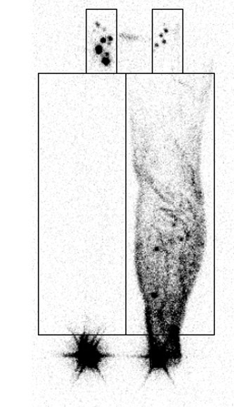
Fig. 1. Lymphoscintigraphy of left leg lymphedema. Dermal backflow is shown in the entire left leg. Written informed consent was obtained for publication of this study and accompanying images.
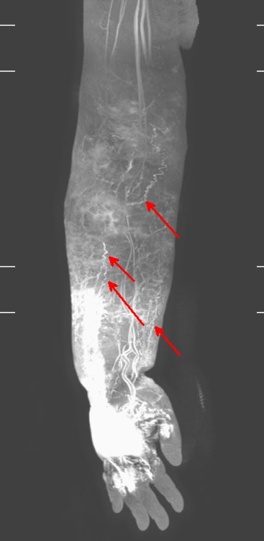
Fig. 2. Magnetic resonance lymphoscintigraphy of the upper arm with lymphedema. Dermal backflow and the tortuous lymphatic vessel (arrows) were found in the fat layer. Written informed consent was obtained for publication of this study and accompanying images.
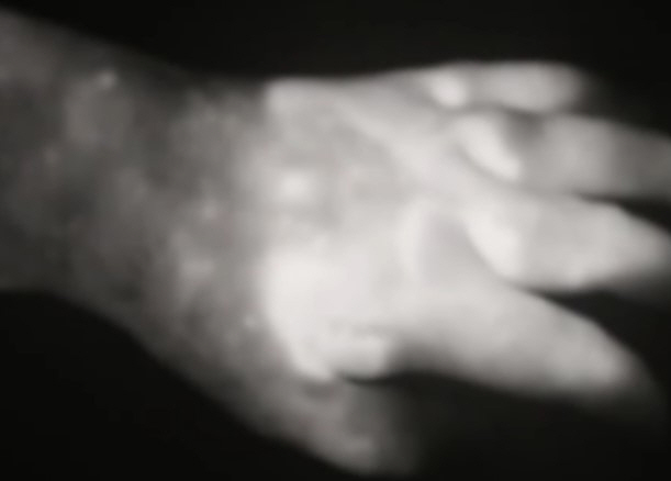
Fig. 3. Indocyanine green lymphography of hand dorsum. There is a dermal backflow. Written informed consent was obtained for publication of this study and accompanying images.

Fig. 4. Duplex color Doppler visualizes cutaneous vein (red arrow) and lymphatic vessel located in deeper layer (green arrow). Written informed consent was obtained for publication of this study and accompanying images.

Fig. 5. Sixty four-years-old female suffered from the left leg for 15 years after ovarian cancer surgery. (A) The patient showed severe swelling of the thigh even after compressive decongestive therapy. (B) Magnetic resonance lymphangiography showed a good functioning lymphatic vessel at the lower leg. (C, D) Three lymphovenous anastomoses were done, and there was good lymphatic flow in indocyanine green lymphangiography intraoperatively by an embedded infrared camera in microscopy. (E) Reduction of thigh circumference was maintained after 6 months of lymphovenous shunting surgery. Written informed consent was obtained for publication of this study and accompanying images.

Fig. 6. Two lymphatic vessels were found in the fat layer after making a skin incision. Fluorescent dye was injected before making a skin incision. These deep lymphatic vessels were sized 0.3 mm and 0.5 mm. Written informed consent was obtained for publication of this study and accompanying images.
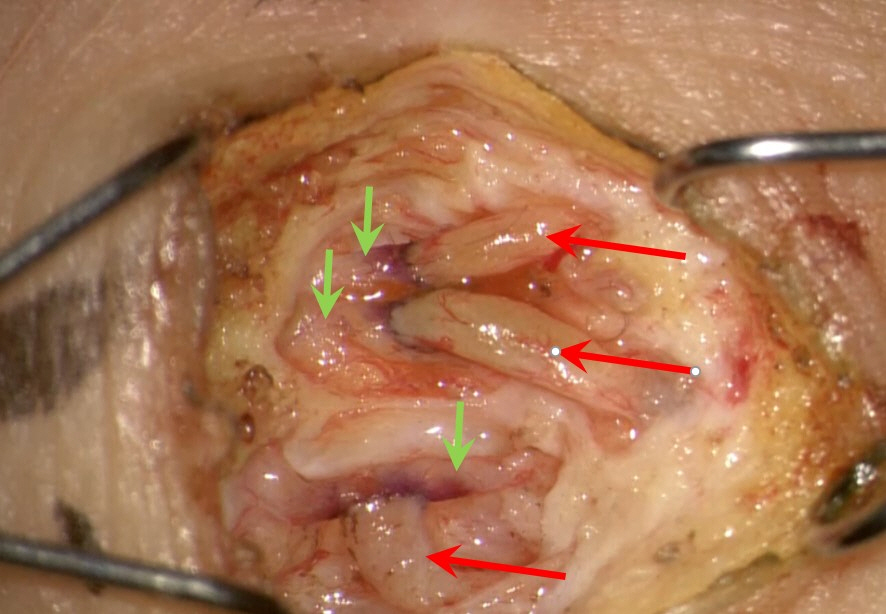
Fig. 7. Three lymphovenous shunting were done through a 1-cm skin incision on the ankle. Veins (red arrows) showed a clear lumen after shunting due to good lymphatic flow to the proximal vein from lymphatic vessels (green arrows). Written informed consent was obtained for publication of this study and accompanying images.

Fig. 8. One vascular lymph node and skin flap, including lymphatic vessel in the deep fat layer, was harvested from the groin. Written informed consent was obtained for publication of this study and accompanying images. Written informed consent was obtained for publication of this study and accompanying images.
Reference
-
1. Warren AG, Brorson H, Borud LJ, Slavin SA. Lymphedema: a comprehensive review. Ann Plast Surg. 2007; 59:464–72.2. Brouillard P, Boon L, Vikkula M. Genetics of lymphatic anomalies. J Clin Invest. 2014; 124:898–904.
Article3. Turpin S, Lambert R. Lymphoscintigraphy of chylous anomalies: chylothorax, chyloperitoneum, chyluria, and lymphangiomatosis-15-year experience in a pediatric setting and review of the literature. J Nucl Med Technol. 2018; 46:123–8.
Article4. Maclellan RA, Greene AK. Lymphedema. Semin Pediatr Surg. 2014; 23:191–7.
Article5. Yao K, Tu H, Li YH, et al. Modified technique of radical inguinal lymphadenectomy for penile carcinoma: morbidity and outcome. J Urol. 2010; 184:546–52.
Article6. Van der Zee AG, Oonk MH, De Hullu JA, et al. Sentinel node dissection is safe in the treatment of early-stage vulvar cancer. J Clin Oncol. 2008; 26:884–9.
Article7. DiSipio T, Rye S, Newman B, Hayes S. Incidence of unilateral arm lymphoedema after breast cancer: a systematic review and meta-analysis. Lancet Oncol. 2013; 14:500–15.
Article8. Becker C, Arrive L, Saaristo A, et al. Surgical treatment of congenital lymphedema. Clin Plast Surg. 2012; 39:377–84.
Article9. Gashev AA, Zawieja DC. Hydrodynamic regulation of lymphatic transport and the impact of aging. Pathophysiology. 2010; 17:277–87.
Article10. Borri M, Gordon KD, Hughes JC, et al. Magnetic resonance imaging-based assessment of breast cancer-related lymphoedema tissue composition. Invest Radiol. 2017; 52:554–61.
Article11. Kim G, Smith MP, Donohoe KJ, Johnson AR, Singhal D, Tsai LL. MRI staging of upper extremity secondary lymphedema: correlation with clinical measurements. Eur Radiol. 2020; 30:4686–94.
Article12. Zampell JC, Aschen S, Weitman ES, et al. Regulation of adipogenesis by lymphatic fluid stasis: part I. Adipogenesis, fibrosis, and inflammation. Plast Reconstr Surg. 2012; 129:825–34.13. Chu SY, Wang SC, Chan WH, et al. Volumetric differences in the suprafascial and subfascial compartments of patients with secondary unilateral lower limb lymphedema. Plast Reconstr Surg. 2020; 145:1528–37.
Article14. Kerchner K, Fleischer A, Yosipovitch G. Lower extremity lymphedema update: pathophysiology, diagnosis, and treatment guidelines. J Am Acad Dermatol. 2008; 59:324–31.15. Badger CM, Peacock JL, Mortimer PS. A randomized, controlled, parallel-group clinical trial comparing multilayer bandaging followed by hosiery versus hosiery alone in the treatment of patients with lymphedema of the limb. Cancer. 2000; 88:2832–7.
Article16. Ezzo J, Manheimer E, McNeely ML, et al. Manual lymphatic drainage for lymphedema following breast cancer treatment. Cochrane Database Syst Rev. 2015; (5):CD003475.
Article17. Shao Y, Zhong DS. Manual lymphatic drainage for breast cancer-related lymphoedema. Eur J Cancer Care (Engl). 2017; 26:10.1111/ecc.12517.
Article18. Ko DS, Lerner R, Klose G, Cosimi AB. Effective treatment of lymphedema of the extremities. Arch Surg. 1998; 133:452–8.
Article19. Jeffs E, Ream E, Shewbridge A, et al. Exploring patient perception of success and benefit in self-management of breast cancer-related arm lymphoedema. Eur J Oncol Nurs. 2016; 20:173–83.
Article20. National Comprehensive Cancer Network (NCCN). NCCN guidelines: clinical practice guidelines in oncology. Survivorship version 2, 2020 [Internet]. Plymouth Meeting (PA): NCCN;2020. Jul. 14. [cited 2021 Jan 12]. Available from: https://www.nccn.org/professionals/physician_gls/default.aspx.21. Shah C, Arthur DW, Wazer D, Khan A, Ridner S, Vicini F. The impact of early detection and intervention of breast cancer-related lymphedema: a systematic review. Cancer Med. 2016; 5:1154–62.
Article22. Lasinski BB, McKillip Thrift K, Squire D, et al. A systematic review of the evidence for complete decongestive therapy in the treatment of lymphedema from 2004 to 2011. PM R. 2012; 4:580–601.
Article23. Cormier JN, Rourke L, Crosby M, Chang D, Armer J. The surgical treatment of lymphedema: a systematic review of the contemporary literature (2004-2010). Ann Surg Oncol. 2012; 19:642–51.
Article24. Campisi C, Bellini C, Campisi C, Accogli S, Bonioli E, Boccardo F. Microsurgery for lymphedema: clinical research and long-term results. Microsurgery. 2010; 30:256–60.
Article25. Stecker MS, Fan CM. Lymphangiography for thoracic duct interventions. Tech Vasc Interv Radiol. 2016; 19:277–85.
Article26. O'Donnell TF Jr, Rasmussen JC, Sevick-Muraca EM. New diagnostic modalities in the evaluation of lymphedema. J Vasc Surg Venous Lymphat Disord. 2017; 5:261–73.27. Notohamiprodjo M, Weiss M, Baumeister RG, et al. MR lymphangiography at 3.0 T: correlation with lymphoscintigraphy. Radiology. 2012; 264:78–87.
Article28. Mitsumori LM, McDonald ES, Wilson GJ, Neligan PC, Minoshima S, Maki JH. MR lymphangiography: how I do it. J Magn Reson Imaging. 2015; 42:1465–77.
Article29. Mitsumori LM, McDonald ES, Neligan PC, Maki JH. Peripheral magnetic resonance lymphangiography: techniques and applications. Tech Vasc Interv Radiol. 2016; 19:262–72.
Article30. Mazzei FG, Gentili F, Guerrini S, et al. MR lymphangiography: a practical guide to perform it and a brief review of the literature from a technical point of view. Biomed Res Int. 2017; 2017:2598358.
Article31. Chen WF, Lensing JN, Bowen M. Indocyanine green lymphography can be falsely negative in early extremity lymphedema. J Plast Reconstr Aesthet Surg. 2020; 73:391–407.
Article32. Narushima M, Yamamoto T, Ogata F, Yoshimatsu H, Mihara M, Koshima I. Indocyanine green lymphography findings in limb lymphedema. J Reconstr Microsurg. 2016; 32:72–9.
Article33. Yamamoto T, Yamamoto N, Doi K, et al. Indocyanine green-enhanced lymphography for upper extremity lymphedema: a novel severity staging system using dermal backflow patterns. Plast Reconstr Surg. 2011; 128:941–7.34. Hayashi A, Visconti G, Yamamoto T, et al. Intraoperative imaging of lymphatic vessel using ultra high-frequency ultrasound. J Plast Reconstr Aesthet Surg. 2018; 71:778–80.
Article35. Hayashi A, Hayashi N, Yoshimatsu H, Yamamoto T. Effective and efficient lymphaticovenular anastomosis using preoperative ultrasound detection technique of lymphatic vessels in lower extremity lymphedema. J Surg Oncol. 2018; 117:290–8.
Article36. Greene AK, Goss JA. Diagnosis and staging of lymphedema. Semin Plast Surg. 2018; 32:12–6.37. International Society of Lymphology. The diagnosis and treatment of peripheral lymphedema: 2013 Consensus Document of the International Society of Lymphology. Lymphology. 2013; 46:1–11.38. Mihara M, Hayashi Y, Hara H, et al. High-accuracy diagnosis and regional classification of lymphedema using indocyanine green fluorescent lymphography after gynecologic cancer treatment. Ann Plast Surg. 2014; 72:204–8.
Article39. Garza RM, Ooi ASH, Falk J, Chang DW. The relationship between clinical and indocyanine green staging in lymphedema. Lymphat Res Biol. 2019; 17:329–33.
Article40. Yamaguchi K, Kimata Y, Yamada K, Suami H. Peripheral venous angle plasty: a new lymphovenous anastomosis technique for lower extremity lymphedema. Plast Reconstr Surg. 2012; 130:233e–235e.41. Torrisi JS, Joseph WJ, Ghanta S, et al. Lymphaticovenous bypass decreases pathologic skin changes in upper extremity breast cancer-related lymphedema. Lymphat Res Biol. 2015; 13:46–53.
Article42. Yamamoto T, Yamamoto N, Numahata T, et al. Navigation lymphatic supermicrosurgery for the treatment of cancer-related peripheral lymphedema. Vasc Endovascular Surg. 2014; 48:139–43.
Article43. AlJindan FK, Lin CY, Cheng MH. Comparison of outcomes between side-to-end and end-to-end lymphovenous anastomoses for early-grade extremity lymphedema. Plast Reconstr Surg. 2019; 144:486–96.
Article44. Patel KM, Lin CY, Cheng MH. From theory to evidence: long-term evaluation of the mechanism of action and flap integration of distal vascularized lymph node transfers. J Reconstr Microsurg. 2015; 31:26–30.
Article45. Cheng MH, Huang JJ, Wu CW, et al. The mechanism of vascularized lymph node transfer for lymphedema: natural lymphaticovenous drainage. Plast Reconstr Surg. 2014; 133:192e–198e.46. Aschen SZ, Farias-Eisner G, Cuzzone DA, et al. Lymph node transplantation results in spontaneous lymphatic reconnection and restoration of lymphatic flow. Plast Reconstr Surg. 2014; 133:301–10.
Article47. Scaglioni MF, Arvanitakis M, Chen YC, Giovanoli P, Chia-Shen Yang J, Chang EI. Comprehensive review of vascularized lymph node transfers for lymphedema: outcomes and complications. Microsurgery. 2018; 38:222–9.
Article48. Koshima I, Narushima M, Mihara M, et al. Lymphadiposal flaps and lymphaticovenular anastomoses for severe leg edema: functional reconstruction for lymph drainage system. J Reconstr Microsurg. 2016; 32:50–5.
Article49. Blum KS, Hadamitzky C, Gratz KF, Pabst R. Effects of autotransplanted lymph node fragments on the lymphatic system in the pig model. Breast Cancer Res Treat. 2010; 120:59–66.
Article50. Tobbia D, Semple J, Baker A, Dumont D, Johnston M. Experimental assessment of autologous lymph node transplantation as treatment of postsurgical lymphedema. Plast Reconstr Surg. 2009; 124:777–86.
Article51. Nicoli F, Constantinides J, Ciudad P, et al. Free lymph node flap transfer and laser-assisted liposuction: a combined technique for the treatment of moderate upper limb lymphedema. Lasers Med Sci. 2015; 30:1377–85.
Article52. Travis EC, Shugg S, McEwan WM. Lymph node grafting in the treatment of upper limb lymphoedema: a clinical trial. ANZ J Surg. 2015; 85:631–5.
Article53. Honkonen KM, Visuri MT, Tervala TV, et al. Lymph node transfer and perinodal lymphatic growth factor treatment for lymphedema. Ann Surg. 2013; 257:961–7.
Article54. Cornelissen AJ, Qiu SS, Lopez Penha T, et al. Outcomes of vascularized versus non-vascularized lymph node transplant in animal models for lymphedema. Review of the literature. J Surg Oncol. 2017; 115:32–6.
Article55. Ito R, Suami H. Overview of lymph node transfer for lymphedema treatment. Plast Reconstr Surg. 2014; 134:548–56.
Article56. Raju A, Chang DW. Vascularized lymph node transfer for treatment of lymphedema: a comprehensive literature review. Ann Surg. 2015; 261:1013–23.57. Greene AK, Voss SD, Maclellan RA. Liposuction for swelling in patients with lymphedema. N Engl J Med. 2017; 377:1788–9.
Article58. Brorson H. Liposuction in lymphedema treatment. J Reconstr Microsurg. 2016; 32:56–65.
Article59. Carl HM, Walia G, Bello R, et al. Systematic review of the surgical treatment of extremity lymphedema. J Reconstr Microsurg. 2017; 33:412–25.
Article60. Ciudad P, Manrique OJ, Adabi K, et al. Combined double vascularized lymph node transfers and modified radical reduction with preservation of perforators for advanced stages of lymphedema. J Surg Oncol. 2019; 119:439–48.
Article61. Garza RM, Chang DW. Lymphovenous bypass for the treatment of lymphedema. J Surg Oncol. 2018; 118:743–9.
Article62. Ito R, Wu CT, Lin MC, Cheng MH. Successful treatment of early-stage lower extremity lymphedema with side-to-end lymphovenous anastomosis with indocyanine green lymphography assisted. Microsurgery. 2016; 36:310–5.
Article63. Gallagher K, Marulanda K, Gray S. Surgical intervention for lymphedema. Surg Oncol Clin N Am. 2018; 27:195–215.
Article64. Bernas M, Thiadens SR, Smoot B, Armer JM, Stewart P, Granzow J. Lymphedema following cancer therapy: overview and options. Clin Exp Metastasis. 2018; 35:547–51.
Article65. Olszewski WL. Lymphovenous microsurgical shunts in treatment of lymphedema of lower limbs: a 45-year experience of one surgeon/one center. Eur J Vasc Endovasc Surg. 2013; 45:282–90.
Article66. Cha HG, Oh TM, Cho MJ, et al. Changing the paradigm: lymphovenous anastomosis in advanced stage lower extremity lymphedema. Plast Reconstr Surg. 2021; 147:199–207.
Article67. Akita S, Mitsukawa N, Kuriyama M, et al. Comparison of vascularized supraclavicular lymph node transfer and lymphaticovenular anastomosis for advanced stage lower extremity lymphedema. Ann Plast Surg. 2015; 74:573–9.
Article
