Interdigitating Dendritic Cell Sarcoma in Upper Arm Misdiagnosed as Ruptured Epidermal Inclusion Cyst
- Affiliations
-
- 1Department of Plastic and Reconstructive Surgery, Jeonbuk National University Medical School, Jeonju, Korea
- KMID: 2513203
- DOI: http://doi.org/10.12790/ahm.20.0031
Abstract
- Interdigitating dendritic cell sarcoma (IDCS) is an extremely rare neoplasm that usually arises in lymph nodes, but also can exist in extranodal sites. This report is about a case study of IDCS in the upper arm skin with axillary lymph node metastasis. A 66-year old woman had a slowly growing mass with tenderness sensation on her right upper arm that was being injected triamcinolone acetonide at local clinic. The presumptive diagnosis was a ruptured epidermal inclusion cyst, and empiric antibiotic therapy was done; however, she had poor respondence to antibiotic therapy. Therefore, magnetic resonance imaging, incisional biopsy, positron emission tomography-computed tomography, and bone scan were performed and a malignant tumor was diagnosed. She received surgical resection and lymph node dissection of the right axilla. No adjuvant chemotherapy was done. Although extremely rare, this case suggests that extranodal IDCS should be considered in differential diagnosis of untreated atypical skin mass and early biopsy should be performed.
Figure
-
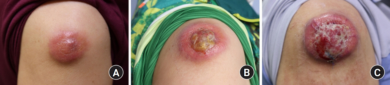
Fig. 1. The tumor enlarged in size during the patient’s following visits. (A) Approximately 3×3-cm sized mass with mild tenderness was located in the subcutaneous layer at the beginning. (B) After 2 months, the patient was referred to the plastic surgery department for more protruding with erythematous color change beside. (C) Another 2 months later, the mass enlarged with ulcerative surface after triamcinolone acetonide injections from the local clinic.
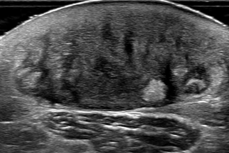
Fig. 2. Ruptured epidermal inclusion body cyst was considered by ultrasonography. The mass was shown in ultrasonography at the lateral aspect of the right arm. The mass is heterogeneously intense with associated vascularity at a measured size of 3.0×1.5×3.0 cm. The image provided the possibility of ruptured epidermal inclusion body cyst due to its feature.
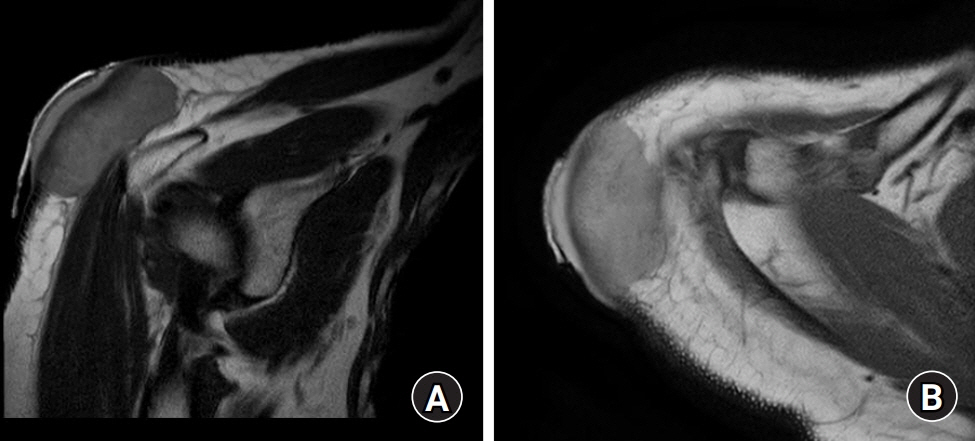
Fig. 3. The mass sited in dermal and subcutaneous layer suggests deltoid fascia involvement. (A) The mass showed low intensity compared to the adjacent subcutaneous fat layer in a T1 image of magnetic resonance imaging (MRI). (B) Whereas the mass showed higher intensity than the surrounding dermal layer providing high suspicion of dermatofibrosarcoma protuberans in a T2 image of MRI.
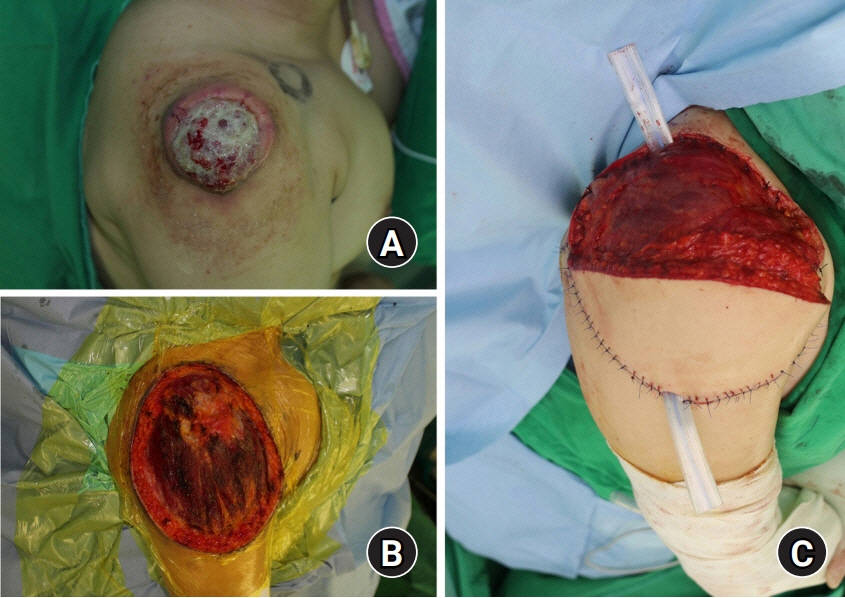
Fig. 4. Intraoperative photographs. (A) Preoperative photo of mass. (B) About 17.5×14.0-cm sized defect on the right shoulder and upper arm. (C) Reconstruction using latissimus dorsi myocutaneous island flap on the lateral side of the defect.

Fig. 5. Microscopic examinations of interdigitating dendritic cell sarcoma (H&E staining, ×100). It was composed of spindle-like, oval-shaped cell with spherical or oval nucleoli.
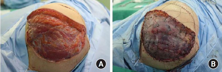
Fig. 6. Intraoperative photographs. (A) The remaining skin and soft tissue defect on the right shoulder. (B) Reconstruction of the defect using split thickness skin graft.
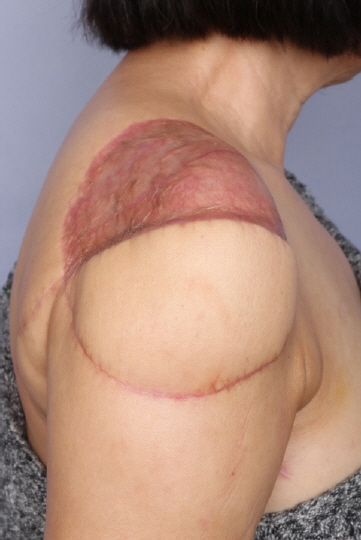
Fig. 7. Nine months after the operation, the reconstruction site was stable and the patient was under rehabilitation therapy.
Reference
-
1. Fonseca R, Yamakawa M, Nakamura S, et al. Follicular dendritic cell sarcoma and interdigitating reticulum cell sarcoma: a review. Am J Hematol. 1998; 59:161–7.
Article2. Han HS, Lee OJ, Lim SN, et al. Extranodal interdigitating dendritic cell sarcoma presenting in the pleura: a case report. J Korean Med Sci. 2011; 26:304–7.
Article3. Rosenberg SA, Niglio SA, Jo VY, Goydos JS. Interdigitating dendritic cell sarcoma presenting in the skin: diagnosis and the role of surgical resection, chemotherapy and radiotherapy in management. Rare Tumors. 2014; 6:5573.
Article4. Zhao C, Xie X, Gai DZ, Wu D, Xin H, Yang T. Interdigitating dendritic cell sarcoma of the spleen with hepatic failure after chemotherapy: a case report. Medicine (Baltimore). 2019; 98:e15535.5. Lee EJ, Hyun DW, Cho HJ, Lee JG. A rare case of interdigitating dendritic cell sarcoma in the nasal cavity. Case Rep Otolaryngol. 2013; 2013:913157.
Article6. Gaertner EM, Tsokos M, Derringer GA, Neuhauser TS, Arciero C, Andriko JA. Interdigitating dendritic cell sarcoma. A report of four cases and review of the literature. Am J Clin Pathol. 2001; 115:589–97.7. Saygin C, Uzunaslan D, Ozguroglu M, Senocak M, Tuzuner N. Dendritic cell sarcoma: a pooled analysis including 462 cases with presentation of our case series. Crit Rev Oncol Hematol. 2013; 88:253–71.
Article8. Han SW, Kim ZS, Kim HM, et al. Interdigitating dendritic cell sarcoma occured alone in axilla. J Korean Surg Soc. 2012; 82:330–4.
Article9. Ohtake H, Yamakawa M. Interdigitating dendritic cell sarcoma and follicular dendritic cell sarcoma: histopathological findings for differential diagnosis. J Clin Exp Hematop. 2013; 53:179–84.10. Wang HT, Xu HY, Zhang R, Liu ZG, Zhang GJ. Interdigitating dendritic cell sarcoma located in the groin: a case report and literature review. J Int Med Res. 2018; 46:4791–9.
Article
- Full Text Links
-
- Actions
-
Cited
- CITED
-
- Close
- Share
-
- Similar articles
-
- A Case of Cutaneous Interdigitating Dendritic Cell Sarcoma on the Chest
- A Ruptured Epidermal Inclusion Cyst in the Breast Presenting as a Recurrent Abscess
- An unusual case of metachronous NK/T cell lymphoma and interdigitating dendritic cell sarcoma
- Interdigitating Reticulum Cell Sarcoma of Lymph Node
- Interdigitating dendritic cell sarcoma occured alone in axilla
