A multidisciplinary approach to restore crown-root fractured maxillary central incisors: orthodontic extrusion and surgical extrusion
- Affiliations
-
- 1Dental Clinic Center, Pusan National University Hospital, Yangsan, Republic of Korea
- 2Department of Periodontology, Dental and Life Science Institute, School of Dentistry, Pusan National University, Yangsan, Republic of Korea
- 3Department of Periodontology and Dental Research Institute, Pusan National University Dental Hospital, Yangsan, Republic of Korea
- KMID: 2512121
- DOI: http://doi.org/10.14368/jdras.2020.36.4.262
Abstract
- To restore a tooth with a fracture line extending below the marginal bone level, a surgical crown lengthening procedure accompanied by ostectomy could be considered to expose the fracture line and reestablish the biologic width. However, this procedure could lead to esthetic failure, especially in the anterior teeth. Therefore, orthodontic extrusion, which elevates the fracture line from within the alveolar socket without sacrificing the supporting bone and gingiva, is recommended. This technique allows for the proper placement of the crown on a sound tooth structure, with the reestablishment of the biologic width. Alternatively, surgical extrusion is an one-step procedure that is simpler and less time-consuming than orthodontic extrusion; placing and adjusting the orthodontic appliance does not require multiple visits. This study presents successful restoration in 2 cases with a crown-tooth root fracture of the maxillary central incisor treated using a multidisciplinary approach through orthodontic extrusion or surgical extrusion followed by successful restoration.
Figure
-

Fig. 1 Pretreatment clinical view and radiograph of case 1. Note fracture line of maxillary left central incisor extended below the buccal gingival after removal of fractured buccal crown fragment (A). Figure (B) represents the initial state which was not removed fractured buccal crown fragment.

Fig. 2 After a 10-week orthodontic extrusion and followed by a 6-week retention phase. Note the coronal migration of the gingiva in the buccal aspect of extruded tooth.
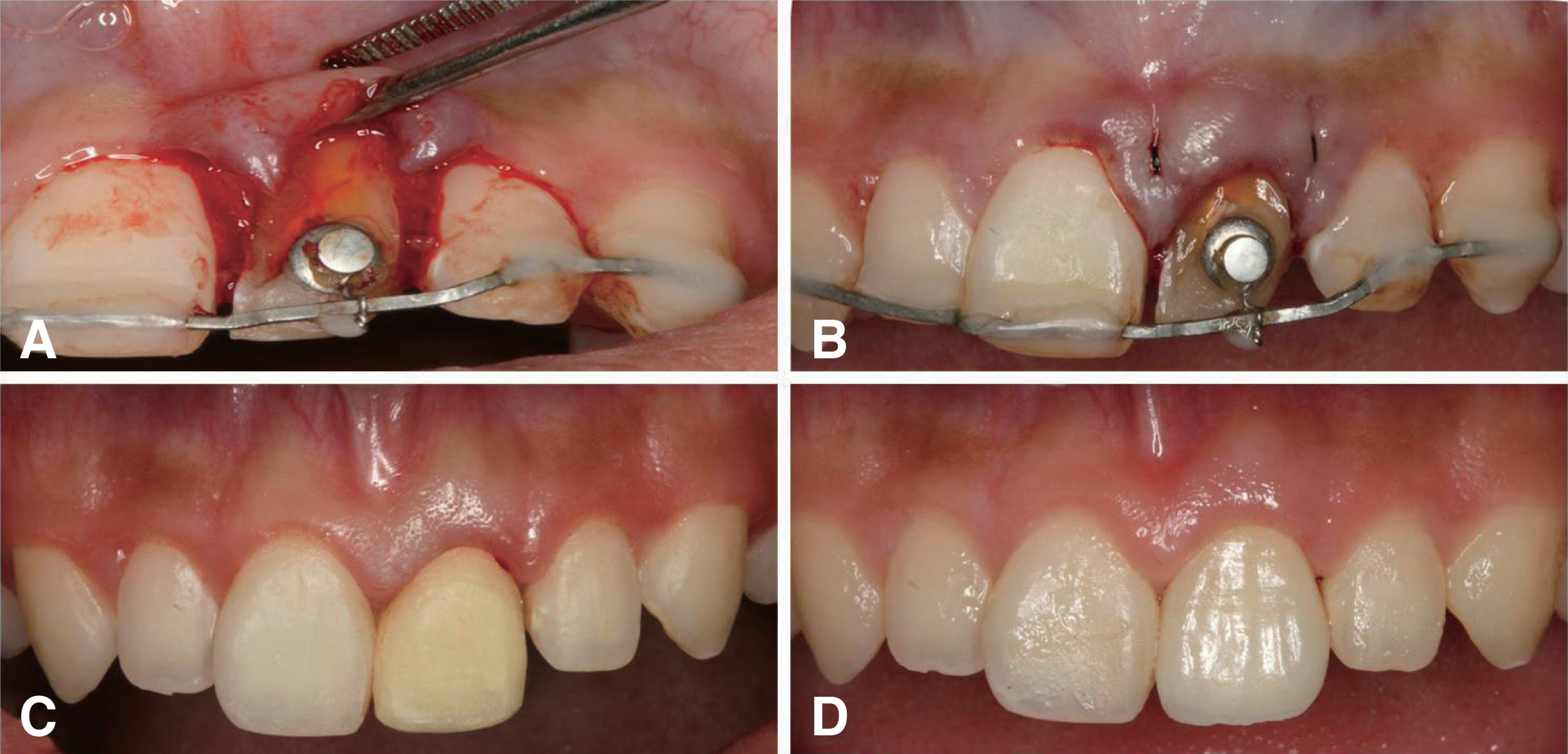
Fig. 3 During surgical clinical crown lengthening, the flap was reflected and osseous resection was performed to achieve adequate distance for biological width with exposure of fracture margin (A). The flap was sutured back to the position (B). Temporary crown was adjusted for marginal gingival adaptation (C). After final prosthetic crown placement (D).

Fig. 4 Pretreatment clinical view and radiograph of case 2. Note fracture lines of maxillary right and left central incisor. Right central incisor was fractured above the gingival line but left central incisor showed the biologic width violation by subgingival crown fracture with complete loss of the crown.

Fig. 5 Buccal view of coronally positioned root splinted with composite resin to maintain tooth stability and sutured (A). One-week after surgical extrusion. Wound healing was uneventful and suture was removed (B). After complement of endodontic treatment and gingivectomy, reconstruction of crown with core and post was done (C). Temporary crown was adjusted for marginal gingival adaptation (D).
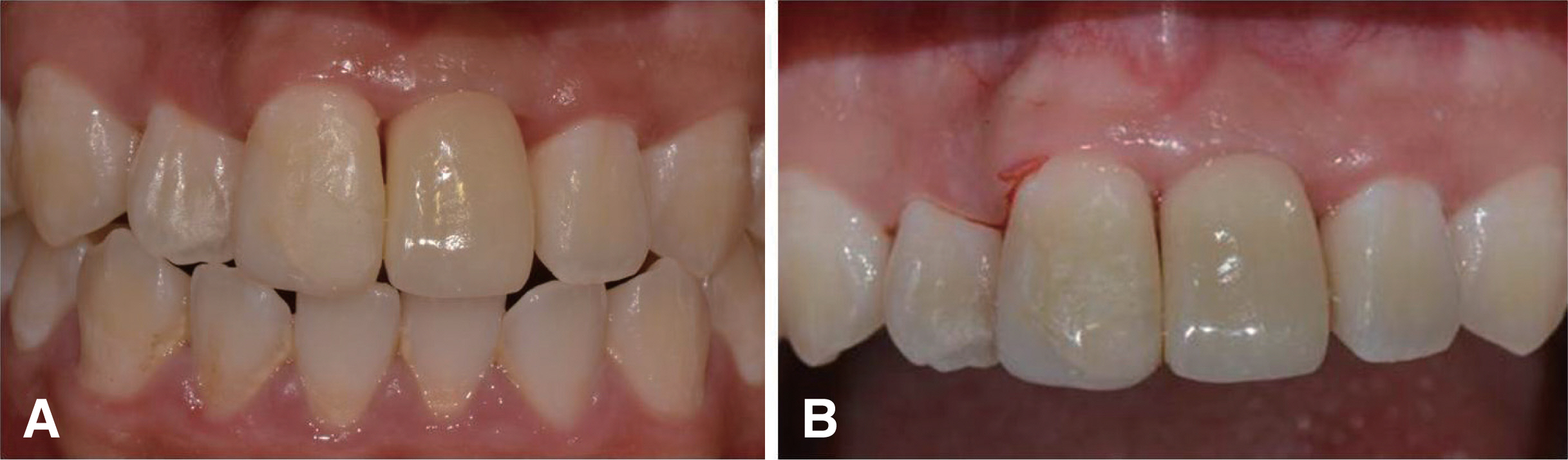
Fig. 6 Clinical view after final prosthetic crown placement (A). Two-year follow-up clinical view showed harmonious soft tissue contour (B).

Fig. 7 Radiograph taken right after surgical extrusion (A), at crown delivery (B), at 1-year follow-up (C) and at 2-year follow-up (D). Note the resolution of periapical radiolucency and there was no signs of root resorption or ankylosis.
Reference
-
References
1. Sorensen JA, Engelman MJ. 1990; Ferrule design and fracture resistance of endodontically treated teeth. J Prosthet Dent. 63:529–36. DOI: 10.1016/0022-3913(90)90070-S. PMID: 2187080.2. Jotkowitz A, Samet N. 2010; Rethinking ferrule - a new approach to an old dilemma. Br Dent J. 209:25–33. DOI: 10.1038/sj.bdj.2010.580. PMID: 20616834.3. Ingber JS, Rose LF, Coslet JG. 1977; The "biologic width" - a concept in periodontics and restorative dentistry. Alpha Omegan. 70:62–5. PMID: 276259.4. Bach N, Baylard JF, Voyer R. 2004; Orthodontic extrusion: Periodontal considerations and applications. J Can Dent Assoc. 70:775–80. PMID: 15588553.5. Çalişkan MK, Türkün M, Gomel M. 1999; Surgical extrusion of crown-root-fractured teeth: a clinical review. Int Endod J. 32:146–51. DOI: 10.1046/j.1365-2591.1999.00199.x. PMID: 10371911.6. Buskin R, Castellon P, Hochstedler JL. 2000; Orthodontic extrusion and orthodontic extraction in preprosthetic treatment using implant therapy. Pract Periodontics Aesthet Dent. 12:213–9. PMID: 11404962.7. Arhun N, Arman A, Ungor M, Erkut S. 2006; A conservative multidisciplinary approach for improved aesthetic results with traumatised anterior teeth. Br Dent J. 201:509–12. DOI: 10.1038/sj.bdj.4814158. PMID: 17057676.8. Palomo F, Kopczyk RA. 1978; Rationale and methods for crown lengthening. J Am Dent Assoc. 96:257–60. DOI: 10.14219/jada.archive.1978.0066. PMID: 272411.9. Malmgren O, Malmgren B, Frykholm A. 1991; Rapid orthodontic extrusion of crown root and cervical root fractured teeth. Endod Dent Traumatol. 7:49–54. DOI: 10.1111/j.1600-9657.1991.tb00183.x. PMID: 1782893.10. Lovdahl PE. 1995; Periodontal management and root extrusion of traumatized teeth. Dent Clin North Am. 39:169–79. PMID: 7890103.11. Smidt A, Lachish-Tandlich M, Venezia E. 2005; Orthodontic extrusion of an extensively broken down anterior tooth: a clinical report. Quintessence Int. 36:89–95. PMID: 15732544.12. Kahnberg KE. 1988; Surgical extrusion of root-fractured teeth - a follow-up study of two surgical methods. Endod Dent Traumatol. 4:85–9. DOI: 10.1111/j.1600-9657.1988.tb00301.x. PMID: 3251760.13. Ottl P, Hahn L, Lauer HC, Fay M. 2002; Fracture characteristics of carbon fibre, ceramic and non-palladium endodontic post systems at monotonously increasing loads. J Oral Rehabil. 29:175–83. DOI: 10.1046/j.1365-2842.2002.00852.x. PMID: 11856397.14. Assif D, Gorfil C. 1994; Biomechanical considerations in restoring endodontically treated teeth. J Prosthet Dent. 71:565–7. DOI: 10.1016/0022-3913(94)90438-3. PMID: 8040817.15. Stern N, Hirshfeld Z. 1973; Principles of preparing endodontically treated teeth for dowel and core restorations. J Prosthet Dent. 30:162–5. DOI: 10.1016/0022-3913(73)90051-6. PMID: 4515670.16. Andreasen JO, Kristerson L. 1981; The effect of limited drying or removal of the periodontal ligament. Periodontal healing after replantation of mature permanent incisors in monkeys. Acta Odontol Scand. 39:1–13. DOI: 10.3109/00016358109162253. PMID: 6943904.17. Kim SH, Tramontina V, Passanezi E. 2004; A new approach using the surgical extrusion procedure as an alternative for the reestablishment of biologic width. Int J Periodontics Restorative Dent. 24:39–45. PMID: 14984144.18. Elkhadem A, Mickan S, Richards D. 2014; Adverse events of surgical extrusion in treatment for crown-root and cervical root fractures: a systematic review of case series/reports. Dent Traumatol. 30:1–14. DOI: 10.1111/edt.12051. PMID: 23796195.19. Das B, Muthu MS. 2013; Surgical extrusion as a treatment option for crown-root fracture in permanent anterior teeth: a systematic review. Dent Traumatol. 29:423–31. DOI: 10.1111/edt.12054. PMID: 23802693.20. Zetu L, Wang HL. 2005; Management of inter-dental/inter-implant papilla. J Clin Periodontol. 32:831–9. DOI: 10.1111/j.1600-051X.2005.00748.x. PMID: 15966894.
- Full Text Links
-
- Actions
-
Cited
- CITED
-
- Close
- Share
-
- Similar articles
-
- Surgical extrusion of a maxillary premolar after orthodontic extrusion: a retrospective study
- Esthetic restoration of subgingival crown-root fractured maxillary anterior tooth using surgical extrusion
- Considerations in the selection of method for clinical crown lengthening
- Esthetic enhancement of a traumatized anterior tooth with a combination of forced eruption and tooth alignment: a case report
- Interdisciplinary rehabilitation of a root-fractured maxillary central incisor: A 12-year follow-up case report
