Gallbladder polyps: evolving approach to the diagnosis and management
- Affiliations
-
- 1Division of Gastroenterology and Hepatology, Department of Internal Medicine, Yeungnam University College of Medicine, Daegu, Korea
- KMID: 2512060
- DOI: http://doi.org/10.12701/yujm.2020.00213
Abstract
- Gallbladder (GB) polyp is a mucosal projection into the GB lumen. With increasing health awareness, GB polyps are frequently found using ultrasonography during health screening. The prevalence of GB polyps ranges between 1.3% and 9.5%. Most patients are asymptomatic and have benign characteristics. Of the nonneoplastic polyps, cholesterol polyps are most common, accounting for 60%–70% of lesions. However, a few polyps have malignant potential. Currently, the guidelines recommend laparoscopic cholecystectomy for polyps larger than 1 cm in diameter due to their malignan potential. The treatment algorithm can be influenced by the size, shape, and numbers of polyps, old age (>50 years), the presence of primary sclerosing cholangitis, and gallstones. This review summarizes the commonly recognized concepts on GB polyps from diagnosis to an algorithm of treatment.
Figure
-
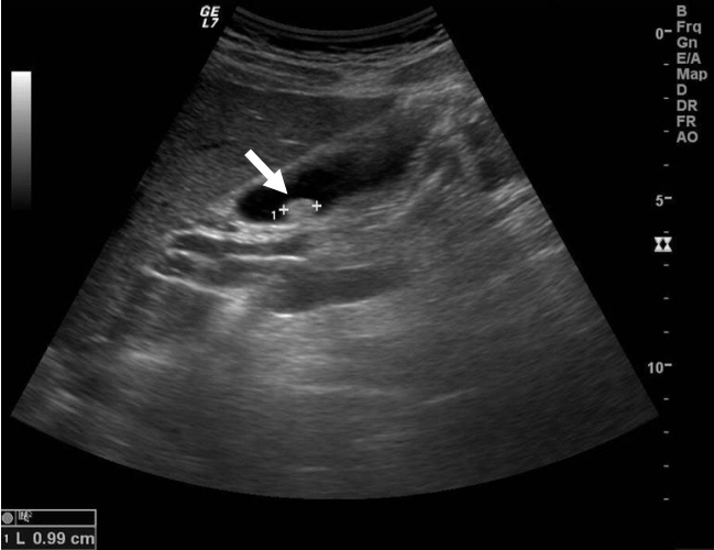
Fig. 1. Ultrasonography shows a polypod lesion (arrow) in the gallbladder. It measured 9.9 mm in maximal diameter and was not mobile regardless of the positional change. Pathologically, it was confirmed as adenoma.
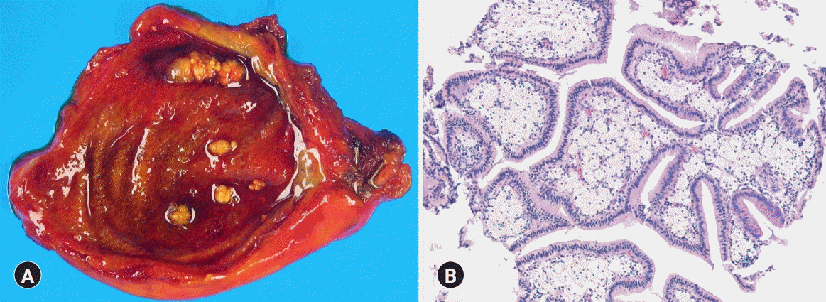
Fig. 2. Cholesterol polyp in a 55-year-old woman. (A) Grossly, the gallbladder shows multiple yellowish polyps in the lumen. (B) Microscopically, cholesterol polyps are characterized by cauliflower architecture. Numerous cholesterol-laden macrophages are present (hematoxylin and eosin stain, x100).
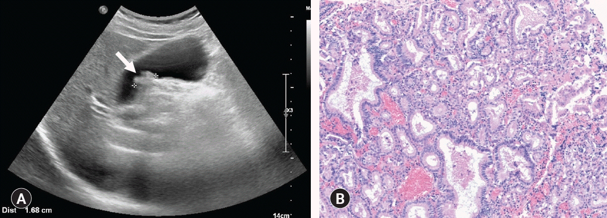
Fig. 3. Adenomatous gallbladder polyp in a 69-year-old man. (A) Ultrasonography shows a sessile polyp (arrow) with a wide base that was immobile and lacks an acoustic shadow. The polyp measured 16.8 mm in maximal diameter. (B) Microscopically, the adenoma is composed of closely packed, pyloric-type glands lined by mucin-containing cuboidal cells (hematoxylin and eosin stain, x100).
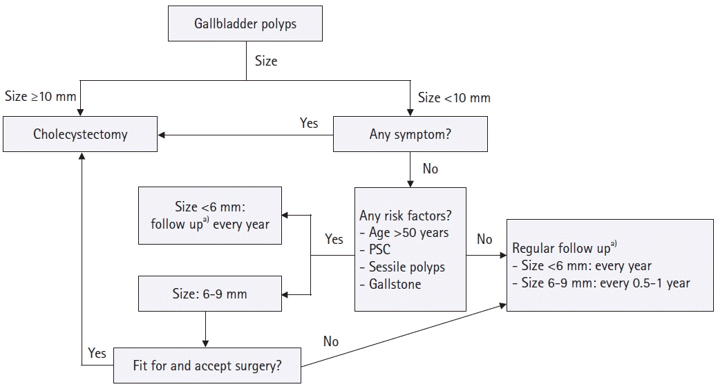
Fig. 4. Algorithm for the management of gallbladder polyps. PSC, primary sclerosing cholangitis. a)During follow up, cholecystectomy is advised if the polyp size increases, however, follow up is unnecessary if the polyp disappears.
Reference
-
References
1. Christensen AH, Ishak KG. Benign tumors and pseudotumors of the gallbladder: report of 180 cases. Arch Pathol. 1970; 90:423–32.2. Moriguchi H, Tazawa J, Hayashi Y, Takenawa H, Nakayama E, Marumo F, et al. Natural history of polypoid lesions in the gall bladder. Gut. 1996; 39:860–2.
Article3. Lin WR, Lin DY, Tai DI, Hsieh SY, Lin CY, Sheen IS, et al. Prevalence of and risk factors for gallbladder polyps detected by ultrasonography among healthy Chinese: analysis of 34 669 cases. J Gastroenterol Hepatol. 2008; 23:965–9.
Article4. Jørgensen T, Jensen KH. Polyps in the gallbladder: a prevalence study. Scand J Gastroenterol. 1990; 25:281–6.5. Segawa K, Arisawa T, Niwa Y, Suzuki T, Tsukamoto Y, Goto H, et al. Prevalence of gallbladder polyps among apparently healthy Japanese: ultrasonographic study. Am J Gastroenterol. 1992; 87:630–3.6. Chen CY, Lu CL, Chang FY, Lee SD. Risk factors for gallbladder polyps in the Chinese population. Am J Gastroenterol. 1997; 92:2066–8.7. Kim SY, Lee HS, Lee YS, Chung KW, Jang BK, Chung WJ, et al. Prevalence and risk factors of gallbladder polyp in adults living in Daegu and Gyeongbuk provinces. Korean J Gastroenterol. 2006; 48:344–50.8. Choi YS, Do JH, Seo SW, Lee SE, Oh HC, Min YJ, et al. Prevalence and risk factors of gallbladder polypoid lesions in a healthy population. Yonsei Med J. 2016; 57:1370–5.
Article9. Mellnick VM, Menias CO, Sandrasegaran K, Hara AK, Kielar AZ, Brunt EM, et al. Polypoid lesions of the gallbladder: disease spectrum with pathologic correlation. Radiographics. 2015; 35:387–99.
Article10. Chattopadhyay D, Lochan R, Balupuri S, Gopinath BR, Wynne KS. Outcome of gall bladder polypoidal lesions detected by transabdominal ultrasound scanning: a nine year experience. World J Gastroenterol. 2005; 11:2171–3.
Article11. Dilek ON, Karasu S, Dilek FH. Diagnosis and treatment of gallbladder polyps: current perspectives. Euroasian J Hepatogastroenterol. 2019; 9:40–8.
Article12. Lee KF, Wong J, Li JC, Lai PB. Polypoid lesions of the gallbladder. Am J Surg. 2004; 188:186–90.
Article13. Zielinski MD, Atwell TD, Davis PW, Kendrick ML, Que FG. Comparison of surgically resected polypoid lesions of the gallbladder to their pre-operative ultrasound characteristics. J Gastrointest Surg. 2009; 13:19–25.
Article14. Azuma T, Yoshikawa T, Araida T, Takasaki K. Differential diagnosis of polypoid lesions of the gallbladder by endoscopic ultrasonography. Am J Surg. 2001; 181:65–70.
Article15. Sadamoto Y, Oda S, Tanaka M, Harada N, Kubo H, Eguchi T, et al. A useful approach to the differential diagnosis of small polypoid lesions of the gallbladder, utilizing an endoscopic ultrasound scoring system. Endoscopy. 2002; 34:959–65.
Article16. Kitano M, Kudo M, Yamao K, Takagi T, Sakamoto H, Komaki T, et al. Characterization of small solid tumors in the pancreas: the value of contrast-enhanced harmonic endoscopic ultrasonography. Am J Gastroenterol. 2012; 107:303–10.
Article17. Kitano M, Sakamoto H, Matsui U, Ito Y, Maekawa K, von Schrenck T, et al. A novel perfusion imaging technique of the pancreas: contrast-enhanced harmonic EUS (with video). Gastrointest Endosc. 2008; 67:141–50.
Article18. Park CH, Chung MJ, Oh TG, Park JY, Bang S, Park SW, et al. Differential diagnosis between gallbladder adenomas and cholesterol polyps on contrast-enhanced harmonic endoscopic ultrasonography. Surg Endosc. 2013; 27:1414–21.
Article19. Choi JH, Seo DW, Choi JH, Park DH, Lee SS, Lee SK, et al. Utility of contrast-enhanced harmonic EUS in the diagnosis of malignant gallbladder polyps (with videos). Gastrointest Endosc. 2013; 78:484–93.
Article20. Golse N, Lewin M, Rode A, Sebagh M, Mabrut JY. Gallbladder adenomyomatosis: diagnosis and management. J Visc Surg. 2017; 154:345–53.
Article21. Gallahan WC, Conway JD. Diagnosis and management of gallbladder polyps. Gastroenterol Clin North Am. 2010; 39:359–67.
Article22. Shapiro RS, Winsberg F. Comet-tail artifact from cholesterol crystals: observations in the postlithotripsy gallbladder and an in vitro model. Radiology. 1990; 177:153–6.
Article23. Mellnick VM, Menias CO, Sandrasegaran K, Hara AK, Kielar AZ, Brunt EM, et al. Polypoid lesions of the gallbladder: disease spectrum with pathologic correlation. Radiographics. 2015; 35:387–99.
Article24. Nishiyama Y, Yamamoto Y, Fukunaga K, Kimura N, Miki A, Sasakawa Y, et al. Dual-time-point 18F-FDG PET for the evaluation of gallbladder carcinoma. J Nucl Med. 2006; 47:633–8.25. Zemour J, Marty M, Lapuyade B, Collet D, Chiche L. Gallbladder tumor and pseudotumor: diagnosis and management. J Visc Surg. 2014; 151:289–300.
Article26. Kim JH, Kim TK, Eun HW, Kim BS, Lee MG, Kim PN, et al. Preoperative evaluation of gallbladder carcinoma: efficacy of combined use of MR imaging, MR cholangiography, and contrast-enhanced dual-phase three-dimensional MR angiography. J Magn Reson Imaging. 2002; 16:676–84.
Article27. Xu A, Hu H. The gallbladder polypoid-lesions conundrum: moving forward with controversy by looking back. Expert Rev Gastroenterol Hepatol. 2017; 11:1071–80.
Article28. Limaiem F, Sassi A, Talbi G, Bouraoui S, Mzabi S. Routine histopathological study of cholecystectomy specimens. Useful? A retrospective study of 1960 cases. Acta Gastroenterol Belg. 2017; 80:365–70.29. Taskin OC, Bellolio E, Dursun N, Seven IE, Roa JC, Araya JC, et al. Non-neoplastic polyps of the gallbladder: a clinicopathologic analysis of 447 cases. Am J Surg Pathol. 2020; 44:467–76.30. Xu A, Zhang Y, Hu H, Zhao G, Cai J, Huang A. Gallbladder polypoid-lesions: what are they and how should they be treated? A single-center experience based on 1446 cholecystectomy patients. J Gastrointest Surg. 2017; 21:1804–12.
Article31. Persley KM. Gallbladder polyps. Curr Treat Options Gastroenterol. 2005; 8:105–8.
Article32. Haradome H, Ichikawa T, Sou H, Yoshikawa T, Nakamura A, Araki T, et al. The pearl necklace sign: an imaging sign of adenomyomatosis of the gallbladder at MR cholangiopancreatography. Radiology. 2003; 227:80–8.
Article33. Susumu S, Matsuo S, Tsutsumi R, Azuma T, Obata S, Hayashi T. Inflammatory polyp of the gallbladder mimicking early polypoid carcinoma. Case Rep Gastroenterol. 2009; 3:255–9.
Article34. Perez-Montiel D, Mucientes F, Spencer L, Klaassen R, Suster S. Polypoid leiomyosarcoma of the gallbladder: study of a case associated with adenomyomatous hyperplasia. Ann Diagn Pathol. 2004; 8:358–63.
Article35. Yang HL, Sun YG, Wang Z. Polypoid lesions of the gallbladder: diagnosis and indications for surgery. Br J Surg. 1992; 79:227–9.
Article36. Adsay NV. Neoplastic precursors of the gallbladder and extrahepatic biliary system. Gastroenterol Clin North Am. 2007; 36:889–900.
Article37. Roa I, de Aretxabala X, Araya JC, Roa J. Preneoplastic lesions in gallbladder cancer. J Surg Oncol. 2006; 93:615–23.
Article38. Albores-Saavedra J, Chable-Montero F, Gonzalez-Romo MA, Ramirez Jaramillo M, Henson DE. Adenomas of the gallbladder: morphologic features, expression of gastric and intestinal mucins, and incidence of high-grade dysplasia/carcinoma in situ and invasive carcinoma. Hum Pathol. 2012; 43:1506–13.
Article39. Trivedi V, Gumaste VV, Liu S, Baum J. Gallbladder cancer: adenoma-carcinoma or dysplasia-carcinoma sequence? Gastroenterol Hepatol (N Y). 2008; 4:735–7.40. Adsay V, Jang KT, Roa JC, Dursun N, Ohike N, Bagci P, et al. Intracholecystic papillary-tubular neoplasms (ICPN) of the gallbladder (neoplastic polyps, adenomas, and papillary neoplasms that are ≥1.0 cm): clinicopathologic and immunohistochemical analysis of 123 cases. Am J Surg Pathol. 2012; 36:1279–301.41. Kiruthiga KG, Kodiatte TA, Burad D, Kurian R, Raju RS, Rymbai ML, et al. Intracholecystic papillary-tubular neoplasms of the gallbladder: a clinicopathological study of 36 cases. Ann Diagn Pathol. 2019; 40:88–93.
Article42. Park JK, Yoon YB, Kim YT, Ryu JK, Yoon WJ, Lee SH, et al. Management strategies for gallbladder polyps: is it possible to predict malignant gallbladder polyps? Gut Liver. 2008; 2:88–94.
Article43. Lee J, Yun M, Kim KS, Lee JD, Kim CK. Risk stratification of gallbladder polyps (1-2 cm) for surgical intervention with 18F-FDG PET/CT. J Nucl Med. 2012; 53:353–8.
Article44. Koh T, Taniguchi H, Yamaguchi A, Kunishima S, Yamagishi H. Differential diagnosis of gallbladder cancer using positron emission tomography with fluorine-18-labeled fluoro-deoxyglucose (FDG-PET). J Surg Oncol. 2003; 84:74–81.
Article45. Babu BI, Dennison AR, Garcea G. Management and diagnosis of gallbladder polyps: a systematic review. Langenbecks Arch Surg. 2015; 400:455–62.
Article46. Heitz L, Kratzer W, Grater T, Schmidberger J; EMIL study group. Gallbladder polyps: a follow-up study after 11 years. BMC Gastroenterol. 2019; 19:42.47. Colecchia A, Larocca A, Scaioli E, Bacchi-Reggiani ML, Di Biase AR, Azzaroli F, et al. Natural history of small gallbladder polyps is benign: evidence from a clinical and pathogenetic study. Am J Gastroenterol. 2009; 104:624–9.
Article48. Csendes A, Burgos AM, Csendes P, Smok G, Rojas J. Late follow-up of polypoid lesions of the gallbladder smaller than 10 mm. Ann Surg. 2001; 234:657–60.
Article49. Wiles R, Thoeni RF, Barbu ST, Vashist YK, Rafaelsen SR, Dewhurst C, et al. Management and follow-up of gallbladder polyps: joint guidelines between the European Society of Gastrointestinal and Abdominal Radiology (ESGAR), European Association for Endoscopic Surgery and other Interventional Techniques (EAES), International Society of Digestive Surgery-European Federation (EFISDS) and European Society of Gastrointestinal Endoscopy (ESGE). Eur Radiol. 2017; 27:3856–66.50. Andren-Sandberg A. Diagnosis and management of gallbladder polyps. N Am J Med Sci. 2012; 4:203–11.
Article51. Myers RP, Shaffer EA, Beck PL. Gallbladder polyps: epidemiology, natural history and management. Can J Gastroenterol. 2002; 16:187–94.
Article52. Kwon W, Jang JY, Lee SE, Hwang DW, Kim SW. Clinicopathologic features of polypoid lesions of the gallbladder and risk factors of gallbladder cancer. J Korean Med Sci. 2009; 24:481–7.
Article53. Bhatt NR, Gillis A, Smoothey CO, Awan FN, Ridgway PF. Evidence based management of polyps of the gall bladder: a systematic review of the risk factors of malignancy. Surgeon. 2016; 14:278–86.
Article54. Cha BH, Hwang JH, Lee SH, Kim JE, Cho JY, Kim H, et al. Pre-operative factors that can predict neoplastic polypoid lesions of the gallbladder. World J Gastroenterol. 2011; 17:2216–22.
Article55. Albores-Saavedra J, Vardaman CJ, Vuitch F. Non-neoplastic polypoid lesions and adenomas of the gallbladder. Pathol Annu. 1993; 28:145–77.56. Aldouri AQ, Malik HZ, Waytt J, Khan S, Ranganathan K, Kummaraganti S, et al. The risk of gallbladder cancer from polyps in a large multiethnic series. Eur J Surg Oncol. 2009; 35:48–51.
Article57. European Association for the Study of the Liver. EASL Clinical Practice Guidelines: management of cholestatic liver diseases. J Hepatol. 2009; 51:237–67.
Article58. Said K, Glaumann H, Bergquist A. Gallbladder disease in patients with primary sclerosing cholangitis. J Hepatol. 2008; 48:598–605.
Article59. van Erp LW, Cunningham M, Narasimman M, Ale Ali H, Jhaveri K, Drenth JP, et al. Risk of gallbladder cancer in patients with primary sclerosing cholangitis and radiographically detected gallbladder polyps. Liver Int. 2020; 40:382–92.
Article60. Torres C, Antonioli D, Odze RD. Polypoid dysplasia and adenomas in inflammatory bowel disease: a clinical, pathologic, and follow-up study of 89 polyps from 59 patients. Am J Surg Pathol. 1998; 22:275–84.
- Full Text Links
-
- Actions
-
Cited
- CITED
-
- Close
- Share
-
- Similar articles
-
- Recent Updates on the Diagnosis and Management of Gallbladder Polyps
- Practical Guidelines for Management of Gallbladder Polyps
- Recent Updates on Management and Follow-up of Gallbladder Polyps
- Recent Updates on Diagnosis, Treatment, and Follow-up of Gallbladder Polyps
- Disparity between ultrasonographic and pathologic findings of laparoscopically removed gallbladder polyps
