Restor Dent Endod.
2020 Nov;45(4):e53. 10.5395/rde.2020.45.e53.
Effect of post space preparation drills on the incidence of root dentin defects
- Affiliations
-
- 1Department of Oral Sciences, School of Dentistry, University of Cuiabá - UNIC, Cuiabá, MT, Brazil
- 2Department of Endodontics, School of Dentistry, Centro Universitário de Anápolis - UniEvangélica, Anápolis, GO, Brazil
- KMID: 2512044
- DOI: http://doi.org/10.5395/rde.2020.45.e53
Abstract
Objectives
This study investigated the incidence of root dentin defects after the use of different post space preparation (PSP) drills.
Materials and Methods
Seventy-two bovine incisors were selected and obtained 14-mmlong root sections. Twelve roots served as controls with no intervention (G1). The 60 root canals remaining were instrumented using the crown-down technique with the ProTaper Next system and obturated using the lateral condensation technique. Specimens were randomly distributed into 5 groups (n = 12) according to the operative steps performed: G2, root canal instrumentation and filling (I+F); G3, I+F and PSP with Gates-Glidden drills; G4, I+F and PSP with Largo-Peeso reamers; G5, I+F and PSP with Exacto drill; and G6, I+F and PSP with WhitePost drill. Roots were sectioned at 3, 6, 9, and 12 mm from the apex, and digital images were captured. The presence of root dentin defects was recorded. Data were analyzed by the χ2 test, with p < 0.05 considered to indicate statistical significance.
Results
Root dentin defects were observed in 39.6% of the root sections. No defects were observed in G1. G5 had significantly more cracks and craze lines than G1, G2, and G3 (p < 0.05), and more fractures than G1, G2, G3, and G4 (p< 0.05). When all root sections were analyzed together, significantly more defects were observed at the 12-mm level than at the 3-mm level (p < 0.05).
Conclusions
PSP drills caused defects in the root dentin. Gates-Glidden drills caused fewer root defects than Largo-Peeso reamers and Exacto drills.
Figure
-
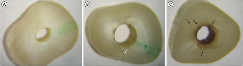
Figure 1 Representative images of root cross-sectional slices and dentin defects: (A) No dentin defects; (B) fracture (white arrow); (C) other defects (black arrows).
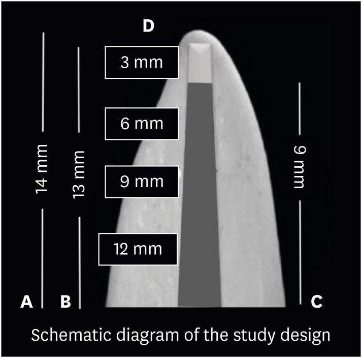
Figure 2 Schematic illustration of the study design: (A) root canal length; (B) working length; (C) post space depth; (D) root cross-sections.
Reference
-
1. Ghoddusi J, Bagherpour A, Mahmudabadi F, Forghani M, Sarmad M. Residual dentin thickness of bifurcated maxillary premolars following two post space preparation methods. Iran Endod J. 2013; 8:94–98. PMID: 23922568.2. Cecchin D, Farina AP, Guerreiro CA, Carlini-Júnior B. Fracture resistance of roots prosthetically restored with intra-radicular posts of different lengths. J Oral Rehabil. 2010; 37:116–122. PMID: 19968767.
Article3. Nergiz I, Schmage P, Ozcan M, Platzer U. Effect of length and diameter of tapered posts on the retention. J Oral Rehabil. 2002; 29:28–34. PMID: 11844029.
Article4. Caputo AA, Standlee JP. Pins and posts--why, when and how. Dent Clin North Am. 1976; 20:299–311. PMID: 1062314.
Article5. Alberto Rubino G, de Miranda Candeiro GT, Gonzales Freire L, Faga Iglecias E, de Mello Lemos É, Luiz Caldeira C, Gavini G. Micro-CT evaluation of gutta-percha removal by two retreatment systems. Iran Endod J. 2018; 13:221–227. PMID: 29707019.6. Çapar ID, Uysal B, Ok E, Arslan H. Effect of the size of the apical enlargement with rotary instruments, single-cone filling, post space preparation with drills, fiber post removal, and root canal filling removal on apical crack initiation and propagation. J Endod. 2015; 41:253–256. PMID: 25433969.
Article7. Kim HC, Lee MH, Yum J, Versluis A, Lee CJ, Kim BM. Potential relationship between design of nickel-titanium rotary instruments and vertical root fracture. J Endod. 2010; 36:1195–1199. PMID: 20630298.
Article8. Arslan H, Karataş E, Çapar ID, Ozsu D, Doğanay E. Effect of ProTaper Universal, Endoflare, Revo-S, HyFlex coronal flaring instruments, and Gates Glidden drills on crack formation. J Endod. 2014; 40:1681–1683. PMID: 25127932.
Article9. Chai H, Tamse A. Fracture mechanics analysis of vertical root fracture from condensation of gutta-percha. J Biomech. 2012; 45:1673–1678. PMID: 22503579.
Article10. Maddalone M, Gagliani M, Citterio CL, Karanxha L, Pellegatta A, Del Fabbro M. Prevalence of vertical root fractures in teeth planned for apical surgery. A retrospective cohort study. Int Endod J. 2018; 51:969–974. PMID: 29478245.
Article11. Ashwinkumar V, Krithikadatta J, Surendran S, Velmurugan N. Effect of reciprocating file motion on microcrack formation in root canals: an SEM study. Int Endod J. 2014; 47:622–627. PMID: 24107320.
Article12. Shemesh H, Bier CA, Wu MK, Tanomaru-Filho M, Wesselink PR. The effects of canal preparation and filling on the incidence of dentinal defects. Int Endod J. 2009; 42:208–213. PMID: 19228210.
Article13. Versiani MA, Souza E, De-Deus G. Critical appraisal of studies on dentinal radicular microcracks in endodontics: methodological issues, contemporary concepts, and future perspectives. Endod Topics. 2015; 33:87–156.14. De-Deus G, Belladonna FG, Souza EM, Silva EJ, Neves AA, Alves H, Lopes RT, Versiani MA. Micro-computed tomographic assessment on the effect of ProTaper Next and Twisted file adaptive systems on dentinal cracks. J Endod. 2015; 41:1116–1119. PMID: 25817212.
Article15. De-Deus G, Belladonna FG, Silva EJ, Souza EM, Carvalhal JC, Perez R, Lopes RT, Versiani MA. Micro-CT assessment of dentinal micro-cracks after root canal filling procedures. Int Endod J. 2017; 50:895–901. PMID: 27689844.16. Aboud LR, Santos BC, Lopes RT, Viana LA, Scelza MF. Effect of aging on dentinal crack formation after treatment and retreatment procedures: a micro-CT study. Braz Dent J. 2018; 29:530–535. PMID: 30517474.
Article17. Aksoy Ç, Keriş EY, Yaman SD, Ocak M, Geneci F, Çelik HH. Evaluation of XP-endo Shaper, Reciproc Blue, and ProTaper Universal systems on dentinal microcrack formation using micro-computed tomography. J Endod. 2019; 45:338–342. PMID: 30803543.
Article18. Shantiaee Y, Dianat O, Mosayebi G, Namdari M, Tordik P. Effect of root canal preparation techniques on crack formation in root dentin. J Endod. 2019; 45:447–452. PMID: 30827767.
Article19. Shemesh H, Wesselink PR, Wu MK. Incidence of dentinal defects after root canal filling procedures. Int Endod J. 2010; 43:995–1000. PMID: 20722755.
Article20. Topçuoğlu HS, Demirbuga S, Tuncay Ö, Pala K, Arslan H, Karataş E. The effects of Mtwo, R-Endo, and D-RaCe retreatment instruments on the incidence of dentinal defects during the removal of root canal filling material. J Endod. 2014; 40:266–270. PMID: 24461416.
Article21. Pedullà E, Genovesi F, Rapisarda S, La Rosa GR, Grande NM, Plotino G, Adorno CG. Effects of 6 single-file systems on dentinal crack formation. J Endod. 2017; 43:456–461. PMID: 28131416.
Article22. Coelho MS, Card SJ, Tawil PZ. Visualization enhancement of dentinal defects by using light-emitting diode transillumination. J Endod. 2016; 42:1110–1113. PMID: 27178248.
Article23. Lim H, Li FC, Friedman S, Kishen A. Residual microstrain in root dentin after canal instrumentation measured with digital moiré interferometry. J Endod. 2016; 42:1397–1402. PMID: 27430943.
Article24. Shemesh H, Lindtner T, Portoles CA, Zaslansky P. Dehydration induces cracking in root dentin irrespective of instrumentation: a two-dimensional and three-dimensional study. J Endod. 2018; 44:120–125. PMID: 29079053.
Article25. Çiçek E, Koçak MM, Sağlam BC, Koçak S. Evaluation of microcrack formation in root canals after instrumentation with different NiTi rotary file systems: a scanning electron microscopy study. Scanning. 2015; 37:49–53. PMID: 25488126.
Article26. Adorno CG, Yoshioka T, Jindan P, Kobayashi C, Suda H. The effect of endodontic procedures on apical crack initiation and propagation ex vivo . Int Endod J. 2013; 46:763–768. PMID: 23402216.
Article27. Borges ÁH, Damião MS, Pereira TM, Filho GS, Miranda-Pedro FL, Luiz de Oliveira da Rosa W, Piva E, Guedes OA. Influence of cervical preflaring on the incidence of root dentin defects. J Endod. 2018; 44:286–291. PMID: 29208400.
Article28. De Bruyne MA, De Moor RJ. SEM analysis of the integrity of resected root apices of cadaver and extracted teeth after ultrasonic root-end preparation at different intensities. Int Endod J. 2005; 38:310–319. PMID: 15876295.
Article29. Rose E, Svec T. An evaluation of apical cracks in teeth undergoing orthograde root canal instrumentation. J Endod. 2015; 41:2021–2024. PMID: 26472677.
Article30. Silva EJ, Carvalho NK, Prado MC, Senna PM, Souza EM, De-Deus G. Bovine teeth can reliably substitute human dentine in an intra-tooth push-out bond strength model? Int Endod J. 2019; 52:1063–1069. PMID: 30697770.
Article31. Fonseca RB, Haiter-Neto F, Carlo HL, Soares CJ, Sinhoreti MA, Puppin-Rontani RM, Correr-Sobrinho L. Radiodensity and hardness of enamel and dentin of human and bovine teeth, varying bovine teeth age. Arch Oral Biol. 2008; 53:1023–1029. PMID: 18675389.
Article32. Adl A, Sedigh-Shams M, Majd M. The effect of using RC prep during root canal preparation on the incidence of dentinal defects. J Endod. 2015; 41:376–379. PMID: 25576205.
Article33. Kansal R, Rajput A, Talwar S, Roongta R, Verma M. Assessment of dentinal damage during canal preparation using reciprocating and rotary files. J Endod. 2014; 40:1443–1446. PMID: 25146029.
Article34. Shori DD, Shenoi PR, Baig AR, Kubde R, Makade C, Pandey S. Stereomicroscopic evaluation of dentinal defects induced by new rotary system: “ProTaper NEXT”. J Conserv Dent. 2015; 18:210–213. PMID: 26069406.
Article35. Ari H, Erdemir A, Belli S. Evaluation of the effect of endodontic irrigation solutions on the microhardness and the roughness of root canal dentin. J Endod. 2004; 30:792–795. PMID: 15505513.
Article
- Full Text Links
-
- Actions
-
Cited
- CITED
-
- Close
- Share
-
- Similar articles
-
- Shaping ability of four rotary nickel-titanium instruments to prepare root canal at danger zone
- Effect of dimethyl sulfoxide on bond durability of fiber posts cemented with etch-and-rinse adhesives
- Post space preparation timing of root canals sealed with AH Plus sealer
- Effect of ultrasonic cleaning on the bond strength of fiber posts in oval canals filled with a premixed bioceramic root canal sealer
- Apical prepration size in infected root canals
