Fibrocartilaginous mesenchymoma with an unusual location in the rib
- Affiliations
-
- 1Department of Pathology, Kosin University Gospel Hospital, Busan, Korea
- KMID: 2511516
- DOI: http://doi.org/10.4132/jptm.2020.10.08
Abstract
- Fibrocartilaginous mesenchymoma is a rare bone tumor, with fewer than 35 cases reported in the literature since 1984. This tumor usually occurs in the long bones of children and adolescents. In the current case, the tumor affected a rib. A 17-year-old boy presented with a mass in the right fifth rib. Radiologic findings revealed an osteolytic mass with cortical destruction and calcification; en bloc resection was performed. The tumor showed three distinct histologic features: bland spindle cell proliferation, benign cartilage nodules, and epiphyseal plate-like enchondral ossification. The pathologic diagnosis was fibrocartilaginous mesenchymoma. The patient remains free of disease 1 year after the surgery. Pathological diagnosis of fibrocartilaginous mesenchymoma can be challenging, especially when the tumor occurs in an unusual site. When any fibro-osseous lesion with a cartilaginous component is encountered, the possibility of fibrocartilaginous mesenchymoma should be considered because of its locally aggressive behavior.
Figure
-
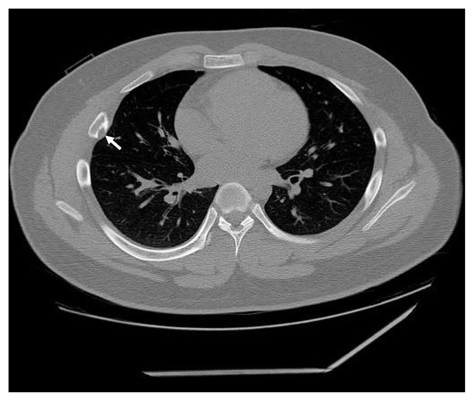
Fig. 1 Computed tomography shows an osteolytic lesion (arrow) with calcification and juxtacortical extension at the posterolateral side of the right 5th rib.

Fig. 2 Histologic findings. (A) Gross examination reveals an ill-defined, eccentrically-located, hard mass with multifocal glistening bluish grey nodules corresponding to cartilage tissue. The cortex had been partially destroyed. (B) The tumor consists of multiple cartilage islands with a fibrocollagenous area (asterisk) and well-formed new bone trabeculae (arrows). (C) Epiphyseal plate-like enchondral ossification is observed at the periphery of the cytologically bland cartilage nodules. (D) A hypocellular fibrocollagenous lesion consisting of spindle and stellate cells without nuclear pleomorphism or mitosis is noted. Reactive woven bone formation with osteoblastic rimming is observed.
Reference
-
References
1. Dahlin DC, Bertoni F, Beabout JW, Campanacci M. Fibrocartilaginous mesenchymoma with low-grade malignancy. Skeletal Radiol. 1984; 12:263–9.
Article2. Gambarotti M, Righi A, Vanel D, et al. Fibrocartilaginous mesenchymoma of bone: a single-institution experience with molecular investigations and a review of the literature. Histopathology. 2017; 71:134–42.
Article3. Saito T, Motoi T, Suehara Y, et al. Fibrocartilaginous mesenchymoma of the tibia with predominant microcystic features: a case report and literature review. Hum Pathol Case Rep. 2019; 16:100288.
Article4. Bulychova IV, Unni KK, Bertoni F, Beabout JW. Fibrocartilagenous mesenchymoma of bone. Am J Surg Pathol. 1993; 17:830–6.
Article5. Czerniak B. Dorfman and Czerniak’s bone tumors. 2nd ed. Philadelphia: Elsevier Saunders;2016. p. 606–14.6. Bhaduri A, Deshpande RB. Fibrocartilagenous mesenchymoma versus fibrocartilagenous dysplasia: are these a single entity? Am J Surg Pathol. 1995; 19:1447–8.7. Lin J, Shulman SC, Steelman CK, et al. Fibrocartilaginous mesenchymoma, a unique osseous lesion: case report with review of the literature. Skeletal Radiol. 2011; 40:1495–9.
Article8. Fletcher CD. WHO classification of tumours of soft tissue and bone tumours. 5th ed. Lyon: IARC Press;2020. p. 470–1.
- Full Text Links
-
- Actions
-
Cited
- CITED
-
- Close
- Share
-
- Similar articles
-
- Rosette-forming epithelioid osteosarcoma in the rib: a rare case of location and morphology
- A Case of Primary Malignant Mesenchymoma of the Heart
- Two cases of Benign Mesenchymoma arising in Thigh and Forearm
- A Case of Nasopharyngeal Mesenchymoma
- A Case of the Malignant Mesenchymoma of the Liver in Childhood
