J Korean Med Sci.
2021 Jan;36(3):e22. 10.3346/jkms.2021.36.e22.
Antimicrobial Resistance and Molecular Analysis of Staphylococcus aureus in Staphylococcal Scalded Skin Syndrome among Children in Korea
- Affiliations
-
- 1Department of Pediatrics, Jeju National University School of Medicine, Jeju, Korea
- 2Department of Pediatrics, Seoul National University Bundang Hospital, Seongnam, Korea
- 3Department of Pediatrics, Seoul National University College of Medicine, Seoul, Korea
- 4Department of Pediatrics, Seoul National University Children's Hospital, Seoul, Korea
- KMID: 2510742
- DOI: http://doi.org/10.3346/jkms.2021.36.e22
Abstract
- Background
Staphylococcal scalded skin syndrome (SSSS) is a skin disease characterized by blistering and desquamation caused by exfoliative toxins (ETs) of Staphylococcus aureus (S. aureus). Although many countries show predominance of methicillin-susceptible S. aureus (MSSA), cases of methicillin-resistant S. aureus (MRSA) have been reported.
Methods
Twenty-six children aged <15 years diagnosed with SSSS from January 2010 to December 2017 from three hospitals were included. S. aureus isolates from cases were analyzed for multilocus sequence types and ETs. Medical records were reviewed for clinical characteristics, treatment, and antimicrobial susceptibility patterns of S. aureus
Results
Among the 26 cases, mean age was 2.3 years. According to skin manifestations patients were classified as generalized (n = 10, 38.5%), intermediate (n = 11, 42.3%), and abortive (n = 5, 19.2%). Among all cases, 96.2% (25/26) were due to MRSA and the macrolideresistance rate was 92.3% (24/26). ST89 (n = 21, 80.8%) was the most prevalent clone, followed by single clones of ST1, ST5, ST72, ST121, and ST1507. The eta gene was detected in one (3.8%) isolate which was MSSA. The etb gene was detected in 14 (53.8%) isolates, all of which were ST89. Nafcillin or first-generation cephalosporin was most commonly prescribed (n=20, 76.9%). Vancomycin was administered in four patients (15.4%) and clindamycin in nine patients (34.6%). Among MRSA cases, there was no difference in duration of treatment when comparing the use of antimicrobials to which the causative bacteria were susceptible or non-susceptible (9.75 vs. 8.07 days, P > 0.05).
Conclusion
S. aureus isolated from children with SSSS in Korea demonstrated a high prevalence of methicillin-resistant ST89 clones that harbored theetb gene. The predominance of MRSA suggests that antibiotics to which MRSA are susceptible may be considered for empirical antibiotic treatment in children with SSSS in Korea. Further studies on the role and effectiveness of systemic antibiotics in SSSS are warranted.
Figure
-
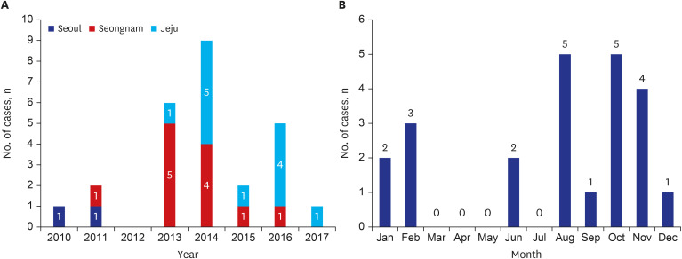
Fig. 1 Timely distribution of children who were diagnosed as staphylococcal scalded skin syndrome. (A) yearly distribution. Seoul, Seongnam and Jeju were cities which participating hospitals were located in, (B) monthly distribution.
Reference
-
1. Leung AK, Barankin B, Leong KF. Staphylococcal-scalded skin syndrome: evaluation, diagnosis, and management. World J Pediatr. 2018; 14(2):116–120. PMID: 29508362.
Article2. Mishra AK, Yadav P, Mishra A. A systemic review on staphylococcal scalded skin syndrome (SSSS): a rare and critical diesease of neonates. Open Microbiol J. 2016; 10(1):150–159. PMID: 27651848.3. Berk DR, Bayliss SJ. MRSA, staphylococcal scalded skin syndrome, and other cutaneous bacterial emergencies. Pediatr Ann. 2010; 39(10):627–633. PMID: 20954609.
Article4. Braunstein I, Wanat KA, Abuabara K, McGowan KL, Yan AC, Treat JR. Antibiotic sensitivity and resistance patterns in pediatric staphylococcal scalded skin syndrome. Pediatr Dermatol. 2014; 31(3):305–308. PMID: 24033633.
Article5. Lamand V, Dauwalder O, Tristan A, Casalegno JS, Meugnier H, Bes M, et al. Epidemiological data of staphylococcal scalded skin syndrome in France from 1997 to 2007 and microbiological characteristics of Staphylococcus aureus associated strains. Clin Microbiol Infect. 2012; 18(12):E514–21. PMID: 23078129.
Article6. Yamaguchi T, Yokota Y, Terajima J, Hayashi T, Aepfelbacher M, Ohara M, et al. Clonal association of Staphylococcus aureus causing bullous impetigo and the emergence of new methicillin-resistant clonal groups in Kansai district in Japan. J Infect Dis. 2002; 185(10):1511–1516. PMID: 11992289.7. Hörner A, Hörner R, Salla A, Nunes MS, Garzon LR, Rampelotto RF, et al. Staphylococcal scalded skin syndrome in a premature newborn caused by methicillin-resistant Staphylococcus aureus: case report. Sao Paulo Med J. 2015; 133(5):450–453. PMID: 26648436.
Article8. Lamanna O, Bongiorno D, Bertoncello L, Grandesso S, Mazzucato S, Pozzan GB, et al. Rapid containment of nosocomial transmission of a rare community-acquired methicillin-resistant Staphylococcus aureus (CA-MRSA) clone, responsible for the Staphylococcal Scalded Skin Syndrome (SSSS). Ital J Pediatr. 2017; 43(1):5. PMID: 28061866.
Article9. Jeon H, Ma SH, Jo HJ, Woo MS, An H, Park H, et al. Long-term persistence of sequence type 89 methicillin-resistant Staphylococcus aureus isolated from cases of staphylococcal scalded skin syndrome in a Korean community. J Med Microbiol. 2016; 65(12):1542–1544. PMID: 27902366.
Article10. Ma SH, Lee YS, Lee SH, Kim HK, Jin JS, Shin EK, et al. Meticillin-resistant Staphylococcus aureus clones with distinct clinical and microbiological features in a Korean community. J Med Microbiol. 2007; 56(Pt 6):866–868. PMID: 17510277.
Article11. Kang JD, Park SD. Reclassification of staphylococcal scalded skin syndrome by clinical analysis of 25 cases. Korean J Dermatol. 2004; 42(4):398–405.12. Johnson WM, Tyler SD, Ewan EP, Ashton FE, Pollard DR, Rozee KR. Detection of genes for enterotoxins, exfoliative toxins, and toxic shock syndrome toxin 1 in Staphylococcus aureus by the polymerase chain reaction. J Clin Microbiol. 1991; 29(3):426–430. PMID: 2037659.
Article13. Arnold JD, Hoek SN, Kirkorian AY. Epidemiology of staphylococcal scalded skin syndrome in the United States: a cross-sectional study, 2010–2014. J Am Acad Dermatol. 2018; 78(2):404–406. PMID: 29332709.
Article14. Neylon O, O'Connell NH, Slevin B, Powell J, Monahan R, Boyle L, et al. Neonatal staphylococcal scalded skin syndrome: clinical and outbreak containment review. Eur J Pediatr. 2010; 169(12):1503–1509. PMID: 20625909.
Article15. Li MY, Hua Y, Wei GH, Qiu L. Staphylococcal scalded skin syndrome in neonates: an 8-year retrospective study in a single institution. Pediatr Dermatol. 2014; 31(1):43–47. PMID: 23557104.
Article16. Wang Z, Feig JL, Mannschreck DB, Cohen BA. Antibiotic sensitivity and clinical outcomes in staphylococcal scalded skin syndrome. Pediatr Dermatol. 2020; 37(1):222–223. PMID: 31626359.
Article17. Chi CY, Wang SM, Lin HC, Liu CC. A clinical and microbiological comparison of Staphylococcus aureus toxic shock and scalded skin syndromes in children. Clin Infect Dis. 2006; 42(2):181–185. PMID: 16355327.
Article18. Kim ES, Song JS, Lee HJ, Choe PG, Park KH, Cho JH, et al. A survey of community-associated methicillin-resistant Staphylococcus aureus in Korea. J Antimicrob Chemother. 2007; 60(5):1108–1114. PMID: 17884831.
Article19. Kim HB, Jang HC, Nam HJ, Lee YS, Kim BS, Park WB, et al. In vitro activities of 28 antimicrobial agents against Staphylococcus aureus isolates from tertiary-care hospitals in Korea: a nationwide survey. Antimicrob Agents Chemother. 2004; 48(4):1124–1127. PMID: 15047511.
Article20. Park SY, Chung DR, Yoo JR, Baek JY, Kim SH, Ha YE, et al. Sequence type 72 community-associated meticillin-resistant Staphylococcus aureus emerged as a predominant clone of nasal colonization in newly admitted patients. J Hosp Infect. 2016; 93(4):386–389. PMID: 26874934.
Article21. Sung JY, Lee J, Choi EH, Lee HJ. Changes in molecular epidemiology of community-associated and health care-associated methicillin-resistant Staphylococcus aureus in Korean children. Diagn Microbiol Infect Dis. 2012; 74(1):28–33. PMID: 22722011.
Article22. Chong YP, Kim ES, Park SJ, Park KH, Kim T, Kim MN, et al. Accessory gene regulator (agr) dysfunction in Staphylococcus aureus bloodstream isolates from South Korean patients. Antimicrob Agents Chemother. 2013; 57(3):1509–1512. PMID: 23254438.
Article23. Kwon JC, Kim SH, Park SH, Choi SM, Lee DG, Choi JH, et al. Molecular epidemiologic analysis of methicillin-resistant Staphylococcus aureus isolates from bacteremia and nasal colonization at 10 intensive care units: multicenter prospective study in Korea. J Korean Med Sci. 2011; 26(5):604–611. PMID: 21532849.
Article24. Kikuta H, Shibata M, Nakata S, Yamanaka T, Sakata H, Akizawa K, et al. Predominant dissemination of PVL-negative CC89 MRSA with SCCmec type II in children with impetigo in Japan. Int J Pediatr. 2011; 2011:143872. PMID: 22187567.25. Shi D, Higuchi W, Takano T, Saito K, Ozaki K, Takano M, et al. Bullous impetigo in children infected with methicillin-resistant Staphylococcus aureus alone or in combination with methicillin-susceptible S. aureus: analysis of genetic characteristics, including assessment of exfoliative toxin gene carriage. J Clin Microbiol. 2011; 49(5):1972–1974. PMID: 21430094.
Article26. Staiman A, Hsu DY, Silverberg JI. Epidemiology of staphylococcal scalded skin syndrome in U.S. children. Br J Dermatol. 2018; 178(3):704–708. PMID: 29077993.
Article27. Yamasaki O, Tristan A, Yamaguchi T, Sugai M, Lina G, Bes M, et al. Distribution of the exfoliative toxin D gene in clinical Staphylococcus aureus isolates in France. Clin Microbiol Infect. 2006; 12(6):585–588. PMID: 16700711.
Article28. Bukowski M, Wladyka B, Dubin G. Exfoliative toxins of Staphylococcus aureus. Toxins (Basel). 2010; 2(5):1148–1165. PMID: 22069631.
Article29. Becker K, Friedrich AW, Lubritz G, Weilert M, Peters G, Von Eiff C. Prevalence of genes encoding pyrogenic toxin superantigens and exfoliative toxins among strains of Staphylococcus aureus isolated from blood and nasal specimens. J Clin Microbiol. 2003; 41(4):1434–1439. PMID: 12682126.
Article30. Nhan TX, Leclercq R, Cattoir V. Prevalence of toxin genes in consecutive clinical isolates of Staphylococcus aureus and clinical impact. Eur J Clin Microbiol Infect Dis. 2011; 30(6):719–725. PMID: 21225304.
Article31. Koosha RZ, Fooladi AA, Hosseini HM, Aghdam EM. Prevalence of exfoliative toxin A and B genes in Staphylococcus aureus isolated from clinical specimens. J Infect Public Health. 2014; 7(3):177–185. PMID: 24637043.
Article32. Mohseni M, Rafiei F, Ghaemi EA. High frequency of exfoliative toxin genes among Staphylococcus aureus isolated from clinical specimens in the north of Iran: Alarm for the health of individuals under risk. Iran J Microbiol. 2018; 10(3):158–165. PMID: 30112153.
- Full Text Links
-
- Actions
-
Cited
- CITED
-
- Close
- Share
-
- Similar articles
-
- Antibiotic resistance of Staphylococcus aureus colonized in children with staphylococcal scalded skin syndrome
- Four Cases of Staphylococcal Scalded Skin Syndrome
- Two Cases of Staphylococcal Scalded Skin Syndrome
- Recent Trends in Methicillin-resistant Staphylococcus aureus Infection and Antibiotic Treatment in Staphylococcal Scalded Skin Syndrome in Patients in Childhood: A Single-center Study
- Sequential Cases of Staphylococcal Scalded Skin Syndrome in Very Low Birth Weight Infants
