J Neurocrit Care.
2020 Nov;13(2):133-136. 10.18700/jnc.200029.
Radiculopathy caused by lumbar epidural varix
- Affiliations
-
- 1Department of Neurology, Jeju National University School of Medicine, Jeju, Republic of Korea
- 2Jeju National University School of Medicine, Jeju, Republic of Korea
- 3Department of Neurosurgery, Jeju National University School of Medicine, Jeju, Republic of Korea
- 4Department of Pathology, Jeju National University School of Medicine, Jeju, Republic of Korea
- KMID: 2509946
- DOI: http://doi.org/10.18700/jnc.200029
Abstract
- Background
Lumbar epidural varix (LEV) is a very rare condition caused by dilatation of the vertebral venous plexus. LEV can result in lumbosacral radiculopathy, which is often mistaken for lumbosacral herniated intervertebral disc (HIVD).
Case Report
A 72-year-old man visited the emergency department (ED) with radiating pain of the right leg that had developed 3 weeks previously. Before the ED visit, he was diagnosed with lumbosacral radiculopathy due to HIVD based on lumbar X-rays at an outpatient clinic. Despite conservative treatment, his symptoms deteriorated. On magnetic resonance imaging at the ED, an epidural cystic mass in the right L5–S1 with multiple HIVDs was observed. The mass was surgically removed, and the histological findings showed a dilated vessel with a thrombus, which led to the final diagnosis of LEV.
Conclusion
In lumbosacral radiculopathy, LEV should be considered as a possibility even if degenerative spine disease is observed on lumbar X-rays.
Keyword
Figure
-
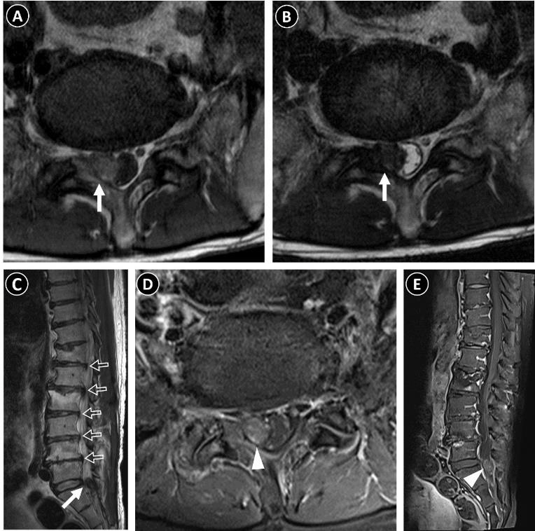
Fig. 1. Preoperative lumbar magnetic resonance images. Axial T1-weighted (A) and T2-weighted (B), and sagittal T2-weighted (C) images show an irregular mass lesion (closed arrows) involving the right facet joint at L5–S1 and the right epidural space at the L5–S1 level. Multiple bulging discs (open arrows) with compression fractures in the T12 and L2 vertebral bodies are also shown on sagittal T2-weighted images (C). Heterogeneous enhancement of the mass is shown on gadolinium-enhanced T1-weighted axial (D) and sagittal images (E) (arrowheads).

Fig. 2. Dilated and tortuous vessel wall (arrows) with an organized thrombus (arrowheads) in the histological examination (H&E ×100).
Reference
-
1. Slin'ko EI, Al-Qashqish II. Surgical treatment of lumbar epidural varices. J Neurosurg Spine. 2006; 5:414–23.2. Endres S. Epidural varicosis as a possible cause of radicular pain: a case report. J Med Case Rep. 2011; 5:537.
Article3. Bursalı A, Akyoldas G, Guvenal AB, Yaman O. Lumbar epidural varix mimicking disc herniation. J Korean Neurosurg Soc. 2016; 59:410–3.
Article4. Choi S, Kim KY, Lee S, Yoon S. Efficacy of enhanced MRI in epidural varix: report of six cases. J Korean Soc Spine Surg. 2006; 13:210–4.5. Gálvez MM, Cordovez JM, Okuma CP, Montoya CM, Asahi TK. Diagnóstico diferencial de hernia discal. Rev Chil Radiologia. 2017; 23:66–76.6. Zimmerman GA, Weingarten K, Lavyne MH. Symptomatic lumbar epidural varices: report of two cases. J Neurosurg. 1994; 80:914–8.7. Paksoy Y, Gormus N. Epidural venous plexus enlargements presenting with radiculopathy and back pain in patients with inferior vena cava obstruction or occlusion. Spine (Phila Pa 1976). 2004; 29:2419–24.
Article8. Hirabayashi Y, Shimizu R, Fukuda H, Saitoh K, Igarashi T. Effects of the pregnant uterus on the extradural venous plexus in the supine and lateral positions, as determined by magnetic resonance imaging. Br J Anaesth. 1997; 78:317–9.
Article9. Gundry CR, Heithoff KB. Epidural hematoma of the lumbar spine: 18 surgically confirmed cases. Radiology. 1993; 187:427–31.
Article10. Gümbel U, Pia HW, Vogelsang H. Lumbosacral vascular anomalies as the cause of ischialgia. Acta Neurochir (Wien). 1969; 20:131–51.11. Tofuku K, Koga H, Yone K, Komiya S. Spontaneous regression of symptomatic lumbar epidural varix: a case report. Spine (Phila Pa 1976). 2007; 32:E147–9.
- Full Text Links
-
- Actions
-
Cited
- CITED
-
- Close
- Share
-
- Similar articles
-
- Lumbar Epidural Varix Mimicking Disc Herniation
- Radiculopathy Caused by Discal Cyst
- Lumbar Epidural Varix Mimicking Perineural Cyst
- Efficacy of Enhanced MRI in Epidural Varix: Report of Six Cases
- Comparison of Transforaminal Epidural Steroid Injection and Lumbar/Caudal Epidural Steroid Injection for the Treatment of Lumbosacral Radiculopathy
