Biological Enhancement of Healing with Kartogenin Injection at the Tendon-to-Bone Interface in a Rat Rotator Cuff Tear Model
- Affiliations
-
- 1Department of Orthopaedic Surgery, Kyungpook National University Hospital, Daegu, Korea
- 2Department of Bio-Fibers and Materials Science, College of Agriculture and Life Science, Kyungpook National University, Daegu, Korea
- KMID: 2509020
- DOI: http://doi.org/10.5763/kjsm.2020.38.4.217
Abstract
- Purpose
The purpose of this study was to investigate the effects of kartogenin (KGN) on the tendon-bone interface in a rat rotator cuff tear model.
Methods
Twenty male rats were divided into two equal groups; group 1 (repair only) and group 2 (KGN single injection). A rat rotator cuff rupture model was prepared, and KGN (500 μM) was injected into the repair site. The specimens were collected after 8 weeks, and biomechanical and histological analyses were performed. We assessed the healing quality of the tendon-to-bone repair site using six aspects of tendon tissue. The histological findings were classified semiquantitatively into four grades (grade 0, the poorest appearance; grade 1, poorer; grade 2, better; and grade 3, marked regeneration).
Results
Group 2 showed a higher mean ultimate load to failure than the control group (group 1: 25.78±31.38 N, group 2: 55.64±36.02 N; p=0.011). On histological analysis, group 2 exhibited a significantly greater total score (group 1: 7.20±2.14, group 2: 9.50±1.84; p=0.019), collagen fiber continuity (group 1: 1.20±0.42, group 2: 1.70±0.48; p=0.024), and collagen fiber density (group 1: 1.50±0.52, group 2: 2.20±0.63; p=0.080). Metachromasia were more intense in group 2 than in the control group.
Conclusion
A single injection of KGN reinforces biomechanical strength and the formation of collagen and fibrocartilage at the tendon-to-bone interface in a rat rotator cuff tear model.
Keyword
Figure
-
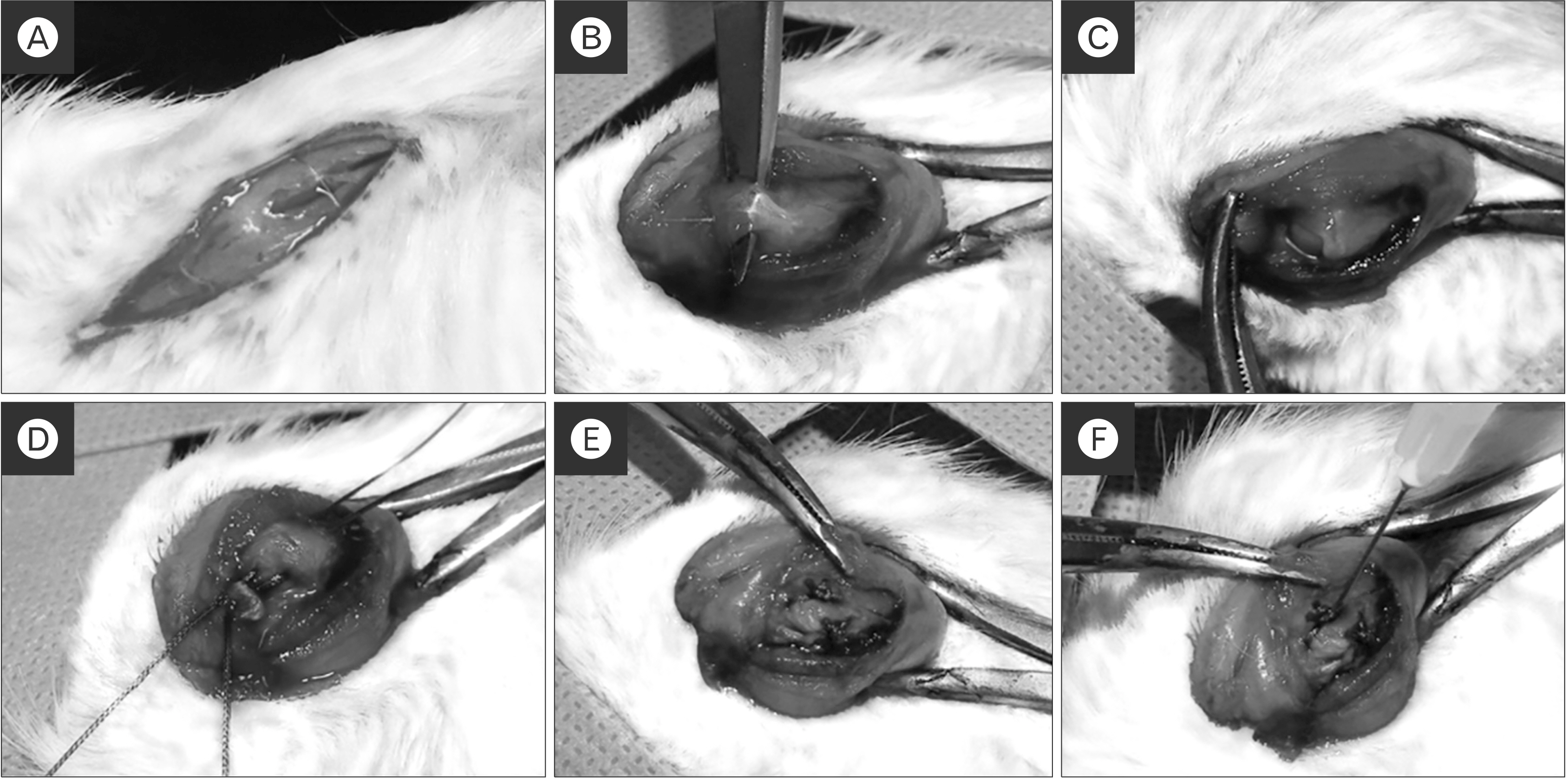
Fig. 1 The animal model and surgical procedures. (A, B) The deltoid muscle split and the supraspinatus tendon was exposed. (C) The supraspinatus tendon was isolated and transected at the end of the tendon insertion site. (D, E) Two bone tunnels were made at the greater tuberosity of the humeral head and single-row repair. The control group received repair only. (F) In the experimental group, kartogenin (500 μM) was injected into the bone-to-tendon interface.

Fig. 2 Histological evaluation at 8 weeks after repair. Group 1 received only supraspinatus repair; group 2 received supraspinatus repair with a single injection of kartogenin. (A) H&E staining, ×50. (B) Masson trichrome staining, ×50. (C) Picrosirius red staining, ×50. Continuity, orientation, and density of collagen fiber showed a higher average grading in group 2 compared with group 1. Group 2 showed better maturation and density of the tendon-to-bone interface structure (arrows) than group 1.
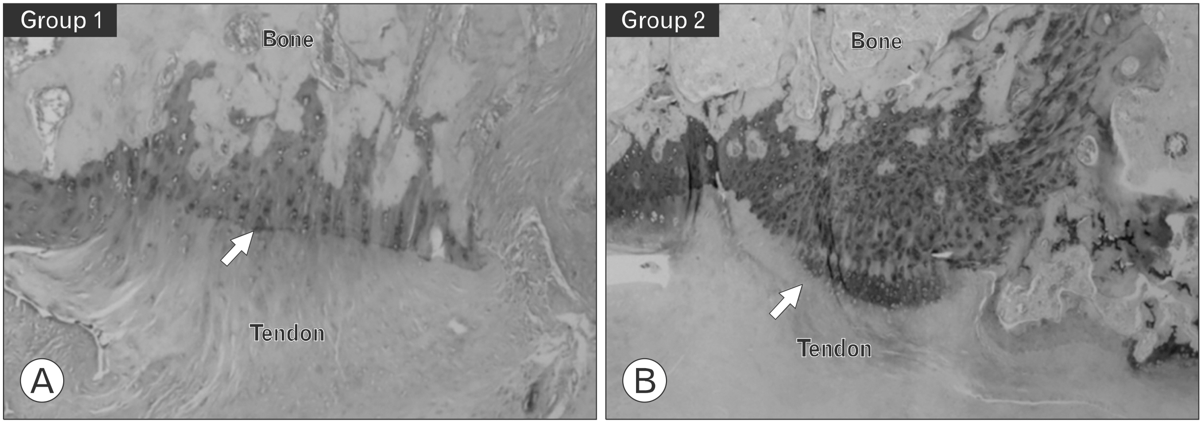
Fig. 3 Histological evaluation at 8 weeks after repair. (A) Group 1 received only supraspinatus repair; (B) group 2 received supraspinatus repair with a single injection of kartogenin. The arrows indicate the tendon-to-bone interface. This area is the supraspinatus enthesis, and fibrocartilaginous transition site and was stained with Toluidine blue (×50). The metachromasia were more intense in the supraspinatus enthesis of group 2 compared to the control group.
Reference
-
1. Yamaguchi K, Ditsios K, Middleton WD, Hildebolt CF, Galatz LM, Teefey SA. 2006; The demographic and morphological features of rotator cuff disease: a comparison of asymptomatic and symptomatic shoulders. J Bone Joint Surg Am. 88:1699–704. DOI: 10.2106/00004623-200608000-00002. PMID: 16882890.2. Wang VM, Wang FC, McNickle AG, et al. 2010; Medial versus lateral supraspinatus tendon properties: implications for double- row rotator cuff repair. Am J Sports Med. 38:2456–63. DOI: 10.1177/0363546510376817. PMID: 20929937. PMCID: PMC3772634.3. Le BT, Wu XL, Lam PH, Murrell GA. 2014; Factors predicting rotator cuff retears: an analysis of 1000 consecutive rotator cuff repairs. Am J Sports Med. 42:1134–42. DOI: 10.1177/0363546514525336. PMID: 24748610.4. Castricini R, Longo UG, De Benedetto M, et al. 2011; Platelet-rich plasma augmentation for arthroscopic rotator cuff repair: a randomized controlled trial. Am J Sports Med. 39:258–65. DOI: 10.1177/0363546510390780. PMID: 21160018.5. Wang LL, Yin XF, Chu XC, Zhang YB, Gong XN. 2018; Platelet-derived growth factor subunit B is required for tendon-bone healing using bone marrow-derived mesenchymal stem cells after rotator cuff repair in rats. J Cell Biochem. 119:8897–908. DOI: 10.1002/jcb.27143. PMID: 30105826.
Article6. Yonemitsu R, Tokunaga T, Shukunami C, et al. 2019; Fibroblast growth factor 2 enhances tendon-to-bone healing in a rat rotator cuff repair of chronic tears. Am J Sports Med. 47:1701–12. DOI: 10.1177/0363546519836959. PMID: 31038985.
Article7. Liu Q, Yu Y, Reisdorf RL, et al. 2019; Engineered tendon- fibrocartilage-bone composite and bone marrow-derived mesenchymal stem cell sheet augmentation promotes rotator cuff healing in a non-weight-bearing canine model. Biomaterials. 192:189–98. DOI: 10.1016/j.biomaterials.2018.10.037. PMID: 30453215.8. Wang C, Hu Q, Song W, Yu W, He Y. 2020; Adipose stem cell- derived exosomes decrease fatty infiltration and enhance rotator cuff healing in a rabbit model of chronic tears. Am J Sports Med. 48:1456–64. DOI: 10.1177/0363546520908847. PMID: 32272021.9. Zhang J, Wang JH. 2014; Kartogenin induces cartilage-like tissue formation in tendon-bone junction. Bone Res. 2:14008. DOI: 10.1038/boneres.2014.8. PMID: 25419468. PMCID: PMC4237211.
Article10. Im GI. 2018; Application of kartogenin for musculoskeletal regeneration. J Biomed Mater Res A. 106:1141–8. DOI: 10.1002/jbm.a.36300. PMID: 29164815.
Article11. Mohan G, Magnitsky S, Melkus G, et al. 2016; Kartogenin treatment prevented joint degeneration in a rodent model of osteoarthritis: a pilot study. J Orthop Res. 34:1780–9. DOI: 10.1002/jor.23197. PMID: 26895619. PMCID: PMC6348064.
Article12. Xu X, Shi D, Shen Y, et al. 2015; Full-thickness cartilage defects are repaired via a microfracture technique and intraarticular injection of the small-molecule compound kartogenin. Arthritis Res Ther. 17:20. DOI: 10.1186/s13075-015-0537-1. PMID: 25641548. PMCID: PMC4376363.
Article13. Zhang J, Yuan T, Zheng N, Zhou Y, Hogan MV, Wang JH. 2017; The combined use of kartogenin and platelet-rich plasma promotes fibrocartilage formation in the wounded rat Achilles tendon entheses. Bone Joint Res. 6:231–44. DOI: 10.1302/2046-3758.64.BJR-2017-0268.R1. PMID: 28450316. PMCID: PMC5415905.
Article14. Kim DH, Min SG, Yoon JP, et al. 2019; Mechanical augmentation with absorbable alginate sheet enhances healing of the rotator cuff. Orthopedics. 42:e104–10. DOI: 10.3928/01477447-20181206-04. PMID: 30540880.
Article15. Johnson K, Zhu S, Tremblay MS, et al. 2012; A stem cell-based approach to cartilage repair. Science. 336:717–21. DOI: 10.1126/science.1215157. PMID: 22491093.
Article16. Kang ML, Ko JY, Kim JE, Im GI. 2014; Intra-articular delivery of kartogenin-conjugated chitosan nano/microparticles for cartilage regeneration. Biomaterials. 35:9984–94. DOI: 10.1016/j.biomaterials.2014.08.042. PMID: 25241157.
Article17. Wang D, Tan H, Lebaschi AH, et al. 2018; Kartogenin enhances collagen organization and mechanical strength of the repaired enthesis in a murine model of rotator cuff repair. Arthroscopy. 34:2579–87. DOI: 10.1016/j.arthro.2018.04.022. PMID: 30037570. PMCID: PMC6371391.
Article18. Huang C, Zhang X, Luo H, et al. 2020; Jul. 7. Effect of kartogenin-loaded gelatin methacryloyl hydrogel scaffold with bone marrow stimulation for enthesis healing in rotator cuff repair. J Shoulder Elbow Surg. [Epub]. https://doi.org/10.1016/. DOI: 10.1016/j.jse.2020.06.013. PMID: 32650072.
Article19. Shi D, Xu X, Ye Y, et al. 2016; Photo-cross-linked scaffold with kartogenin-encapsulated nanoparticles for cartilage regeneration. ACS Nano. 10:1292–9. DOI: 10.1021/acsnano.5b06663. PMID: 26757419.
Article20. Hu Q, Ding B, Yan X, et al. 2017; Polyethylene glycol modified PAMAM dendrimer delivery of kartogenin to induce chondrogenic differentiation of mesenchymal stem cells. Nanomedicine. 13:2189–98. DOI: 10.1016/j.nano.2017.05.011. PMID: 28579434.
Article
- Full Text Links
-
- Actions
-
Cited
- CITED
-
- Close
- Share
-
- Similar articles
-
- Current and Future Strategies to Enhance Healing at the Tendon-To-Bone Interface of a Rotator Cuff Tear
- Usefulness of Multiphase Scaffolds for Improving Tendon to Bone Healing for Rotator Cuff Tears in Shoulder
- Biological Characteristics of Rotator Cuff Tendon
- Can the Rotator Cuff Tear Be Treated with Atelocollagen?
- Selective Serotonin Reuptake Inhibitor Promotes Bone-Tendon Interface Healing in a Rotator Cuff Tear Rat Model
