KLF4 Regulates Goblet Cell Differentiation in BMI1+ Reserve Intestinal Stem Cell Lineage during Homeostasis
- Affiliations
-
- 1Department of Medicine, Renaissance School of Medicine, Stony Brook University, Stony Brook, NY, USA
- 2Department of Gastroenterology and Metabolism, Nagoya City University Graduate School of Medical Sciences, Nagoya, Japan
- 3Department of Physiology and Biophysics, Renaissance School of Medicine, Stony Brook University, Stony Brook, NY, USA
- KMID: 2508916
- DOI: http://doi.org/10.15283/ijsc20048
Abstract
- Krüppel-like factor 4 (KLF4) is a zinc-finger transcription factor, expressed in villus cells of the intestinal epithelium, that promotes cellular differentiation and tissue homeostasis. Previous studies suggest that BMI1+ cells represent secretory progenitors with reserve intestinal stem cell (rISC) activity. However, it has not been elucidated how KLF4 contributes to crypt regeneration originated from BMI1+ rISC lineage during homeostasis. In this study, Bmi1-CreER ;Rosa26eYFP (Bmi1Ctrl ) and Bmi1-CreER ;Rosa26eYFP ;Klf4fl/fl (Bmi1△Klf4 ) mice were injected with tamoxifen to label BMI1+ cells and their lineage and to delete Klf4. During homeostasis, MUC2+ goblet cells appeared in the BMI1+ cell lineage 2, 3 and 7 days after tamoxifen administration. After Klf4 deletion in BMI1+ cells, the number of KLF4+ and MUC2+ cells in eYFP+ cells decreased in Bmi1△Klf4 mice compared with Bmi1Ctrl mice. Thus, KLF4 was positively correlated with goblet cell differentiation in BMI1+ cell derived lineage. In ex-vivo analysis, organoids derived from single eYFP+ cells of Bmi1Ctrl mice contained MUC2-expressing cells that co-expressed KLF4. On the other hand, organoids derived from Klf4-deleted eYFP+ cells from Bmi1△Klf4 mice showed reduced number of MUC2-expressing cells. In conclusion, these results suggest that KLF4 regulates goblet cell differentiation in BMI1+ ISC-derived lineage during homeostasis.
Figure
-
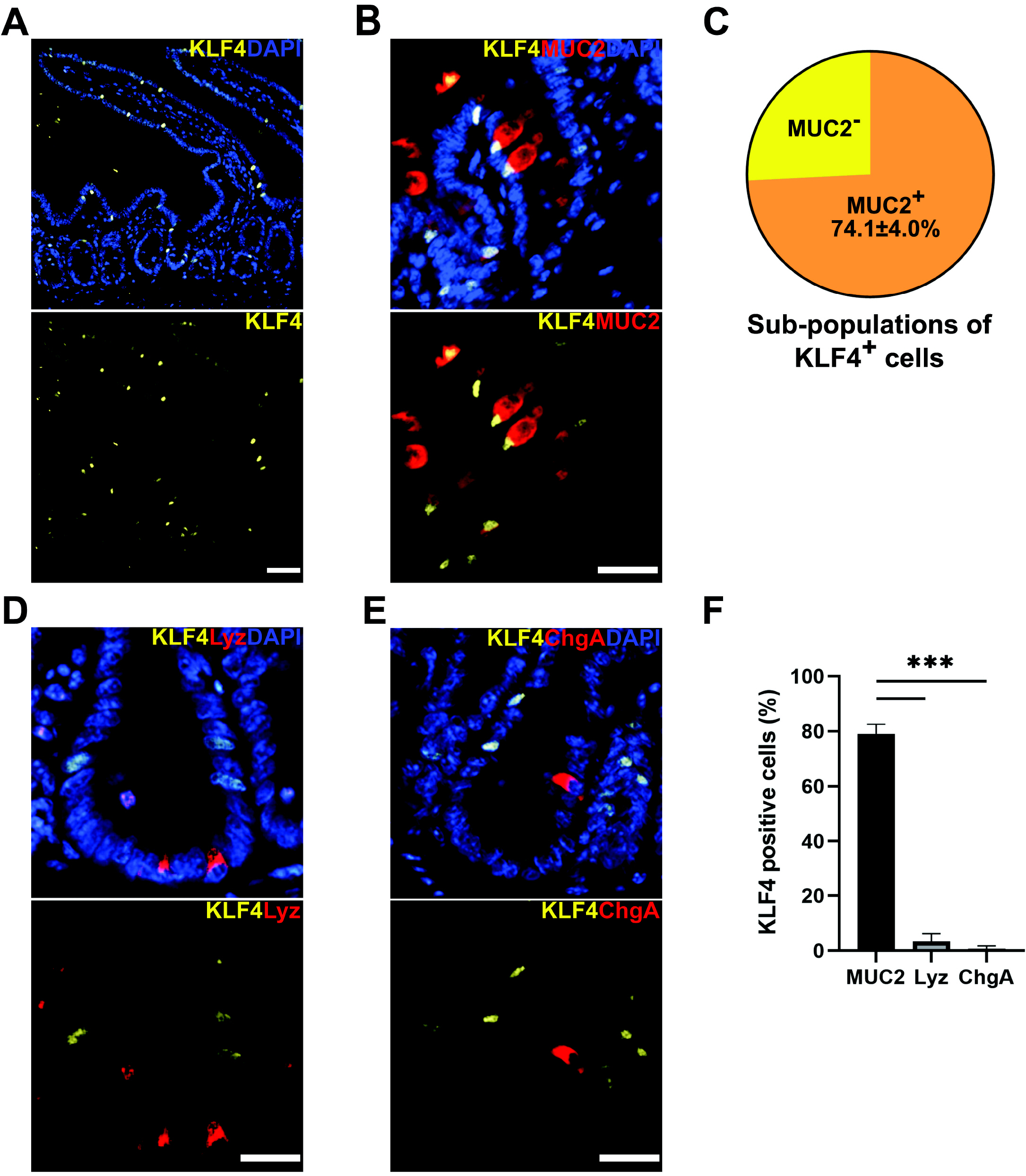
Fig. 1 Relationship between KLF4 expression and markers of differentiated cells in proximal small intestine. (A) Immunostaining of Bmi1Ctrl mouse intestine. KLF4 (yellow) was expressed mainly in the terminally differentiated epithelial cells and was also expressed in a subpopulation of the cells in the crypt. Scale bar, 50 μm. (B) Immunostaining for MUC2 (goblet cell marker, red) and KLF4 (yellow). MUC2 was overlapped with KLF4 in the crypt of proximal small intestine. Scale bar, 25 μm. (C) MUC2 was co-expressed in 74.1±4.0% of KLF4+ cells. KLF4+ cells were collected from a total of 50 crypts per Bmi1Ctrl mouse (n=3). (D) Immunostaining for lysozyme (Paneth cell marker, red) and KLF4 (yellow). Scale bar, 25 μm. (E) Immunostaining for chromogranin A (enteroendocrine cell marker, red) and KLF4 (yellow). Scale bar, 25 μm. (F) Co-expression rate of KLF4+ in MUC2+ cells was significantly higher than Lyz+ and ChgA+ cells (79.0±2.0%, 3.3±1.7%, and 0.7±0.7%, respectively). ***p<0.001 MUC2+, lysozyme+, and chromogranin A+ cells were collected from a total of 150, 100 and 60 crypts from 3 Bmi1Ctrl mice, respectively (n=3 for each group). Lyz, lysozyme; ChgA, chromogranin A.
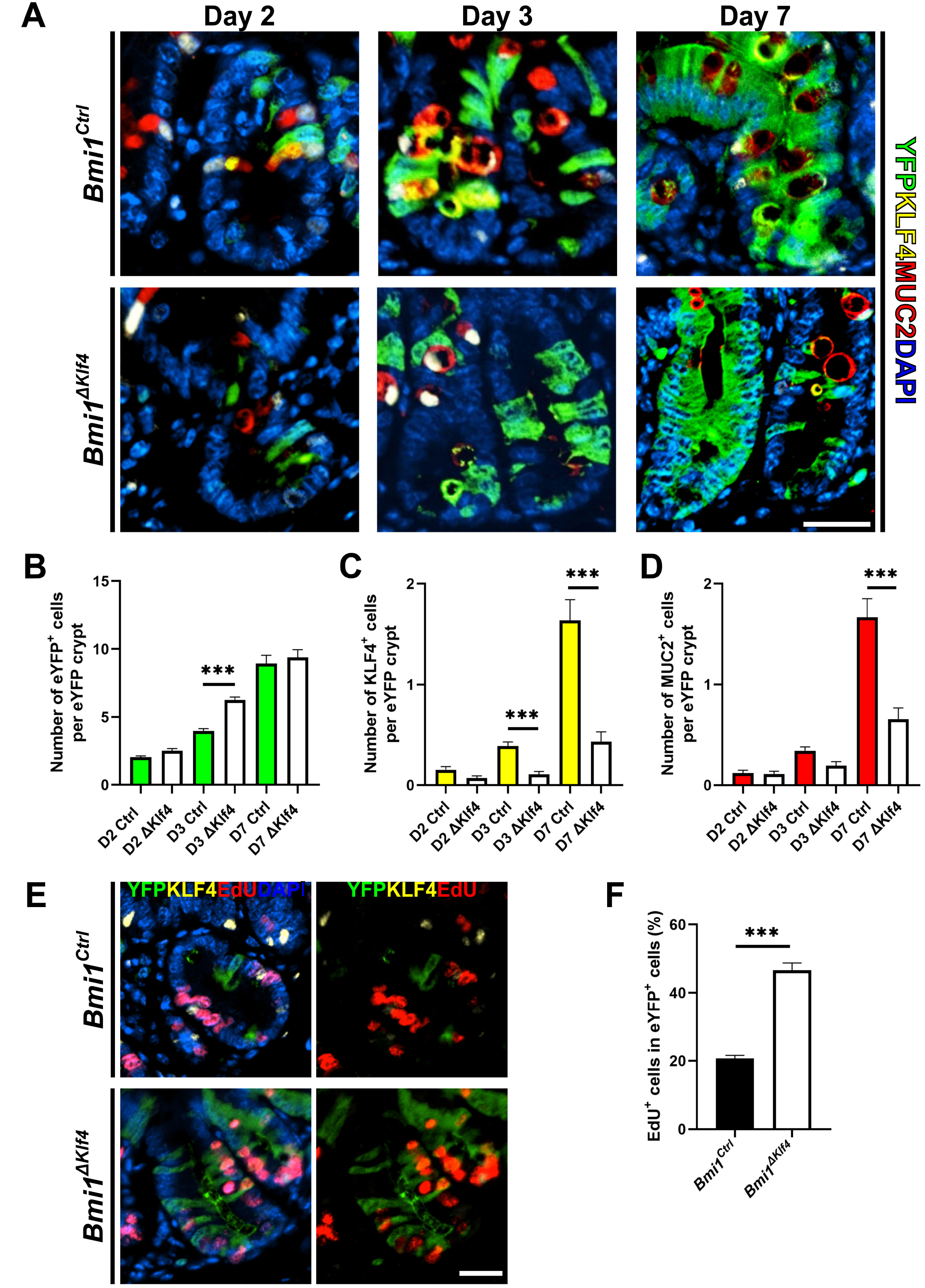
Fig. 2 KLF4 modulates goblet cell differentiation in Bmi1 cell lineage during homeostasis. (A) Immunostaining for eYFP (green), MUC2 (red) and KLF4 (yellow). Representative image of proximal small intestinal crypt at day 2, 3 and 7 following tamoxifen injection. Goblet cell differentiation is involved in Bmi1-eYFP cell lineage. Scale bar, 25 μm. (B), (C) and (D) The number of eYFP, MUC2 and KLF4 positive cells per eYFP+ including crypt increased as the time following tamoxifen administration in both groups. Data were collected from at least 20 crypts per mouse (n=3 each group). Dunn’s multiple comparisons test with Kruskal-Wallis test. (B) The number of eYFP+ cells was significantly higher in Bmi1ΔKlf4 than in Bmi1Ctrl at day 3 following tamoxifen administration. (C) The number of KLF4+ cells was significantly higher in Bmi1Ctrl than in Bmi1ΔKlf4 at day 7 following tamoxifen administration (p<0.001). (D) The number of MUC2+ cells was significantly lower in Bmi1ΔKlf4 than in Bmi1Ctrl at day 3 and 7 following tamoxifen administration (p<0.001). (E) Immunostaining for eYFP (green), KLF4 (yellow) and EdU (red) for the proximal small intestinal crypts at day 3 following tamoxifen administration. Scale bar, 25 μm. (F) At day 3 after tamoxifen injection, EdU positivity in eYFP+ cells of Bmi1ΔKlf4 was significantly higher than Bmi1Ctrl (46.7±2.1% and 20.7±0.9%, respectively). Student t test. EYFP+ cells were collected from 50 crypts per mouse (n=3 each group). ***p<0.001.

Fig. 3 KLF4 is positively correlated with goblet cell differentiation. (A) The pie chart shows the subpopula-tions of eYFP+ cells distinguished by co-staining with KLF4 and/or MUC2. A total of at 150∼260 eYFP+ cells were counted from each mouse at day 7 after tamoxifen administration (n=3 each group). (B) KLF4 and MUC2 were significantly positively correlated (Spearman correlation= 0.9429, p<0.01).
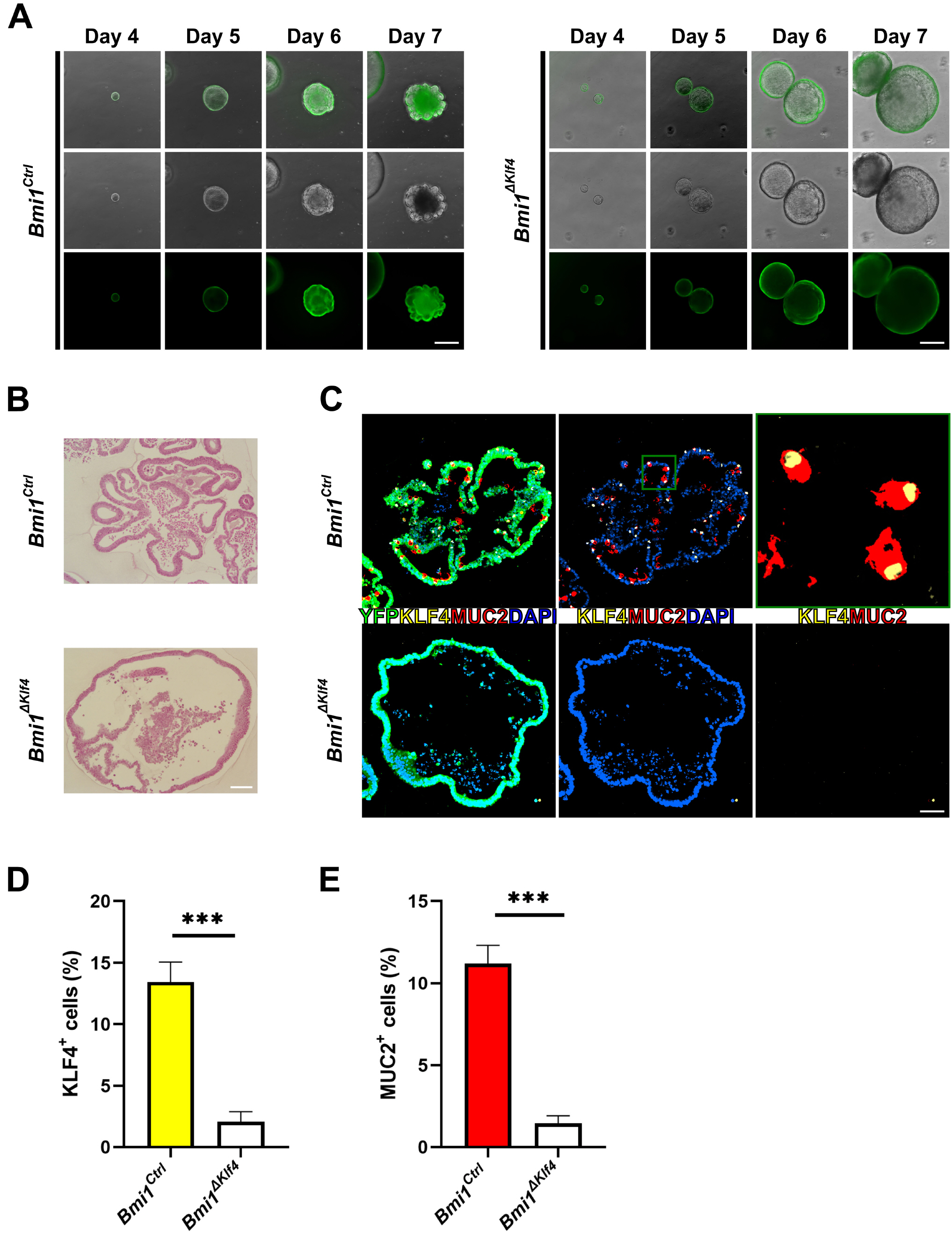
Fig. 4 Organoids derived from single FACS sorted eYFP+ cells of Bmi1Ctrl and Bmi1ΔKlf4 mice. (A) Representative time course images of cultured organoids derived from Bmi1Ctrl and Bmi1ΔKlf4 mice. Upper panels, merged image of bright-field and fluorescent image; middle panel, bright field image; lower panel, fluorescent image. Scale bars, 500 μm. (B) Hematoxylin and eosin staining at culture day 7. Scale bars, 100 μm. (C) Cultured organoids were collected at day 7. Immunostaining for eYFP (green), MUC2 (red), KLF4 (yellow). Left panel merged image for eYFP, MUC2, KLF4 and DAPI (blue); middle panel, merged image for MUC2, KLF4 and DAPI; right panel merged image for MUC2 and KLF4. Upper right panel is magnified image of green square in upper middle panel. In Bmi1Ctrl mice derived organoids, 73.9% of MUC2+ cells were co-expressed with KLF4. Scale bars, 50 μm. (D) and (E) Percentage of KLF4+ and MUC2+ cells were significantly higher in Bmi1Ctrl organoid than Bmi1ΔKlf4 at culture day 7 (Bmi1Ctrl vs. Bmi1ΔKlf4, 11.2±1.1% vs. 1.4±0.5% and 13.4±1.6% vs 2.1±0.8% for KLF4 and MUC2, respectively). Cells were analyzed from 10 sections for each group and 10∼200 cells were collected from each section. Organoids were pooled from 3 mice for each group. ***p<0.001.
Reference
-
References
1. Barker N, van Es JH, Kuipers J, Kujala P, van den Born M, Cozijnsen M, Haegebarth A, Korving J, Begthel H, Peters PJ, Clevers H. 2007; Identification of stem cells in small intestine and colon by marker gene Lgr5. Nature. 449:1003–1007. DOI: 10.1038/nature06196. PMID: 17934449.
Article2. Clevers H. 2013; The intestinal crypt, a prototype stem cell compartment. Cell. 154:274–284. DOI: 10.1016/j.cell.2013.07.004. PMID: 23870119.
Article3. Kim CK, Yang VW, Bialkowska AB. 2017; The role of intestinal stem cells in epithelial regeneration following radiation-induced gut injury. Curr Stem Cell Rep. 3:320–332. DOI: 10.1007/s40778-017-0103-7. PMID: 29497599. PMCID: PMC5818549.
Article4. Sangiorgi E, Capecchi MR. 2008; Bmi1 is expressed in vivo in intestinal stem cells. Nat Genet. 40:915–920. DOI: 10.1038/ng.165. PMID: 18536716. PMCID: PMC2906135.
Article5. Tian H, Biehs B, Warming S, Leong KG, Rangell L, Klein OD, de Sauvage FJ. 2011; A reserve stem cell population in small intestine renders Lgr5-positive cells dispensable. Nature. 478:255–259. DOI: 10.1038/nature10408. PMID: 21927002. PMCID: PMC4251967.
Article6. Yan KS, Chia LA, Li X, Ootani A, Su J, Lee JY, Su N, Luo Y, Heilshorn SC, Amieva MR, Sangiorgi E, Capecchi MR, Kuo CJ. 2012; The intestinal stem cell markers Bmi1 and Lgr5 identify two functionally distinct populations. Proc Natl Acad Sci U S A. 109:466–471. DOI: 10.1073/pnas.1118857109. PMID: 22190486. PMCID: PMC3258636.
Article7. Noah TK, Donahue B, Shroyer NF. 2011; Intestinal development and differentiation. Exp Cell Res. 317:2702–2710. DOI: 10.1016/j.yexcr.2011.09.006. PMID: 21978911. PMCID: PMC3210330.
Article8. Muñoz J, Stange DE, Schepers AG, van de Wetering M, Koo BK, Itzkovitz S, Volckmann R, Kung KS, Koster J, Radulescu S, Myant K, Versteeg R, Sansom OJ, van Es JH, Barker N, van Oudenaarden A, Mohammed S, Heck AJ, Clevers H. 2012; The Lgr5 intestinal stem cell signature: robust expression of proposed quiescent '+4' cell markers. EMBO J. 31:3079–3091. DOI: 10.1038/emboj.2012.166. PMID: 22692129. PMCID: PMC3400017.
Article9. Ghaleb AM, Yang VW. 2017; Krüppel-like factor 4 (KLF4): what we currently know. Gene. 611:27–37. DOI: 10.1016/j.gene.2017.02.025. PMID: 28237823. PMCID: PMC5391259.
Article10. Kuruvilla JG, Kim CK, Ghaleb AM, Bialkowska AB, Kuo CJ, Yang VW. 2016; Krüppel-like factor 4 modulates development of BMI1(+) intestinal stem cell-derived lineage following γ-radiation-induced gut injury in mice. Stem Cell Reports. 6:815–824. DOI: 10.1016/j.stemcr.2016.04.014. PMID: 27237377. PMCID: PMC4911500.
Article11. Ghaleb AM, Aggarwal G, Bialkowska AB, Nandan MO, Yang VW. 2008; Notch inhibits expression of the Krüppel-like factor 4 tumor suppressor in the intestinal epithelium. Mol Cancer Res. 6:1920–1927. DOI: 10.1158/1541-7786.MCR-08-0224. PMID: 19074836. PMCID: PMC2628949.
Article12. Ghaleb AM, McConnell BB, Kaestner KH, Yang VW. 2011; Altered intestinal epithelial homeostasis in mice with intestine-specific deletion of the Krüppel-like factor 4 gene. Dev Biol. 349:310–320. DOI: 10.1016/j.ydbio.2010.11.001. PMID: 21070761. PMCID: PMC3022386.
Article13. Imajo M, Ebisuya M, Nishida E. 2015; Dual role of YAP and TAZ in renewal of the intestinal epithelium. Nat Cell Biol. 17:7–19. DOI: 10.1038/ncb3084. PMID: 25531778.
Article14. Kim CK, Saxena M, Maharjan K, Song JJ, Shroyer KR, Bialkowska AB, Shivdasani RA, Yang VW. 2020; Krüppel-like factor 5 regulates stemness, lineage specification, and regeneration of intestinal epithelial stem cells. Cell Mol Gastroenterol Hepatol. 9:587–609. DOI: 10.1016/j.jcmgh.2019.11.009. PMID: 31778829. PMCID: PMC7078555.
Article15. Miyoshi H, Stappenbeck TS. 2013; In vitro expansion and genetic modification of gastrointestinal stem cells in spheroid cul-ture. Nat Protoc. 8:2471–2482. DOI: 10.1038/nprot.2013.153. PMID: 24232249. PMCID: PMC3969856.
Article16. Flandez M, Guilmeau S, Blache P, Augenlicht LH. 2008; KLF4 regulation in intestinal epithelial cell maturation. Exp Cell Res. 314:3712–3723. DOI: 10.1016/j.yexcr.2008.10.004. PMID: 18977346. PMCID: PMC2652355.
Article17. Itzkovitz S, Lyubimova A, Blat IC, Maynard M, van Es J, Lees J, Jacks T, Clevers H, van Oudenaarden A. 2011; Single-molecule transcript counting of stem-cell markers in the mouse intestine. Nat Cell Biol. 14:106–114. DOI: 10.1038/ncb2384. PMID: 22119784. PMCID: PMC3292866.
Article18. Yan KS, Gevaert O, Zheng GXY, Anchang B, Probert CS, Larkin KA, Davies PS, Cheng ZF, Kaddis JS, Han A, Roelf K, Calderon RI, Cynn E, Hu X, Mandleywala K, Wilhelmy J, Grimes SM, Corney DC, Boutet SC, Terry JM, Belgrader P, Ziraldo SB, Mikkelsen TS, Wang F, von Furstenberg RJ, Smith NR, Chandrakesan P, May R, Chrissy MAS, Jain R, Cartwright CA, Niland JC, Hong YK, Carrington J, Breault DT, Epstein J, Houchen CW, Lynch JP, Martin MG, Plevritis SK, Curtis C, Ji HP, Li L, Henning SJ, Wong MH, Kuo CJ. 2017; Intestinal enteroendocrine lineage cells possess homeostatic and injury-inducible stem cell activity. Cell Stem Cell. 21:78–90.e6. DOI: 10.1016/j.stem.2017.06.014. PMID: 28686870. PMCID: PMC5642297.
Article19. Schneider H, Pelaseyed T, Svensson F, Johansson MEV. 2018; Study of mucin turnover in the small intestine by in vivo labeling. Sci Rep. 8:5760. DOI: 10.1038/s41598-018-24148-x. PMID: 29636525. PMCID: PMC5893601.
Article20. Gersemann M, Becker S, Kübler I, Koslowski M, Wang G, Herrlinger KR, Griger J, Fritz P, Fellermann K, Schwab M, Wehkamp J, Stange EF. 2009; Differences in goblet cell differentiation between Crohn's disease and ulcerative colitis. Differentiation. 77:84–94. DOI: 10.1016/j.diff.2008.09.008. PMID: 19281767.
Article21. Katz JP, Perreault N, Goldstein BG, Lee CS, Labosky PA, Yang VW, Kaestner KH. 2002; The zinc-finger transcription factor Klf4 is required for terminal differentiation of goblet cells in the colon. Development. 129:2619–2628. PMID: 12015290. PMCID: PMC2225535.
Article
- Full Text Links
-
- Actions
-
Cited
- CITED
-
- Close
- Share
-
- Similar articles
-
- Effect of BMI1 Knockdown on Cell Proliferation, Apoptosis, Invasiveness, and Migration of U251 Glioma Cells
- Proteomics analysis of tumor-infiltrated T cell reveals CD127+ and KLRG1+ memory CD8+ T cells control immunotherapy efficacy in hepatocellular carcinoma
- Human Placenta-Derived ECM Supports Tri-Lineage Differentiation of Human Induced Pluripotent Stem Cells
- Stem Cells in Colorectal Cancer: New Potential Therapeutic Target
- Biopsy Findings of Conjunotival Goblet Cell Densities Around The Pterygium
