Histological assessment of the human heart valves and its relationship with age
- Affiliations
-
- 1PhD Degree Program in Anatomy, Department of Anatomy, Faculty of Medicine, Chiang Mai University, Chiang Mai, Thailand
- 2Department of Anatomy, Faculty of Medicine, Chiang Mai University, Chiang Mai, Thailand
- 3Department of Pathology, Faculty of Medicine, Chiang Mai University, Chiang Mai, Thailand
- 4Excellence in Osteology Research and Training Center (ORTC), Chiang Mai University, Chiang Mai, Thailand
- KMID: 2507638
- DOI: http://doi.org/10.5115/acb.20.093
Abstract
- The human heart valves are complex anatomical structures consisting of leaflets with many supporting structures. With advancing age, the microstructure of the components of the valves can change. Knowledge and understanding of the anatomical relationships between the different components of the heart valve structures and their relationship with age is crucial for the development and progression of treatment of valvular disease. The purpose of this study was to determine histological changes of the components of the heart valves and their relationship with age. Fifty hearts taken from cadavers were included to examine the histology of the tricuspid, mitral, pulmonary, and aortic valves. All specimens were stained with Elastic Van Gieson, and picrosirius red to enable the evaluation of elastic and collagen fibers, respectively. There was a gradual increase in elastic and collagen fibers with advancing age, particularly over 40 years, in all valve types. In the case of tricuspid and mitral valves increases in collagen and elastic fibers were observed starting in the fifth decade. Elastic fiber fragmentation was observed in specimens over 50 years. In the case of the pulmonary and the aortic valves, collagen fibers were denser and more irregular in the sixth to seventh decades when compared to younger ages while elastic fibers were significantly increased in the sixth decade. In addition, an increase in fat deposition had an association with aging. These findings provide additional basic knowledge in age-related morphological changes of the heart valves and will increase understanding concerning valvular heart diseases and treatment options.
Keyword
Figure
-
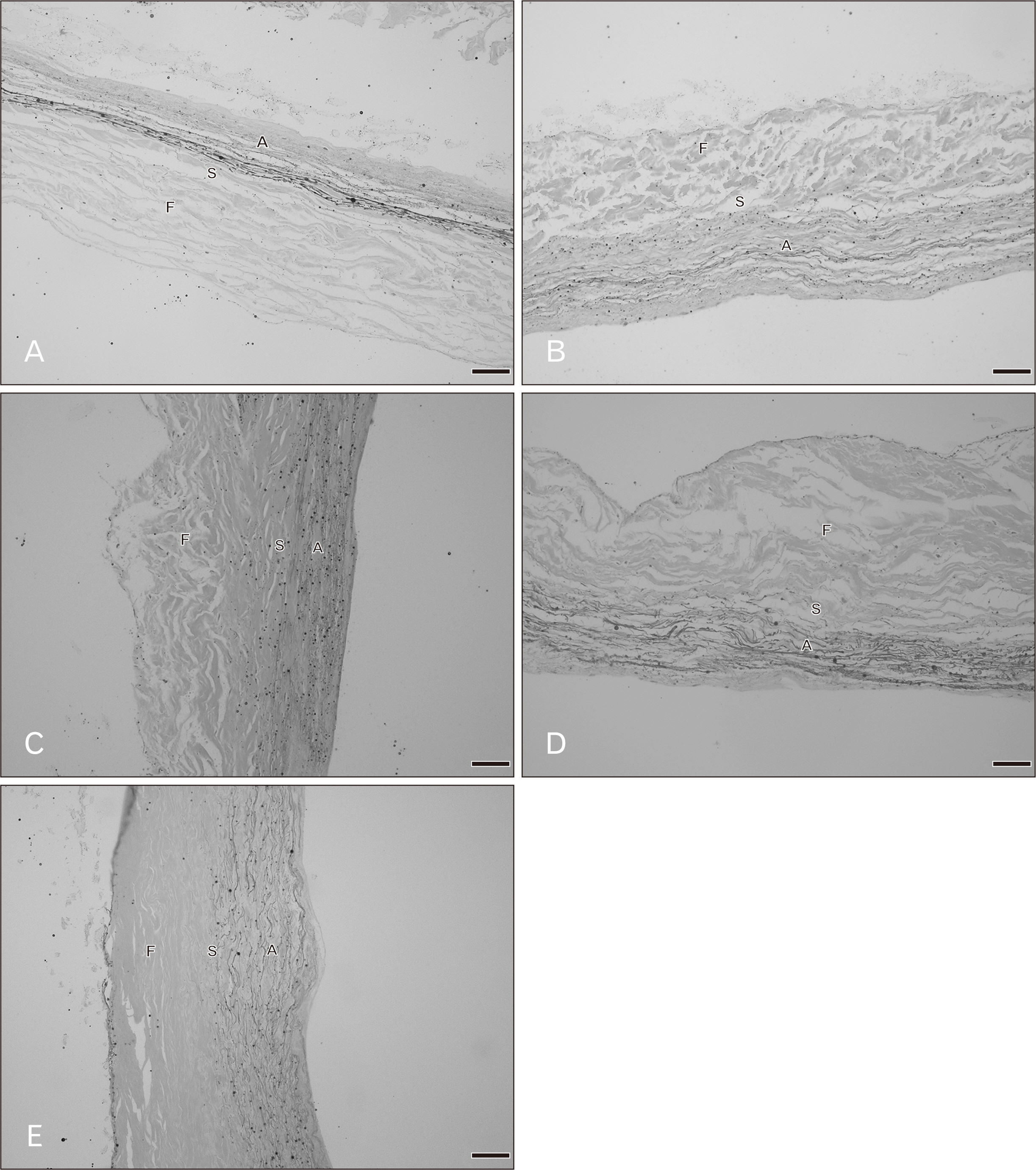
Fig. 1 Radial histological sections of the tricuspid valve in various age groups with Elastic Van Gieson staining. (A) A 20–29 years, (B) 40–49 years, (C) 50–59 years, (D) 60–69 years, (E) 70–79 years. A, atrialis; F, fibrosa; S, spongiosa. Scale Bars=50 μm (A–E).
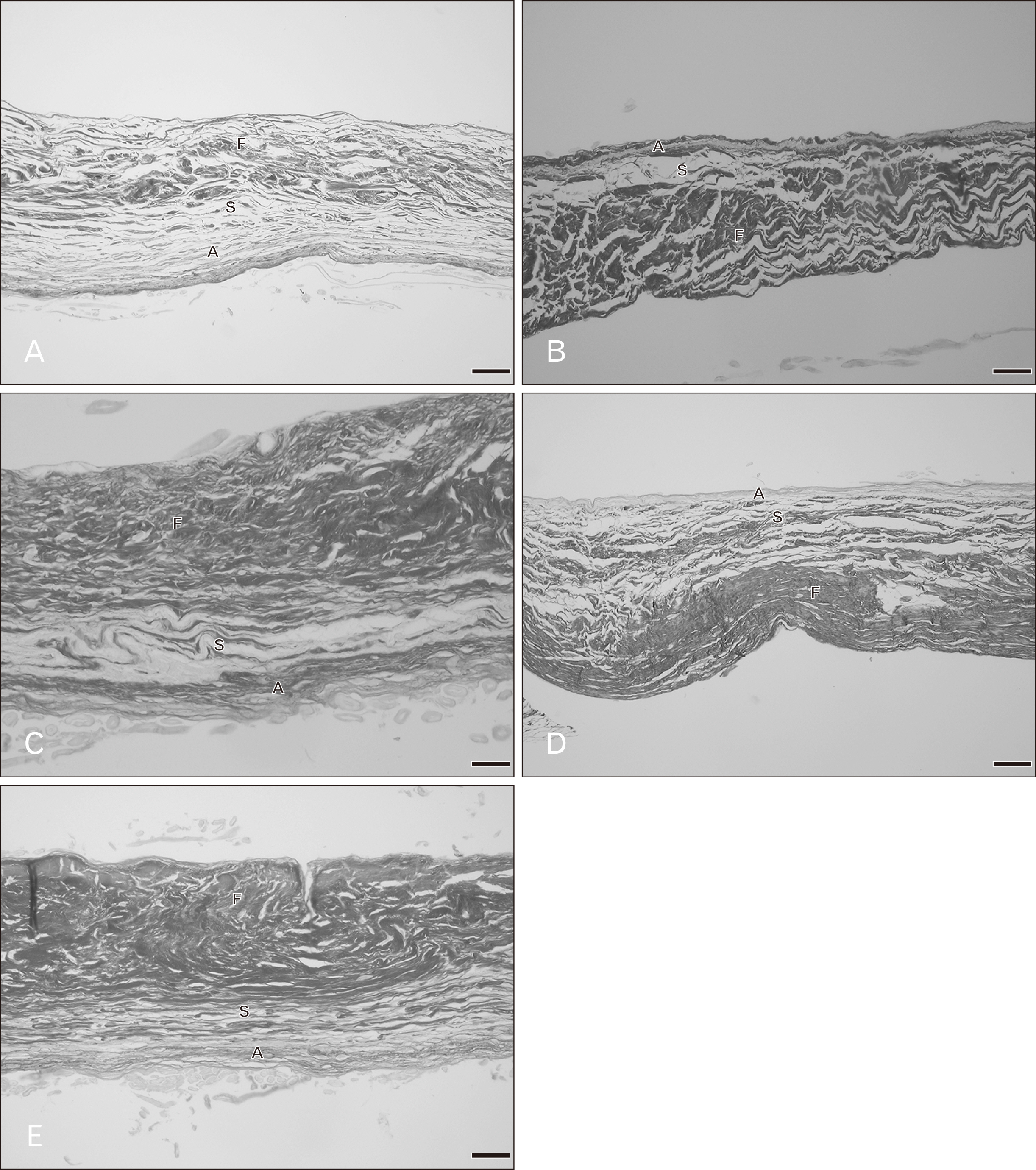
Fig. 2 Radial histological sections of the tricuspid valve in various age groups with picrosirius red staining. (A) A 20–29 years, (B) 40–49 years, (C) 50–59 years, (D) 60–69 years, (E) 70–79 years. A, atrialis; F, fibrosa; S, spongiosa. Scale Bars=50 μm (A–E).

Fig. 3 Radial histological sections of the mitral valve in various age groups with Elastic Van Gieson staining. (A) A 20–29 years, (B) 40–49 years, (C) 50–59 years, (D) 60–69 years, (E) 80–89 years. A, atrialis; F, fibrosa; S, spongiosa; V, ventricularis. Scale Bars=50 μm (A–E).
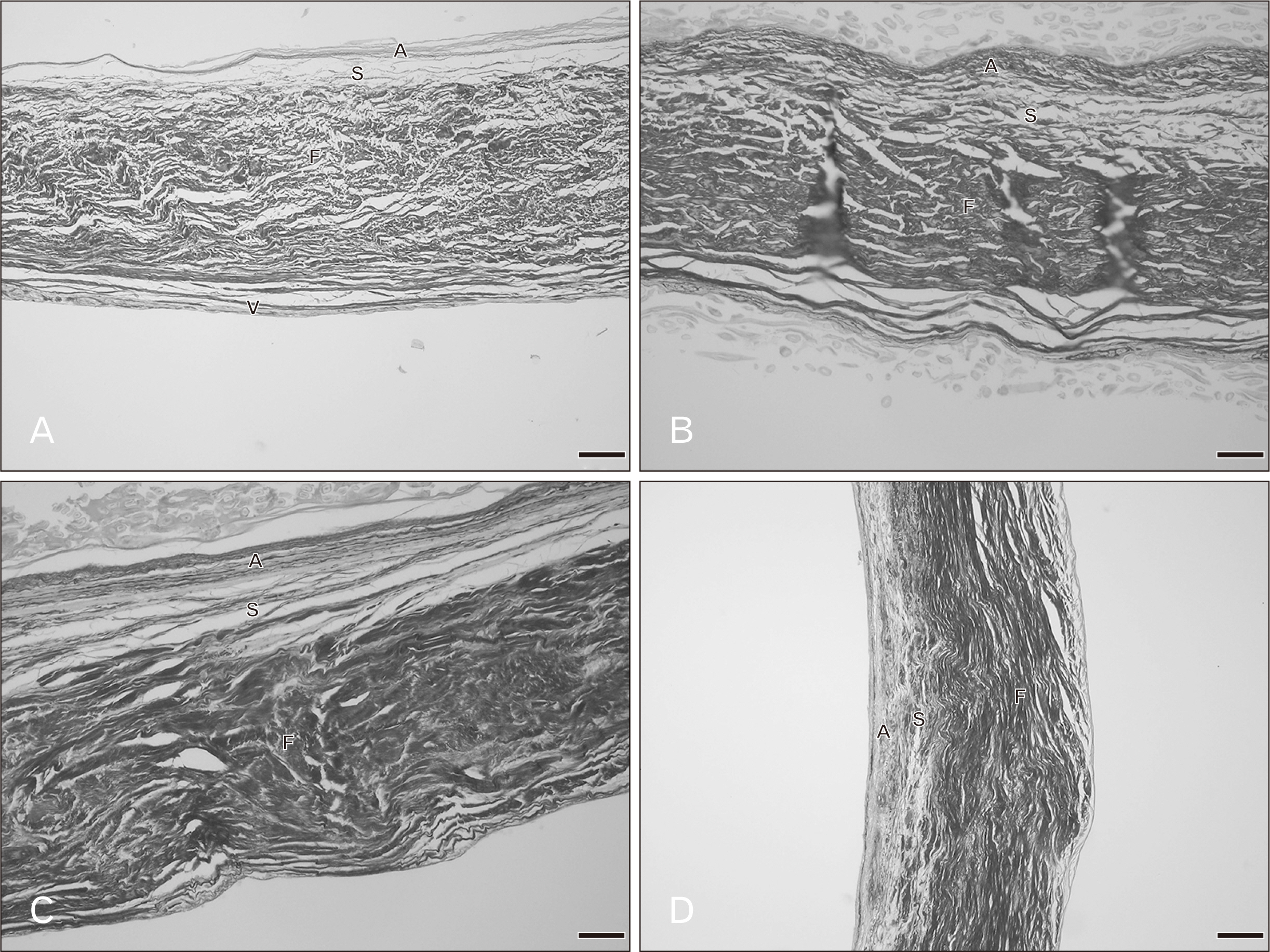
Fig. 4 Radial histological sections of the mitral valve in various age groups with picrosirius red staining. (A) A 20–29 years, (B) 40–49 years, (C) 60–69 years, (D) 80–89 years. A, atrialis; F, fibrosa; S, spongiosa; V, ventricularis. Scale Bars=50 μm (A), 20 μm (B–D).
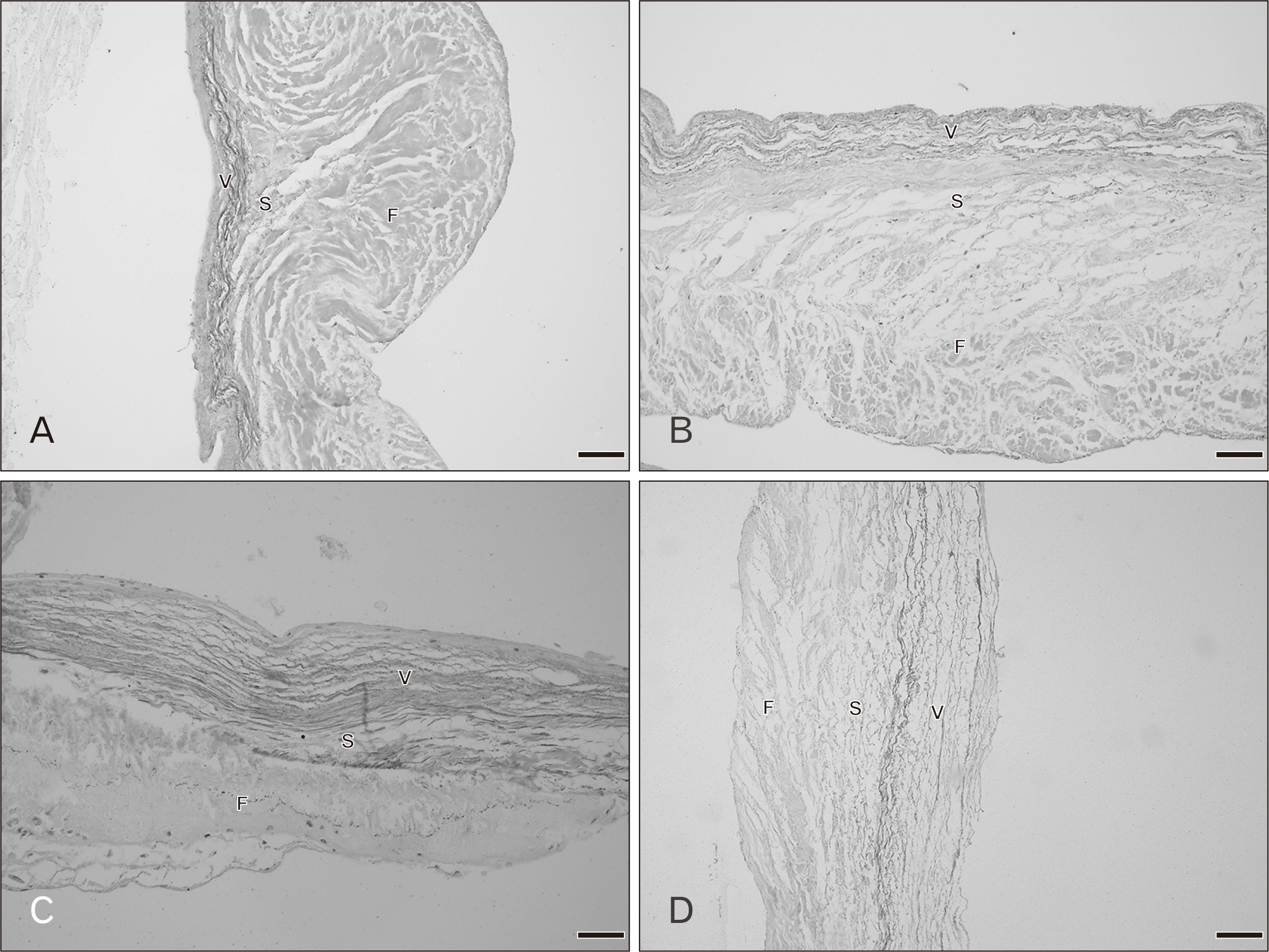
Fig. 5 Radial histological sections of the pulmonary valve in various age groups with Elastic Van Gieson staining. (A) A 30–39 years, (B) 50–59 years, (C) 60–69 years, (D) 70–79 years. F, fibrosa; S, spongiosa; V, ventricularis. Scale Bars=50 μm (A–D).
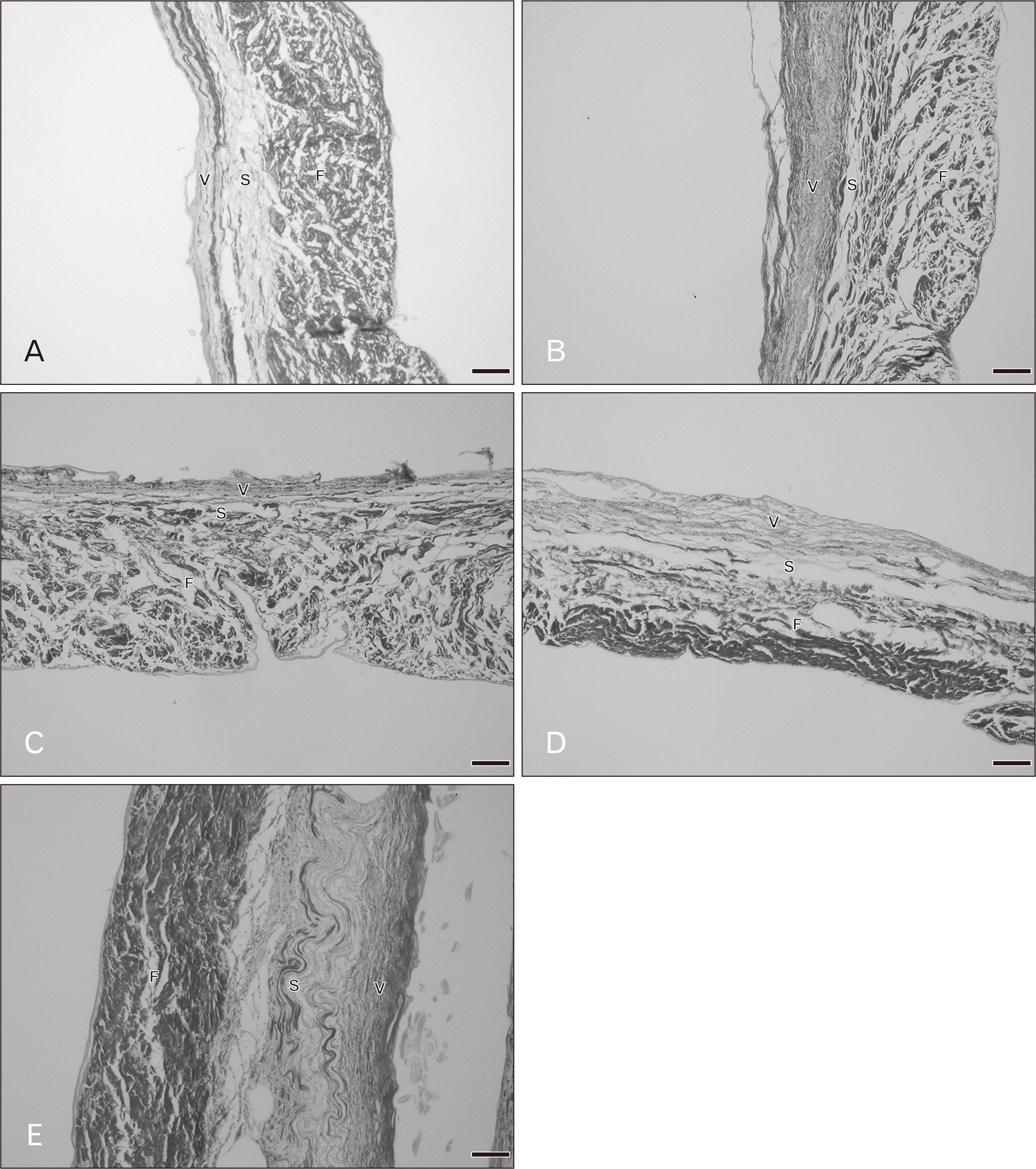
Fig. 6 Radial histological sections of the pulmonary valve in various age groups with picrosirius red staining. (A) A 20–29 years, (B) 40–49 years, (C) 50–59 years, (D) 60–69 years, (E) 70–79 years. F, fibrosa; S, spongiosa; V, ventricularis. Scale Bars=20 μm (A), 50 μm (B–E).
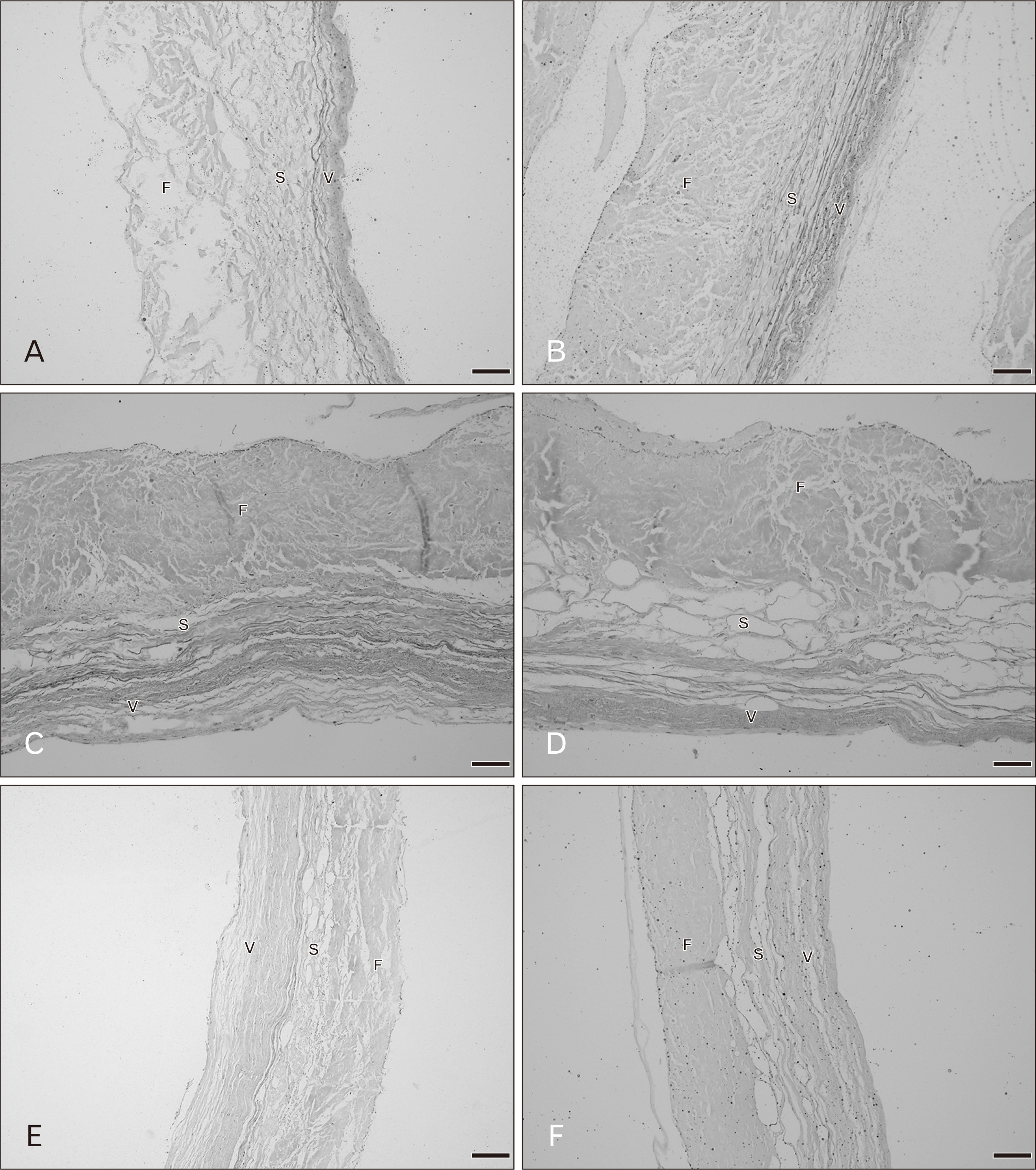
Fig. 7 Radial histological sections of the aortic valve in various age groups with Elastic Van Gieson staining. (A) A 20–29 years, (B) 40–49 years, (C) 50–59 years, (D) 60–69 years, (E) 70–79 years, (F) 80–89 years. F, fibrosa; S, spongiosa; V, ventricularis. Scale Bars=50 μm (A–F).
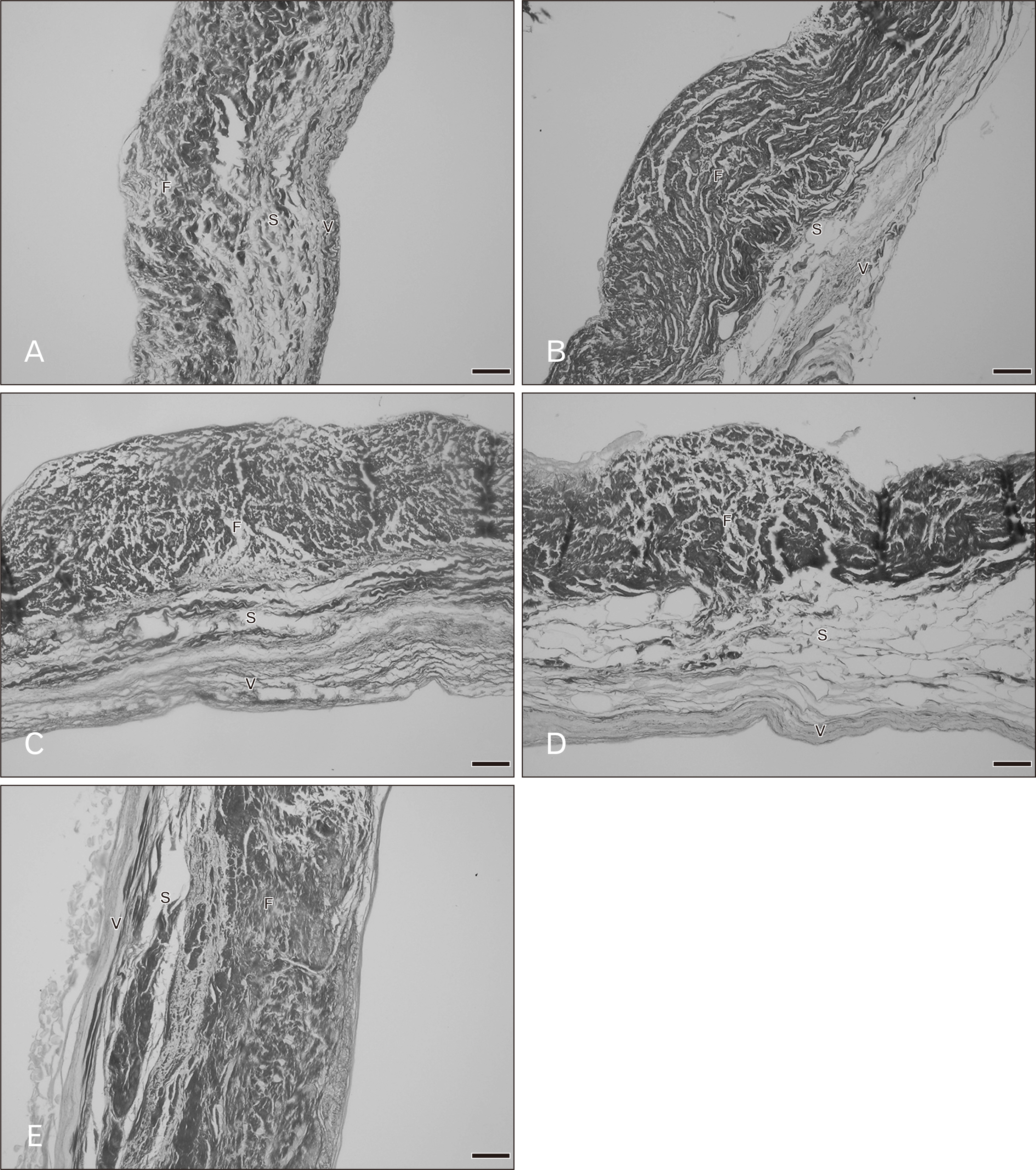
Fig. 8 Radial histological sections of the aortic valve in various age groups with picrosirius red staining. (A) A 20–29 years, (B) 40–49 years, (C) 50–59 years, (D) 60–69 years, (E) 70–79 years. F, fibrosa; S, spongiosa; V, ventricularis. Scale Bars=100 μm (A), 50 μm (B–E).
Cited by 2 articles
-
Raymond de Vieussens (1641–1715): connoisseur of cardiologic anatomy and pathological forms thereof
Sanjib Kumar Ghosh, Ananya Priya, Ravi Kant Narayan
Anat Cell Biol. 2021;54(4):417-423. doi: 10.5115/acb.21.108.Histological observations of age-related changes in the epiglottis associated with decreased deglutition function in older adults
Masamitsu Serikawa, Kimiharu Ambe, Akinobu Usami
Anat Cell Biol. 2023;56(3):374-381. doi: 10.5115/acb.23.081.
Reference
-
References
1. Hinton RB, Yutzey KE. 2011; Heart valve structure and function in development and disease. Annu Rev Physiol. 73:29. DOI: 10.1146/annurev-physiol-012110-142145. PMID: 20809794.
Article2. Misfeld M, Sievers HH. 2007; Heart valve macro- and microstructure. Philos Trans R Soc Lond B Biol Sci. 362:1421. DOI: 10.1098/rstb.2007.2125. PMID: 17581807. PMCID: PMC2440405.
Article3. Anderson RH, Ho SY, Becker AE. 2000; Anatomy of the human atrioventricular junctions revisited. Anat Rec. 260:81. DOI: 10.1002/1097-0185(20000901)260:1<81::AID-AR90>3.0.CO;2-3. PMID: 10967539. PMCID: PMC7309280.
Article4. Mendelson K, Schoen FJ. 2006; Heart valve tissue engineering: concepts, approaches, progress, and challenges. Ann Biomed Eng. 34:1799. DOI: 10.1007/s10439-006-9163-z. PMID: 17053986. PMCID: PMC1705506.
Article5. Mozaffarian D, Benjamin EJ, Go AS, Arnett DK, Blaha MJ, Cushman M, Das SR, de Ferranti S, Després JP, Fullerton HJ, Howard VJ, Huffman MD, Isasi CR, Jiménez MC, Judd SE, Kissela BM, Lichtman JH, Lisabeth LD, Liu S, Mackey RH, Magid DJ, McGuire DK, Moy CS, Mohler ER 3rd, Muntner P, Mussolino ME, Nasir K, Neumar RW, Nichol G, Palaniappan L, Pandey DK, Reeves MJ, Rodriguez CJ, Rosamond W, Sorlie PD, Stein J, Towfighi A, Turan TN, Virani SS, Woo D, Yeh RW, Turner MB. 2016; Heart Disease and Stroke Statistics-2016 update: a report from the American Heart Association. Circulation. 133:e38–360. DOI: 10.1161/CIR.0000000000000366. PMID: 26811276.6. Benjamin EJ, Blaha MJ, Chiuve SE, Cushman M, Das SR, Deo R, de Ferranti SD, Floyd J, Fornage M, Gillespie C, Isasi CR, Jiménez MC, Jordan LC, Judd SE, Lackland D, Lichtman JH, Lisabeth L, Liu S, Longenecker CT, Mackey RH, Matsushita K, Mozaffarian D, Mussolino ME, Nasir K, Neumar RW, Palaniappan L, Pandey DK, Thiagarajan RR, Reeves MJ, Ritchey M, Rodriguez CJ, Roth GA, Rosamond WD, Sasson C, Towfighi A, Tsao CW, Turner MB, Virani SS, Voeks JH, Willey JZ, Wilkins JT, Wu JH, Alger HM, Wong SS, Muntner P. 2017; Heart Disease and Stroke Statistics-2017 update: a report from the American Heart Association. Circulation. 135:e146–603. DOI: 10.1161/CIR.0000000000000485. PMID: 28874428.
Article7. Nichols M, Townsend N, Scarborough P, Rayner M. 2013; Cardiovascular disease in Europe: epidemiological update. Eur Heart J. 34:3028. DOI: 10.1093/eurheartj/eht356. PMID: 24014390.
Article8. Iung B, Vahanian A. 2011; Epidemiology of valvular heart disease in the adult. Nat Rev Cardiol. 8:162. DOI: 10.1038/nrcardio.2010.202. PMID: 21263455.
Article9. Schoen FJ. 2018; Morphology, clinicopathologic correlations, and mechanisms in heart valve health and disease. Cardiovasc Eng Technol. 9:126. DOI: 10.1007/s13239-016-0277-7. PMID: 27502286.
Article10. Athavale S, Deopujari R, Sinha U, Lalwani R, Kotgirwar S. 2017; Is tricuspid valve really tricuspid? Anat Cell Biol. 50:1. DOI: 10.5115/acb.2017.50.1.1. PMID: 28417048.
Article11. Gupta C, Shetti VR, Manju BVM. 2013; Dimensions of the human adult mitral valve in the embalmed cadaver. J Morphol Sci. 30:6–10.12. Gross L, Kugel MA. 1931; Topographic anatomy and histology of the valves in the human heart. Am J Pathol. 7:445–74.13. Pham T, Sulejmani F, Shin E, Wang D, Sun W. 2017; Quantification and comparison of the mechanical properties of four human cardiac valves. Acta Biomater. 54:345. DOI: 10.1016/j.actbio.2017.03.026. PMID: 28336153.
Article14. Parvin Nejad S, Blaser MC, Santerre JP, Caldarone CA, Simmons CA. 2016; Biomechanical conditioning of tissue engineered heart valves: too much of a good thing? Adv Drug Deliv Rev. 96:161. DOI: 10.1016/j.addr.2015.11.003. PMID: 26555371.
Article15. Hasan A, Ragaert K, Swieszkowski W, Selimović S, Paul A, Camci-Unal G, Mofrad MR, Khademhosseini A. 2014; Biomechanical properties of native and tissue engineered heart valve constructs. J Biomech. 47:1949–63. DOI: 10.1016/j.jbiomech.2013.09.023. PMID: 24290137. PMCID: PMC6028870.
Article16. Kim KM, Valigorsky JM, Mergner WJ, Jones RT, Pendergrass RF, Trump BF. 1976; Aging changes in the human aortic valve in relation to dystrophic calcification. Hum Pathol. 7:47. DOI: 10.1016/S0046-8177(76)80005-6. PMID: 1244310.
Article17. Balguid A, Rubbens MP, Mol A, Bank RA, Bogers AJ, van Kats JP, de Mol BA, Baaijens FP, Bouten CV. 2007; The role of collagen cross-links in biomechanical behavior of human aortic heart valve leaflets--relevance for tissue engineering. Tissue Eng. 13:1501–11. DOI: 10.1089/ten.2006.0279. PMID: 17518750.
Article18. van Geemen D, Soares AL, Oomen PJ, Driessen-Mol A, Janssen-van den Broek MW, van den Bogaerdt AJ, Bogers AJ, Goumans MJ, Baaijens FP, Bouten CV. 2016; Age-dependent changes in geometry, tissue composition and mechanical properties of fetal to adult cryopreserved human heart valves. PLoS One. 11:e0149020. DOI: 10.1371/journal.pone.0149020. PMID: 26867221. PMCID: PMC4750936.
Article19. McDonald PC, Wilson JE, McNeill S, Gao M, Spinelli JJ, Rosenberg F, Wiebe H, McManus BM. 2002; The challenge of defining normality for human mitral and aortic valves: geometrical and compositional analysis. Cardiovasc Pathol. 11:193–209. DOI: 10.1016/S1054-8807(01)00102-8. PMID: 12140125.20. Aikawa E, Whittaker P, Farber M, Mendelson K, Padera RF, Aikawa M, Schoen FJ. 2006; Human semilunar cardiac valve remodeling by activated cells from fetus to adult: implications for postnatal adaptation, pathology, and tissue engineering. Circulation. 113:1344–52. DOI: 10.1161/CIRCULATIONAHA.105.591768. PMID: 16534030.21. Sacks MS, David Merryman W, Schmidt DE. 2009; On the biomechanics of heart valve function. J Biomech. 42:1804. DOI: 10.1016/j.jbiomech.2009.05.015. PMID: 19540499.
Article22. Sell S, Scully RE. 1965; Aging changes in the aortic and mitral valves. Histologic and histochemical studies, with observations on the pathogenesis of calcific aortic stenosis and calcification of the mitral annulus. Am J Pathol. 46:345. PMID: 14270114. PMCID: PMC1920370.23. Guyton AC. 1986. Textbook of medical physiology. 8th ed. Saunders;Philadelphia:24. Waller BF. 1988; The old-age heart: normal aging changes which can produce or mimic cardiac disease. Clin Cardiol. 11:513. DOI: 10.1002/clc.4960110802. PMID: 3048829.
Article25. Pomerance A. 1967; Ageing changes in human heart valves. Br Heart J. 29:222. DOI: 10.1136/hrt.29.2.222. PMID: 6020817.
Article26. Donnelly KB. 2008; Cardiac valvular pathology: comparative pathology and animal models of acquired cardiac valvular diseases. Toxicol Pathol. 36:204. DOI: 10.1177/0192623307312707. PMID: 18474943.
Article27. Somers P, Knaapen M, Mistiaen W. 2006; Histopathology of calcific aortic valve stenosis. Acta Cardiol. 61:557. DOI: 10.2143/AC.61.5.2017772. PMID: 17117757.
Article28. Angelini A, Basso C, Grassi G, Casarotto D, Thiene G. 1994; Surgical pathology of valve disease in the elderly. Aging (Milano). 6:225. DOI: 10.1007/BF03324247. PMID: 7880871.
Article29. Warren BA, Yong JL. 1997; Calcification of the aortic valve: its progression and grading. Pathology. 29:360. DOI: 10.1080/00313029700169315. PMID: 9423215.
Article
