An Unusual Presentation of a Solid Pseudopapillary Tumor of the Pancreas Mimicking Adenocarcinoma
- Affiliations
-
- 1Department of Internal Medicine, Wonkwang University College of Medicine and Hospital, Iksan, Korea
- 2Institute of Wonkwang Medical Science, Wonkwang University College of Medicine and Hospital, Iksan, Korea
- 3Department of Pathology, Wonkwang University College of Medicine and Hospital, Iksan, Korea
- KMID: 2507597
- DOI: http://doi.org/10.5946/ce.2019.158
Abstract
- Solid pseudopapillary tumors of the pancreas are rare and typically occur in young women. Compared with pancreatic adenocarcinoma, solid pseudopapillary tumors are characterized by notable indolent biological behavior associated with a favorable prognosis. Despite their large size, these tumors rarely metastasize. Even in cases of hepatic metastasis, most lesions are usually solitary in distribution and are amenable to resection. We report a case of a 55-year-old man with a small solid pseudopapillary tumor (≤3-cm diameter) mimicking a pancreatic adenocarcinoma, with multiple hepatic metastases. The diagnosis was confirmed by endoscopic ultrasound-guided fine-needle biopsy using a 22-G core needle. Unfortunately, rapid tumor progression led to patient mortality 5 months after diagnosis. To our knowledge, this is the first case report that describes a small solid pseudopapillary tumor of the pancreas with multiple hepatic metastasis and poor prognosis in a patient who was diagnosed with this condition at the time of initial diagnosis.
Figure
-
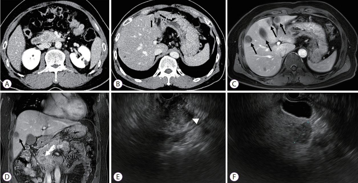
Fig. 1. Abdomen computed tomography scan showing (A) a 2.5 cm low attenuating mass (black open arrow) in the pancreatic head and (B) a less-enhancing lesion in the hepatic lobe (black arrows). Magnetic resonance imaging scans (arterial phase of an axial T-1 weighted image) showing ill-defined low signal intensity in the pancreatic head and (C) multiple hepatic lesions (black arrows). (D) Delayed phase of a coronal T-1 weighted image showing heterogenous echogenic lesions (white arrow) in the pancreatic head without main pancreatic duct dilatation or pancreatic parenchymal atrophy. An ill-defined low signal intensity (black arrow) is observed in the right hepatic lobe. Endoscopic ultrasound showing (E) an ill-defined mixed echogenic mass in the pancreatic head and (F) irregular margins with low, dense lesions in the right hepatic lobes.

Fig. 2. Histopathological examination of a biopsy specimen showing (A) monomorphic cells with delicate papillary fronds (white arrows) without a fibrovascular core (black circles; hematoxylin & eosin stain, ×200). (B) Neoplastic cells are diffusely and strongly stained with beta-catenin (immunohistochemical staining, ×200).
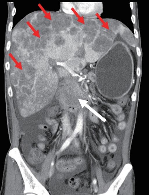
Fig. 3. Abdominal computed tomography scan showing an enlarged pancreatic mass (white arrow), multiple hepatic masses (red arrows), and massive ascites.
Reference
-
1. Estrella JS, Li L, Rashid A, et al. Solid pseudopapillary neoplasm of the pancreas: clinicopathologic and survival analyses of 64 cases from a single institution. Am J Surg Pathol. 2014; 38:147–157.2. Kosmahl M, Seada LS, Jänig U, Harms D, Klöppel G. Solid-pseudopapillary tumor of the pancreas: its origin revisited. Virchows Arch. 2000; 436:473–480.
Article3. Klimstra DS, Wenig BM, Heffess CS. Solid-pseudopapillary tumor of the pancreas: a typically cystic carcinoma of low malignant potential. Semin Diagn Pathol. 2000; 17:66–80.4. Machado MC, Machado MA, Bacchella T, Jukemura J, Almeida JL, Cunha JE. Solid pseudopapillary neoplasm of the pancreas: distinct patterns of onset, diagnosis, and prognosis for male versus female patients. Surgery. 2008; 143:29–34.
Article5. Baek JH, Lee JM, Kim SH, et al. Small (<or=3 cm) solid pseudopapillary tumors of the pancreas at multiphasic multidetector CT. Radiology. 2010; 257:97–106.6. Watanabe Y, Okamoto K, Okada K, Aikawa M, Koyama I, Yamaguchi H. A case of aggressive solid pseudopapillary neoplasm: comparison of clinical and pathologic features with non-aggressive cases. Pathol Int. 2017; 67:202–207.
Article7. Sperti C, Berselli M, Pasquali C, Pastorelli D, Pedrazzoli S. Aggressive behaviour of solid-pseudopapillary tumor of the pancreas in adults: a case report and review of the literature. World J Gastroenterol. 2008; 14:960–965.
Article8. Tang LH, Aydin H, Brennan MF, Klimstra DS. Clinically aggressive solid pseudopapillary tumors of the pancreas: a report of two cases with components of undifferentiated carcinoma and a comparative clinicopathologic analysis of 34 conventional cases. Am J Surg Pathol. 2005; 29:512–519.9. Takahashi Y, Fukusato T, Aita K, et al. Solid pseudopapillary tumor of the pancreas with metastases to the lung and liver. Pathol Int. 2005; 55:792–796.
Article10. Wang WB, Zhang TP, Sun MQ, Peng Z, Chen G, Zhao YP. Solid pseudopapillary tumor of the pancreas with liver metastasis: clinical features and management. Eur J Surg Oncol. 2014; 40:1572–1577.
Article11. Gomez P, Yorke R, Ayala AG, Ro JY. Solid-pseudopapillary neoplasm of pancreas with long delayed liver metastasis. Ann Diagn Pathol. 2012; 16:380–384.
Article12. Kim EY. Role of repeated endoscopic ultrasound-guided fine needle aspiration for inconclusive initial cytology result. Clin Endosc. 2013; 46:540–542.
Article13. Ohara Y, Oda T, Hashimoto S, et al. Pancreatic neuroendocrine tumor and solid-pseudopapillary neoplasm: key immunohistochemical profiles for differential diagnosis. World J Gastroenterol. 2016; 22:8596–8604.
Article14. Kato T, Egawa N, Kamisawa T, et al. A case of solid pseudopapillary neoplasm of the pancreas and tumor doubling time. Pancreatology. 2002; 2:495–498.
Article15. Menon SS, Guruvayoorappan C, Sakthivel KM, Rasmi RR. Ki-67 protein as a tumour proliferation marker. Clin Chim Acta. 2019; 491:39–45.
Article16. Yang F, Yu X, Bao Y, Du Z, Jin C, Fu D. Prognostic value of Ki-67 in solid pseudopapillary tumor of the pancreas: Huashan experience and systematic review of the literature. Surgery. 2016; 159:1023–1031.
Article17. Fried P, Cooper J, Balthazar E, Fazzini E, Newall J. A role for radiotherapy in the treatment of solid and papillary neoplasms of the pancreas. Cancer. 1985; 56:2783–2785.
Article18. Maffuz A, Bustamante Fde T, Silva JA, Torres-Vargas S. Preoperative gemcitabine for unresectable, solid pseudopapillary tumour of the pancreas. Lancet Oncol. 2005; 6:185–186.
Article19. Morikawa T, Onogawa T, Maeda S, et al. Solid pseudopapillary neoplasms of the pancreas: an 18-year experience at a single Japanese institution. Surg Today. 2013; 43:26–32.
Article20. Kang CM, Kim KS, Choi JS, Kim H, Lee WJ, Kim BR. Solid pseudopapillary tumor of the pancreas suggesting malignant potential. Pancreas. 2006; 32:276–280.
Article
- Full Text Links
-
- Actions
-
Cited
- CITED
-
- Close
- Share
-
- Similar articles
-
- Solid Pseudopapillary Tumor of the Pancreas in Child: A Case Report
- A Case of Solid Pseudopapillary Tumor of the Pancreas Presenting as a Submucosal Tumor with Hemorrhage
- A First Case of Solid Pseudopapillary Tumor of the Pancreas in an Old Man in South Korea
- Solid pseudopapillary tumor with hepatic metastasis
- The Growth of an Extrapancreatic Solid Pseudopapillary Tumor from the Greater Omentum: A Case Report
