Clinical Results of Various Treatments for Retinal Capillary Hemangioma
- KMID: 2507403
- DOI: http://doi.org/10.3341/kjo.2019.0075
Abstract
- Purpose
We report the clinical outcomes of retinal capillary hemangioma (RCH) after the application of various treatments.
Methods
We performed a retrospective chart analysis of eight eyes treated for RCH between August 2009 and January 2018. During the follow-up period, the status and progression of the RCHs were checked by fundus photography, fluorescein angiography, and optical coherence tomography, and additional treatments were applied when necessary.
Results
Three of the five patients had bilateral RCH, and two had unilateral RCH. Six eyes received laser photocoagulation; two eyes received cryotherapy, and one eye received intravitreal Avastin injection. Three eyes each had intravitreal triamcinolone injection, subtenon triamcinolone injection, and intravitreal dexamethasone injection to control inflammation. Also, two patients took oral prednisolone, and one patient used prednisolone eye drops to control inflammation. Two eyes underwent vitrectomy and scleral buckling due to deterioration of the epiretinal membrane and vitreal traction, respectively. As a result of those treatments, the tumors were stable in five of the eight eyes. However, one eye is now in a pre-phthisis state, and one patient who refused treatment showed progression of the tumor, epiretinal membrane, and traction.
Conclusions
Because RCHs vary in size, the degree of inflammation, and symptoms, this disorder should be actively treated on a case-by-case basis. Fluorescein angiography should be used periodically to determine recurrence of the tumor or inflammation, and the appropriate treatment should be repeated as necessary. Moreover, regular systemic screening tests for von Hippel-Lindau disease should be performed in RCH patients to ensure that they have no abnormalities other than in the eye.
Figure
-
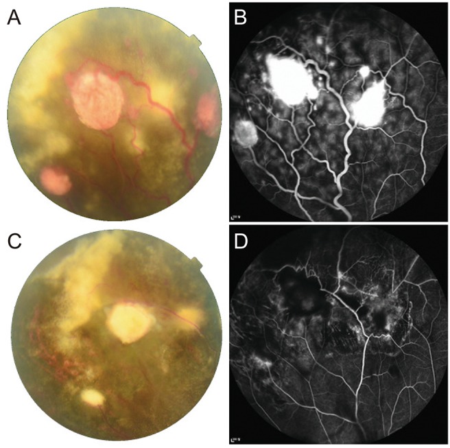
Fig. 1 Clinical outcome of a case 1 patient. (A) Fundus photographs and (B) fluorescein angiography of the right eye of a 29-year-old patient at the initial visit. Three round, orange retinal masses <1.5 disc diameters with dilated feeder vessels and exudations were observed in the peripheral retina. Leakage of fluorescence associated with the lesions was revealed by fluorescein angiography. (C) The size of the masses and exudates decreased after two laser photocoagulation treatments. (D) Leakage around the lesions also decreased after two laser photocoagulation treatments.
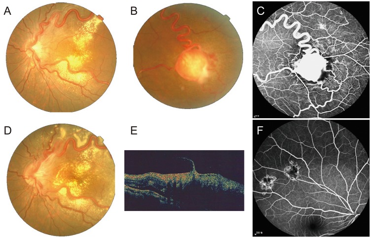
Fig. 2 Clinical outcome of a case 2 patient. (A,B) Fundus photographs and (C) fluorescein angiography (FA) of the left eye of a 13-year-old patient before treatment. A tumor of 2.5 disc diameters with dilated vessels was found in the temporal retina, and exudates were deposited around the vessels. Leakage of fluorescence associated with the lesions was revealed by FA. Although laser photocoagulation was repeated, (D) fundus photographs and (E) optical coherence tomography showed vitreous traction with an epiretinal membrane, and tumor progression was not completely attenuated. (F) The retinal masses in the right eye were stable on FA after four laser photocoagulation treatments.
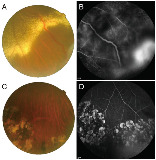
Fig. 3 Clinical outcome of a case 3 patient. (A) Fundus photograph and (B) fluorescein angiography (FA) of the left eye of a 35-year-old patient before cryotherapy. There were exudations with dilated feeder vessels, and (B) FA revealed leakage from the lesion. Seven years after cryotherapy and additional laser photocoagulation treatments, a (C) fundus photograph and (D) FA showed no dilated vessels, exudates, or leakage around the mass.
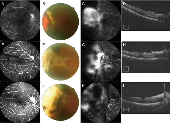
Fig. 4 Clinical outcome of a case 4 patient. (A) The fluorescein angiography (FA) of the right eye of a 33-year-old patient at the first visit revealed inflammation throughout the retina. (B) Fundus photography, (C) FA, and (D) optical coherence tomography showed a mass of 3 disc diameters before treatment. After two cryotherapy treatments, (E) the inflammation disappeared in the FA, but retinal traction worsened in (F) the fundus photograph, (G) FA, and (H) optical coherence tomography. (I–L) After scleral bucking, the right eye reached a stable state.
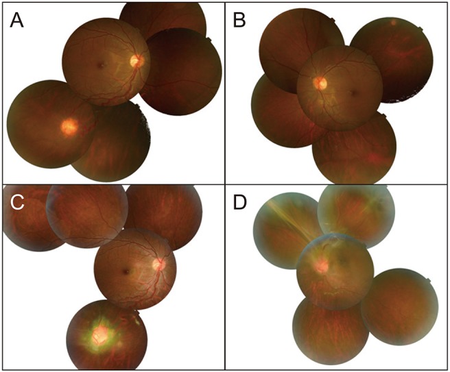
Fig. 5 Clinical outcome of a case 5 patient. (A,B) Fundus photographs of a 13-year-old patient at the initial visit. The retinal tumors were in the right inferotemporal peripheral retina and the left superotemporal retina. Nine years later, without any treatment, (C) the tumor in the right eye became enlarged, and (D) retinal traction and ERM had progressed in the left eye.
Reference
-
1. Singh AD, Shields CL, Shields JA. von Hippel-Lindau disease. Surv Ophthalmol. 2001; 46:117–142. PMID: 11578646.
Article2. Lamiell JM, Salazar FG, Hsia YE. von Hippel-Lindau disease affecting 43 members of a single kindred. Medicine (Baltimore). 1989; 68:1–29. PMID: 2642584.
Article3. Shields JA, Shields CL. Intraocular tumors: an atlas and textbook. 2nd ed. Philadelphia: Lippincott Williams & Wilkins;2008. p. 368–369.4. Maher ER, Yates JR, Harries R, et al. Clinical features and natural history of von Hippel-Lindau disease. Q J Med. 1990; 77:1151–1163. PMID: 2274658.
Article5. Webster AR, Maher ER, Moore AT. Clinical characteristics of ocular angiomatosis in von Hippel-Lindau disease and correlation with germline mutation. Arch Ophthalmol. 1999; 117:371–378. PMID: 10088816.
Article6. Ridley M, Green J, Johnson G. Retinal angiomatosis: the ocular manifestations of von Hippel-Lindau disease. Can J Ophthalmol. 1986; 21:276–283. PMID: 3801976.7. Annesley WH Jr, Leonard BC, Shields JA, Tasman WS. Fifteen year review of treated cases of retinal angiomatosis. Trans Sect Ophthalmol Am Acad Ophthalmol Otolaryngol. 1977; 83:OP446–OP453.8. Singh AD, Damato BE. Clinical ophthalmic oncology: basic principles. Cham: Springer;2019. p. 99–105.9. Lee DS, Kim YY, Kim SY, Kim SD. Treatment of retinal detachment occurring in two cases of von Hippel-Lindau syndrome. J Korean Ophthalmol Soc. 2001; 42:1636–1640.10. Baek SK, Jin SY, Lee YH, Lee TG. A case report of primary vitrectomy in multiple bilateral retinal capillary hemangiomas accompanying epiretinal membrane. J Korean Ophthalmol Soc. 2014; 55:928–935.
Article11. Whitson JT, Welch RB, Green WR. Von Hippel-Lindau disease: case report of a patient with spontaneous regression of a retinal angioma. Retina. 1986; 6:253–259. PMID: 3554423.12. Oosterhuis JA, Rubinstein K. Haemangioma at the optic disc. Ophthalmologica. 1972; 164:362–374. PMID: 5033057.
Article13. Sachdeva R, Dadgostar H, Kaiser PK, et al. Verteporfin photodynamic therapy of six eyes with retinal capillary haemangioma. Acta Ophthalmol. 2010; 88:e334–e340. PMID: 20946329.
Article14. Fong AH, Li KK, Wong D. Intravitreal ranibizumab, photodynamic therapy, and vitreous surgery for the treatment of juxtapapillary retinal capillary hemangioma. Graefes Arch Clin Exp Ophthalmol. 2011; 249:625–627. PMID: 20676670.
Article15. Kremer I, Gilad E, Ben-Sira I. Juxtapapillary exophytic retinal capillary hemangioma treated by yellow krypton (568 nm) laser photocoagulation. Ophthalmic Surg. 1988; 19:743–747. PMID: 3194109.
Article16. Gaudric A, Krivosic V, Duguid G, et al. Vitreoretinal surgery for severe retinal capillary hemangiomas in von hippel-lindau disease. Ophthalmology. 2011; 118:142–149. PMID: 20801520.
Article17. McDonald HR, Schatz H, Johnson RN, et al. Vitrectomy in eyes with peripheral retinal angioma associated with traction macular detachment. Ophthalmology. 1996; 103:329–335. PMID: 8594522.
Article18. Johnson MW, Flynn HW Jr, Gass JD. Pars plana vitrectomy and direct diathermy for complications of multiple retinal angiomas. Ophthalmic Surg. 1992; 23:47–50. PMID: 1574268.
Article19. Chew EY, Schachat AP. Capillary hemangioblastoma of the retina and von Hippel-Lindau disease. In : Ryan SJ, editor. Retina. 5th ed. London: Saunders/Elsevier;2013. p. 2156–2163.20. Lee HJ, Kang SH, Kim HC. Three cases of retinal capillary hemangioma presenting with retinal detachment. J Korean Ophthalmol Soc. 2003; 44:1936–1942.21. Carr RE, Noble KG. Retinal angiomatosis. Ophthalmology. 1980; 87:956–959. PMID: 7413159.22. Singh A, Shields J, Shields C. Solitary retinal capillary hemangioma: hereditary (von Hippel-Lindau disease) or nonhereditary? Arch Ophthalmol. 2001; 119:232–234. PMID: 11176984.23. Hinz BJ, Schachat AP. Capillary hemangioma of the retina and von Hippel-Lindau disease. In : Ryan SJ, Hinton DR, Schachat AP, editors. Retina. 4th ed. St. Louis: Mosby;2006. p. 615–624.24. Kim HM, Park KH, Woo SJ. Massive exudative retinal detachment following photodynamic therapy and intravitreal bevacizumab injection in retinal capillary hemangioma. Korean J Ophthalmol. 2015; 29:143–145. PMID: 25829835.
Article25. Schmidt D, Natt E, Neumann HP. Long-term results of laser treatment for retinal angiomatosis in von Hippel-Lindau disease. Eur J Med Res. 2000; 5:47–58. PMID: 10720563.26. Shields JA. The expanding role of laser photocoagulation for intraocular tumors. The 1993 H. Christian Zweng Memorial Lecture. Retina. 1994; 14:310–322. PMID: 7817024.27. Lane CM, Turner G, Gregor ZJ, Bird AC. Laser treatment of retinal angiomatosis. Eye (Lond). 1989; 3:33–38. PMID: 2591596.
Article28. Amoils SP, Smith TR. Cryotherapy of angiomatosis retinae. Arch Ophthalmol. 1969; 81:689–691. PMID: 5781743.
Article29. Shields JA. Response of retinal capillary hemangioma to cryotherapy. Arch Ophthalmol. 1993; 111:551. PMID: 8470991.
Article30. Welch RB. Von Hippel-Lindau disease: the recognition and treatment of early angiomatosis retinae and the use of cryosurgery as an adjunct to therapy. Trans Am Ophthalmol Soc. 1970; 68:367–424. PMID: 5535648.31. Singh AD, Nouri M, Shields CL, et al. Treatment of retinal capillary hemangioma. Ophthalmology. 2002; 109:1799–1806. PMID: 12359597.
Article32. Watzke RC. Cryotherapy for retinal angiomatosis: a clinicopathologic report. Arch Ophthalmol. 1974; 92:399–401. PMID: 4429468.
Article33. Los M, Aarsman CJ, Terpstra L, et al. Elevated ocular levels of vascular endothelial growth factor in patients with von Hippel-Lindau disease. Ann Oncol. 1997; 8:1015–1022. PMID: 9402176.
Article34. Ach T, Thiemeyer D, Hoeh AE, et al. Intravitreal bevacizumab for retinal capillary haemangioma: longterm results. Acta Ophthalmol. 2010; 88:e137–e138. PMID: 19681788.
Article35. Chelala E, Dirani A, Fadlallah A. Intravitreal anti-VEGF injection for the treatment of progressive juxtapapillary retinal capillary hemangioma: a case report and mini review of the literature. Clin Ophthalmol. 2013; 7:2143–2146. PMID: 24204117.36. Atebara NH. Retinal capillary hemangioma treated with verteporfin photodynamic therapy. Am J Ophthalmol. 2002; 134:788–790. PMID: 12429270.
Article
- Full Text Links
-
- Actions
-
Cited
- CITED
-
- Close
- Share
-
- Similar articles
-
- Three Cases of Retinal Capillary Hemangioma Presenting with Retinal Detachment
- Solitary Retinal Capillary Hemangioma Treated with Cryotherapy
- Cutis Marmorata Telangiectatica Congenita: A Rare Clinical Manifestation of Capillary Hemangioma?
- Capillary Hemangioma of Testis
- Retinal Capillary Hemangioma Treated with Verteporfin Photodynamic Therapy and Intravitreal Triamcinolone Acetonide
