Korean J Orthod.
2020 Sep;50(5):336-345. 10.4041/kjod.2020.50.5.336.
Treatment modalities for Korean patients with unilateral hemifacial microsomia according to Pruzansky–Kaban types and growth stages
- Affiliations
-
- 1Department of Orthodontics, Dental Research Institute, School of Dentistry, Seoul National University, Seoul, Korea
- 2Department of Plastic and Reconstructive Surgery, Seoul National University Hospital, Seoul, Korea
- 3Department of Orthodontics, School of Dentistry, Seoul National University, Seoul, Korea
- 4Private Practice, Pohang, Korea
- 5Department of Plastic and Reconstructive Surgery, College of Medicine, Seoul National University, Seoul, Korea
- 6Department of Oral and Maxillofacial Surgery, School of Dentistry, Seoul National University, Seoul, Korea
- 7Private Practice, Seoul, Korea
- KMID: 2506468
- DOI: http://doi.org/10.4041/kjod.2020.50.5.336
Abstract
Objective
To investigate the treatment modalities (Tx-Mods) for patients with unilateral hemifacial microsomia (UHFM) according to Pruzansky–Kaban types and growth stages.
Methods
The samples consisted of 82 Korean UHFM patients. Tx-Mods were defined as follows: Tx-Mod-1, growth observation due to mild facial asymmetry; Tx-Mod-2, unilateral functional appliance; TxMod-3, fixed orthodontic treatment; Tx-Mod-4, growth observation due to a definite need for surgical intervention; Tx-Mod-5, unilateral mandibular or bimaxillary distraction osteogenesis (DO); Tx-Mod-6, maxillary fixation using LeFort I osteotomy and mandibular DO/sagittal split ramus osteotomy; TxMod-7, orthognathic surgery; and Tx-Mod-8, costochondral grafting. The type and frequency of Tx-Mod, the number of patients who underwent surgical procedures, and the number of surgeries that each patient underwent, were investigated. Results: The degree of invasiveness and complexity of Tx-Mod increased, with an increase in treatment stage and Pruzansky–Kaban type (initial < final; [I, IIa] < [IIb, III], all p < 0.001). The percentage of patients who underwent surgical procedures increased up to 4.2 times, with an increase in the Pruzansky–Kaban type (I, 24.1%; IIa, 47.1%; IIb, 84.4%; III, 100%; p < 0.001).However, the mean number of surgical procedures that each patient underwent showed a tendency of increase according to the Pruzansky–Kaban types (I, n = 1.1; IIa, n = 1.5; IIb, n = 1.6; III, n = 2.3; p > 0.05).
Conclusions
These findings might be used as basic guidelines for successful treatment planning and prognosis prediction in UHFM patients.
Figure
-
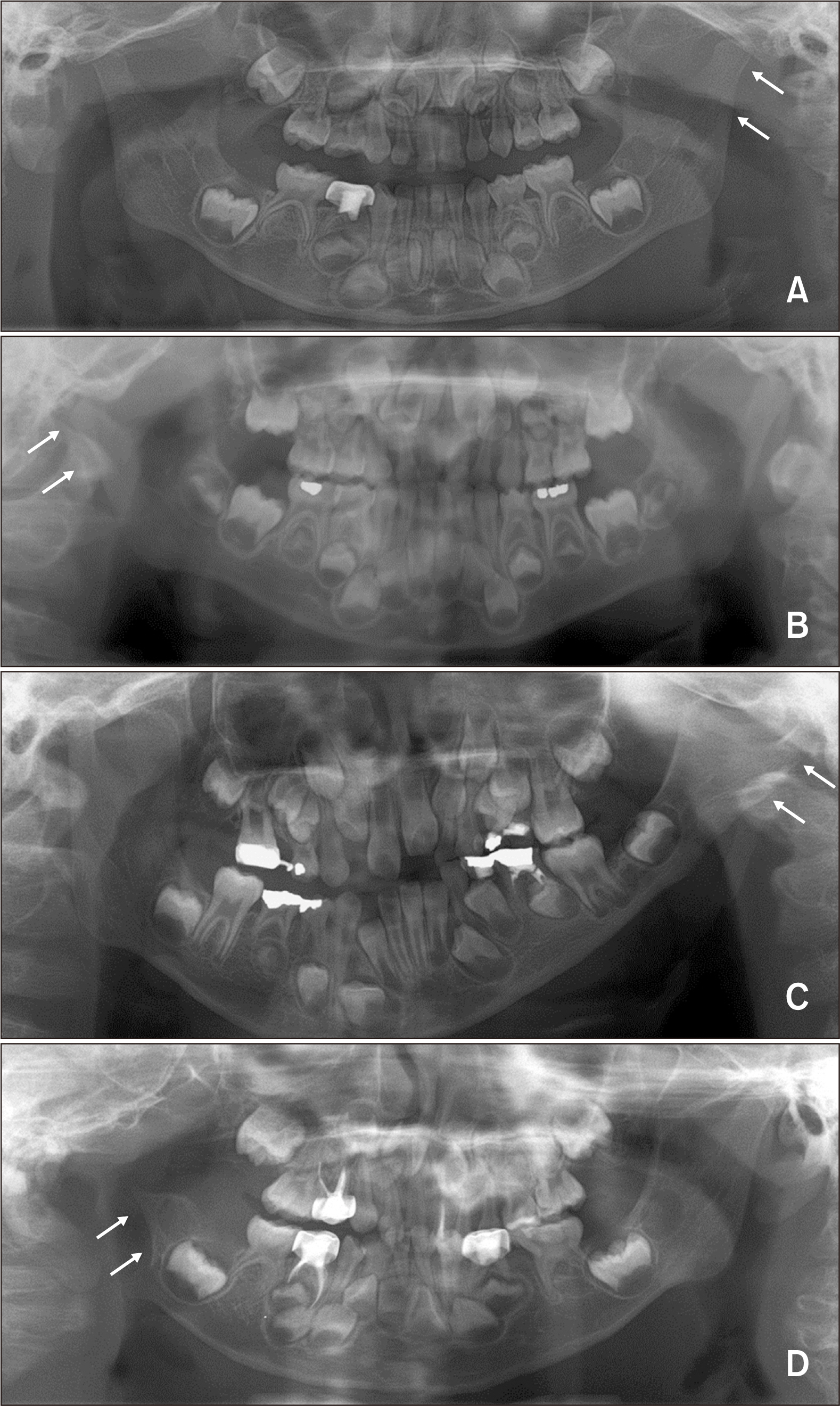
Figure 1 The Pruzansky–Kaban classification for hemifacial microsomia. A, Type I, the ramus/condyle complex has a normal shape but small size. B, Type IIa, the ramus/condyle complex is hypoplastic and abnormally shaped although the glenoid fossa is placed at the right position and the temporomandibular joint is functional. C, Type IIb, the glenoid fossa is placed at the inferiorly, medially, and anteriorly altered position with a severely hypoplastic ramus/condyle complex. D, Type III, complete absence of the ramus/condyle complex and the glenoid fossa. Arrow indicates the involvement side.
Reference
-
1. Grabb WC. 1965; The first and second branchial arch syndrome. Plast Reconstr Surg. 36:485–508. DOI: 10.1097/00006534-196511000-00001. PMID: 5320180.
Article2. Poswillo D. 1975; Hemorrhage in development of the face. Birth Defects Orig Artic Ser. 11:61–81. PMID: 813794.3. Figueroa AA, Pruzansky S. 1982; The external ear, mandible and other components of hemifacial microsomia. J Maxillofac Surg. 10:200–11. DOI: 10.1016/S0301-0503(82)80042-8. PMID: 6961179.
Article4. Hartsfield JK. 2007; Review of the etiologic heterogeneity of the oculo-auriculo-vertebral spectrum (hemifacial microsomia). Orthod Craniofac Res. 10:121–8. DOI: 10.1111/j.1601-6343.2007.00391.x. PMID: 17651128.
Article5. Kaban LB, Moses MH, Mulliken JB. 1986; Correction of hemifacial microsomia in the growing child: a follow-up study. Cleft Palate J. 23 Suppl 1:50–2. PMID: 3545546.6. Cousley RR, Calvert ML. 1997; Current concepts in the understanding and management of hemifacial microsomia. Br J Plast Surg. 50:536–51. DOI: 10.1016/S0007-1226(97)91303-5. PMID: 9422952.
Article7. Choi JY, Hwang KG, Baek SH, Lee JH, Kim TW, Kim MJ, et al. 2001; Original sagittal split osteotomy revisited for mandibular distraction. J Craniomaxillofac Surg. 29:165–73. DOI: 10.1054/jcms.2001.0211. PMID: 11465256.
Article8. McCarthy JG, Katzen JT, Hopper R, Grayson BH. 2002; The first decade of mandibular distraction: lessons we have learned. Plast Reconstr Surg. 110:1704–13. DOI: 10.1097/00006534-200212000-00013. PMID: 12447053.
Article9. Ohtani J, Hoffman WY, Vargervik K, Oberoi S. 2012; Team management and treatment outcomes for patients with hemifacial microsomia. Am J Orthod Dentofacial Orthop. 141(4 Suppl):S74–81. DOI: 10.1016/j.ajodo.2011.12.015. PMID: 22449602.
Article10. Kim S, Seo YJ, Choi TH, Baek SH. 2012; New approach for the surgico-orthodontic treatment of hemifacial microsomia. J Craniofac Surg. 23:957–63. DOI: 10.1097/SCS.0b013e31824dfa09. PMID: 22777431.
Article11. Heike CL, Hing AV, Aspinall CA, Bartlett SP, Birgfeld CB, Drake AF, et al. 2013; Clinical care in craniofacial microsomia: a review of current management recommendations and opportunities to advance research. Am J Med Genet C Semin Med Genet. 163C:271–82. DOI: 10.1002/ajmg.c.31373. PMID: 24132932.
Article12. Suh J, Choi TH, Baek SH, Kim JC, Kim S. 2013; Mandibular distraction in unilateral craniofacial microsomia: longitudinal results until the completion of growth. Plast Reconstr Surg. 132:1244–52. DOI: 10.1097/PRS.0b013e3182a48cf9. PMID: 24165605.13. Pluijmers BI, Caron CJ, Dunaway DJ, Wolvius EB, Koudstaal MJ. 2014; Mandibular reconstruction in the growing patient with unilateral craniofacial microsomia: a systematic review. Int J Oral Maxillofac Surg. 43:286–95. DOI: 10.1016/j.ijom.2013.11.001. PMID: 24332589.
Article14. Yang IH, Chung JH, Yim S, Cho IS, Lim SW, Kim K, et al. 2020; Distribution and phenotypes of hemifacial microsomia and its association with other anomalies. Korean J Orthod. 50:33–41. DOI: 10.4041/kjod.2020.50.1.33. PMID: 32042718. PMCID: PMC6995827.
Article15. Mulliken JB, Kaban LB. 1987; Analysis and treatment of hemifacial microsomia in childhood. Clin Plast Surg. 14:91–100. DOI: 10.1016/S0094-1298(20)30700-8. PMID: 3816041.
Article16. Baek SH, Kim S. 2005; The determinants of successful distraction osteogenesis of the mandible in hemifacial microsomia from longitudinal results. J Craniofac Surg. 16:549–58. DOI: 10.1097/01.SCS.0000159939.85745.A8. PMID: 16077297.
Article17. van de Lande LS, Pluijmers BI, Caron CJJM, Wolvius EB, Dunaway DJ, Koudstaal MJ, et al. 2018; Surgical correction of the midface in craniofacial microsomia. Part 1: a systematic review. J Craniomaxillofac Surg. 46:1427–35. DOI: 10.1016/j.jcms.2018.05.043. PMID: 29907434.
Article18. Pluijmers BI, Caron CJJM, van de Lande LS, Schaal S, Mathijssen IM, Wolvius EB, et al. 2019; Surgical correction of craniofacial microsomia: evaluation of interventions in 565 patients at three major craniofacial units. Plast Reconstr Surg. 143:1467–76. DOI: 10.1097/PRS.0000000000005554. PMID: 31033829.19. Huisinga-Fischer CE, Vaandrager JM, Prahl-Andersen B. 2003; Longitudinal results of mandibular distraction osteogenesis in hemifacial microsomia. J Craniofac Surg. 14:924–33. DOI: 10.1097/00001665-200311000-00017. PMID: 14600637.
Article20. Nagy K, Kuijpers-Jagtman AM, Mommaerts MY. 2009; No evidence for long-term effectiveness of early osteodistraction in hemifacial microsomia. Plast Reconstr Surg. 124:2061–71. DOI: 10.1097/PRS.0b013e3181bcf2a4. PMID: 19952663.21. Vento AR, LaBrie RA, Mulliken JB. 1991; The O.M.E.N.S. classification of hemifacial microsomia. Cleft Palate Craniofac J. 28:68–76. discussion 77DOI: 10.1597/1545-1569_1991_028_0068_tomens_2.3.co_2. PMID: 1848447. PMCID: PMC3476795.
Article22. Xu S, Zhang Z, Tang X, Yin L, Liu W, Shi L. 2015; The influence of gender and laterality on the incidence of hemifacial microsomia. J Craniofac Surg. 26:384–7. DOI: 10.1097/SCS.0000000000001336. PMID: 25723655.
Article
- Full Text Links
-
- Actions
-
Cited
- CITED
-
- Close
- Share
-
- Similar articles
-
- Distraction osteogenesis in patients with hemifacial microsomia
- Clinical study of Simultaneous Correction of Bone and Soft Tissue Deformities in Hemifacial Microsmia
- Three-dimensional functional unit analysis of hemifacial microsomia mandible-a preliminary report
- The use of distraction osteogenesis to treat hemifacial microsomia: a case report
- Distribution and phenotypes of hemifacial microsomia and its association with other anomalies
