Activation of AMPK by Telmisartan Decreases Basal and PDGF-stimulated VSMC Proliferation via Inhibiting the mTOR/p70S6K Signaling Axis
- Affiliations
-
- 1Department of Pharmacology, Yeungnam University College of Medicine, Daegu, Korea
- 2Department of Molecular Medicine, Ewha Womans University College of Medicine, Seoul, Korea
- KMID: 2506085
- DOI: http://doi.org/10.3346/jkms.2020.35.e289
Abstract
- Background
Telmisartan, an angiotensin II type 1 receptor blocker (ARB), is widely used to treat hypertension by blocking the renin-angiotensin-aldosterone system. Although abnormal proliferation of vascular smooth muscle cells (VSMCs) is a well-established contributor to the development of various vascular diseases, such as atherosclerosis, the effect of telmisartan on VSMC proliferation and its mechanism of action have not been fully revealed. Herein, we investigated the molecular mechanism whereby telmisartan inhibits rat VSMC proliferation.
Methods
We measured VSMC proliferation by MTT assay, and performed inhibitor studies and western blot analyses using basal and platelet-derived growth factor (PDGF)-stimulated rat VSMCs. To elucidate the role of AMP-activated protein kinase (AMPK), we introduced dominant-negative (dn)-AMPKα1 gene into VSMCs.
Results
Telmisartan decreased VSMC proliferation, which was accompanied by decreased phosphorylations of mammalian target of rapamycin (mTOR) at Ser2448 (p-mTOR-Ser2448 ) and p70 S6 kinase (p70S6K) at Thr389 (p-p70S6K-Thr389 ) in dose- and time-dependent manners. Telmisartan dose- and time-dependently increased phosphorylation of AMPK at Thr172 (p-AMPK-Thr172 ). Co-treatment with compound C, a specific AMPK inhibitor, or ectopic expression of the dn-AMPKα1 gene, significantly reversed telmisartan-inhibited VSMC proliferation, p-mTOR-Ser2448 and p-p70S6K-Thr389 levels. Among the ARBs tested (including losartan and fimasartan), only telmisartan increased p-AMPK-Thr172 and decreased p-mTOR-Ser2448 , p-p70S6K-Thr389 , and VSMC proliferation. Furthermore, GW9662, a specific and irreversible peroxisome proliferator-activated receptor γ (PPARγ) antagonist, did not affect any of the telmisartan-induced changes. Finally, telmisartan also exhibited inhibitory effects on VSMC proliferation by increasing p-AMPK-Thr172 and decreasing p-mTOR-Ser2448 and p-p70S6K-Thr389 in a PDGF-induced in vitro atherosclerosis model.
Conclusion
These results demonstrated that telmisartan-activated AMPK inhibited basal and PDGF-stimulated VSMC proliferation, at least in part, by downregulating the mTOR/p70S6K signaling axis in a PPARγ-independent manner. These observations suggest that telmisartan could be used to treat arterial narrowing diseases such as atherosclerosis and restenosis.
Keyword
Figure
-
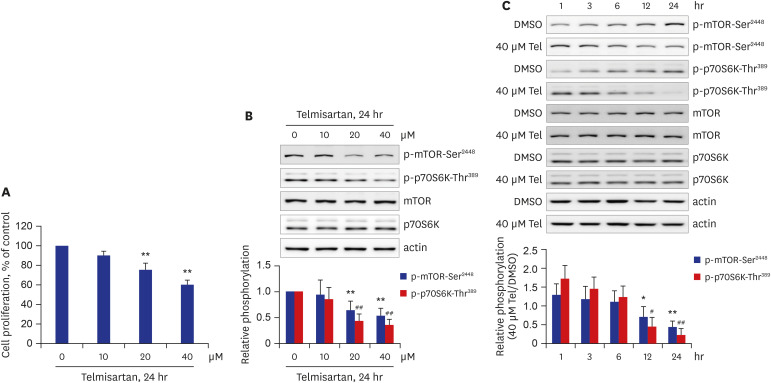
Fig. 1 Telmisartan decreases rat VSMC proliferation by inhibiting p-mTOR-Ser2448 and p-p70S6K-Thr389. (A) VSMC proliferation was measured by MTT assay as described in the METHODS. VSMCs were treated with various doses of telmisartan (0, 10, 20, or 40 μM) for 24 hours. (B) Cells were treated as described above, and then p-mTOR-Ser2448 and p-p70S6K-Thr389 levels were detected using western blot analyses. Nitrocellulose membranes were re-probed with antibodies against mTOR, p70S6K, or β-actin to assess equal sample loading. (C) VSMCs were treated with 40 µM telmisartan or vehicle (DMSO) for the indicated times (0, 1, 3, 6, 12, or 24 hours), and the subsequent p-mTOR-Ser2448 and p-p70S6K-Thr389 levels were detected using western blot analyses as described in Fig. 1B. (B, C) Using western blot results, densitometry was performed to quantitate p-mTOR-Ser2448 and p-p70S6K-Thr389 levels relative to those of total mTOR and p70S6K, respectively. All experiments were performed at least four times independently, and the blots shown are representative of at least four experiments (n = 4). Bar graphs depict mean fold alterations below the control level (± standard deviation).Differences were considered statistically significant at *P < 0.05, #P < 0.05, **P < 0.01, and ##P < 0.01.
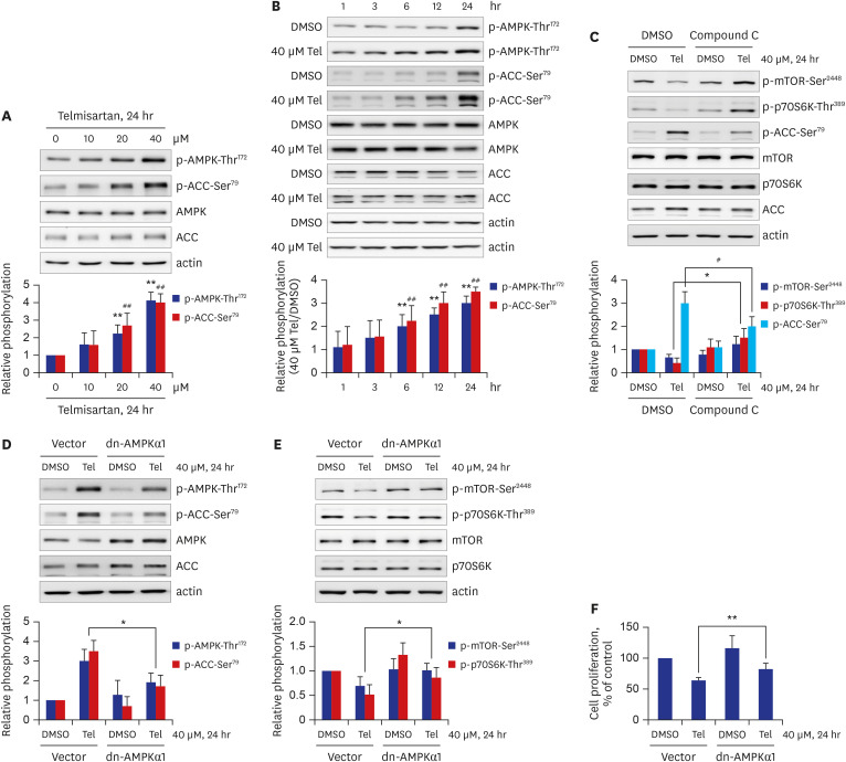
Fig. 2 Telmisartan-activated AMPK inhibits VSMC proliferation by decreasing mTOR/p70S6K signaling pathway. (A) VSMCs were treated with various doses of telmisartan (0, 10, 20, or 40 μM) for 24 hours followed by p-AMPK-Thr172 and p-ACC-Ser79 detection using western blot analyses as described in Fig. 1B. (B) VSMCs were treated with 40 µM telmisartan or vehicle (DMSO) for the indicated times (0, 1, 3, 6, 12, or 24 hours), and the p-AMPK-Thr172 and p-ACC-Ser79 levels were detected using western blot analyses as described in Fig. 1B. (C) VSMCs were treated with 40 µM telmisartan or vehicle (DMSO) in the absence or presence of 10 µM compound C for 24 hours followed by p-mTOR-Ser2448, p-p70S6K-Thr389, and p-ACC-Ser79 detection using western blot analyses as described in Fig. 1B. (D, E) VSMCs were transfected with rat dn-AMPKα1 (D157A) gene or empty vector before treatment with 40 µM telmisartan or vehicle (DMSO) for 24 hours. The resulting p-AMPK-Thr172, p-ACC-Ser79, p-mTOR-Ser2448, and p-p70S6K-Thr389 levels were detected using western blot analyses as described in Fig. 1B. (F) Rat dn-AMPKα1 (D157A) gene- or empty vector-transfected VSMCs were treated with 40 µM telmisartan or vehicle (DMSO) for 24 hours and the subsequent cell proliferation was assessed by MTT assay as described in the METHODS. All experiments were independently performed at least four times, and the blots shown are representative of at least four experiments (n = 4). Bar graphs depict mean fold alterations above/below the control levels (± standard deviation).Differences were considered statistically significant at *P < 0.05, #P < 0.05, **P < 0.01, and ##P < 0.01.
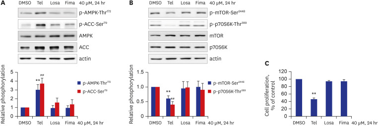
Fig. 3 Among the tested ARBs, only telmisartan induces p-AMPK-Thr172 and inhibits mTOR/p70S6K signaling pathway and VSMC proliferation. (A, B) VSMCs were treated with various ARBs (telmisartan, losartan, or fimasartan; all at a dose of 40 μM) for 24 hours, and then p-AMPK-Thr172, p-ACC-Ser79, p-mTOR-Ser2448, and p-p70S6K-Thr389 levels were assessed using western blot analyses as described in Fig. 1B. (C) VSMCs were treated as described above, and cell proliferation was evaluated by MTT assay as described in the METHODS. All experiments were independently performed at least four times, and the blots shown are representative of at least four experiments (n = 4). Bar graphs depict mean fold alterations above/below the control level (± standard deviation).Differences were considered statistically significant at **P < 0.01 and ##P < 0.01.
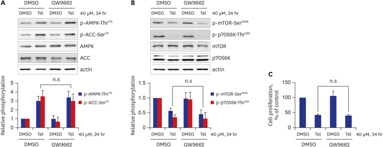
Fig. 4 Telmisartan-inhibited VSMC proliferation is mediated by a PPARγ-independent pathway. (A, B) VSMCs were treated with 40 µM telmisartan or vehicle (DMSO) in the absence or presence of 5 µM GW9662 for 24 hours, and subsequent p-AMPK-Thr172, p-ACC-Ser79, p-mTOR-Ser2448, and p-p70S6K-Thr389 levels were determined using western blot analyses as described in Fig. 1B. (C) VSMCs were treated as described above, and cell proliferation was measured by MTT assay as described in the METHODS. All experiments were independently performed at least four times, and the blots shown are representative of at least four experiments (n = 4). Bar graphs depict mean fold alterations above/below the control level (± standard deviation).n.s = not significant.
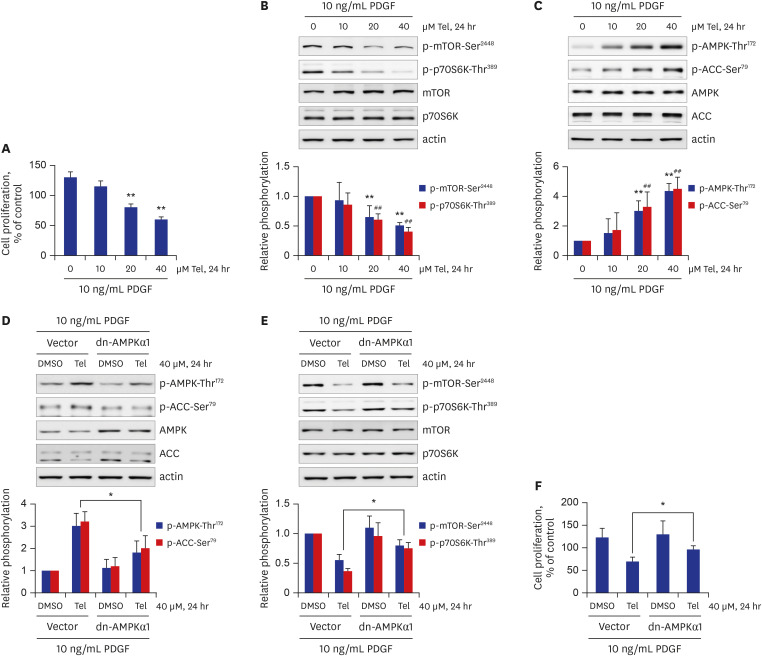
Fig. 5 Telmisartan inhibits PDGF-stimulated VSMC proliferation through AMPK-mediated inhibition of the mTOR/p70S6K signaling pathway. (A) VSMCs were treated with various doses of telmisartan (0, 10, 20, or 40 μM) in the presence of 10 ng/mL PDGF for 24 hours, and subsequent cell proliferation was measured by MTT assay as described in the METHODS. (B, C) VSMCs were treated as described above, and the p-mTOR-Ser2448, p-p70S6K-Thr389, p-AMPK-Thr172, and p-ACC-Ser79 levels were determined using western blot analyses as described in Fig. 1B. (D, E) VSMCs were transfected with rat dn-AMPKα1 (D157A) gene or empty vector, and then treated with 40 µM telmisartan or vehicle (DMSO) in the presence of 10 ng/mL PDGF for 24 hours. The p-AMPK-Thr172, p-ACC-Ser79, p-mTOR-Ser2448, and p-p70S6K-Thr389 levels were detected using western blot analyses as described in Fig. 1B. (F) Rat dn-AMPKα1 (D157A) gene- or empty vector-transfected VSMCs were treated with 40 µM telmisartan or vehicle (DMSO) in the presence of 10 ng/mL PDGF for 24 hours, and subsequent cell proliferation was assessed by MTT assay as described in the METHODS. All experiments were independently performed at least four times, and the blots shown are representative of at least four experiments (n = 4). Bar graphs depict mean fold alterations above/below the control levels (± standard deviation).Differences were considered statistically significant at *P < 0.05, **P < 0.01, and ##P < 0.01.
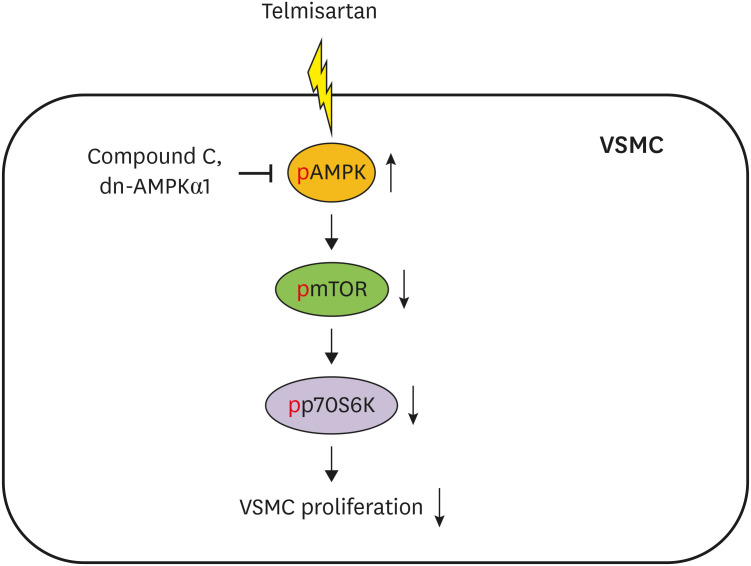
Fig. 6 Schematic illustration of telmisartan inhibition of VSMC proliferation. Telmisartan, not losartan or fimasartan, increases p-AMPK-Thr172 in a PPARγ-independent manner. Increased AMPK activity downregulates p-mTOR-Ser2448 and p-p70S6K-Thr389 levels. Finally, the telmisartan-elevated AMPK activity inhibits VSMC proliferation by decreasing mTOR/p70S6K signaling axis.
Reference
-
1. Benson SC, Pershadsingh HA, Ho CI, Chittiboyina A, Desai P, Pravenec M, et al. Identification of telmisartan as a unique angiotensin II receptor antagonist with selective PPARgamma-modulating activity. Hypertension. 2004; 43(5):993–1002. PMID: 15007034.2. Song KH, Park JH, Jo I, Park JY, Seo J, Kim SA, et al. Telmisartan attenuates hyperglycemia-exacerbated VCAM-1 expression and monocytes adhesion in TNFα-stimulated endothelial cells by inhibiting IKKβ expression. Vascul Pharmacol. 2016; 78:43–52. PMID: 26455386.
Article3. Hwang YJ, Cho DH. Activation of AMPK/proteasome/MLCK degradation signaling axis by telmisartan inhibits VSMC contractility and vessel contraction. Biochem Biophys Res Commun. 2020; 524(4):853–860. PMID: 32046856.
Article4. Lacolley P, Regnault V, Nicoletti A, Li Z, Michel JB. The vascular smooth muscle cell in arterial pathology: a cell that can take on multiple roles. Cardiovasc Res. 2012; 95(2):194–204. PMID: 22467316.
Article5. Rivard A, Andrés V. Vascular smooth muscle cell proliferation in the pathogenesis of atherosclerotic cardiovascular diseases. Histol Histopathol. 2000; 15(2):557–571. PMID: 10809377.6. Yamamoto K, Ohishi M, Ho C, Kurtz TW, Rakugi H. Telmisartan-induced inhibition of vascular cell proliferation beyond angiotensin receptor blockade and peroxisome proliferator-activated receptor-gamma activation. Hypertension. 2009; 54(6):1353–1359. PMID: 19822796.7. Destro M, Cagnoni F, Dognini GP, Galimberti V, Taietti C, Cavalleri C, et al. Telmisartan: just an antihypertensive agent? A literature review. Expert Opin Pharmacother. 2011; 12(17):2719–2735. PMID: 22077832.
Article8. Michel MC, Foster C, Brunner HR, Liu L. A systematic comparison of the properties of clinically used angiotensin II type 1 receptor antagonists. Pharmacol Rev. 2013; 65(2):809–848. PMID: 23487168.
Article9. Miura S, Karnik SS, Saku K. Review: angiotensin II type 1 receptor blockers: class effects versus molecular effects. J Renin Angiotensin Aldosterone Syst. 2011; 12(1):1–7. PMID: 20603272.
Article10. Papadopoulos N, Lennartsson J. The PDGF/PDGFR pathway as a drug target. Mol Aspects Med. 2018; 62:75–88. PMID: 29137923.
Article11. Shawky NM, Segar L. Sulforaphane inhibits platelet-derived growth factor-induced vascular smooth muscle cell proliferation by targeting mTOR/p70S6kinase signaling independent of Nrf2 activation. Pharmacol Res. 2017; 119:251–264. PMID: 28212891.
Article12. Dong X, Hu H, Fang Z, Cui J, Liu F. CTRP6 inhibits PDGF-BB-induced vascular smooth muscle cell proliferation and migration. Biomed Pharmacother. 2018; 103:844–850. PMID: 29710500.
Article13. Gomez D, Owens GK. Smooth muscle cell phenotypic switching in atherosclerosis. Cardiovasc Res. 2012; 95(2):156–164. PMID: 22406749.
Article14. Fuster V, Badimon L, Badimon JJ, Chesebro JH. The pathogenesis of coronary artery disease and the acute coronary syndromes (1). N Engl J Med. 1992; 326(4):242–250. PMID: 1727977.15. Schwartz SM, deBlois D, O'Brien ER. The intima. Soil for atherosclerosis and restenosis. Circ Res. 1995; 77(3):445–465. PMID: 7641318.16. Dzau VJ, Braun-Dullaeus RC, Sedding DG. Vascular proliferation and atherosclerosis: new perspectives and therapeutic strategies. Nat Med. 2002; 8(11):1249–1256. PMID: 12411952.
Article17. Hong MK. Restenosis following coronary angioplasty: current status. Korean J Intern Med (Korean Assoc Intern Med). 2001; 16(2):51–55.18. Li Z, Li Y, Jia Y, Ding B, Yu J. Rab1A knockdown represses proliferation and promotes apoptosis in gastric cancer cells by inhibition of mTOR/p70S6K pathway. Arch Biochem Biophys. 2020; 685:108352. PMID: 32240637.
Article19. Li J, Liu W, Hao H, Wang Q, Xue L. Rapamycin enhanced the antitumor effects of doxorubicin in myelogenous leukemia K562 cells by downregulating the mTOR/p70S6K pathway. Oncol Lett. 2019; 18(3):2694–2703. PMID: 31404320.
Article20. Lu QB, Wan MY, Wang PY, Zhang CX, Xu DY, Liao X, et al. Chicoric acid prevents PDGF-BB-induced VSMC dedifferentiation, proliferation and migration by suppressing ROS/NFκB/mTOR/P70S6K signaling cascade. Redox Biol. 2018; 14:656–668. PMID: 29175753.
Article21. Morita M, Gravel SP, Hulea L, Larsson O, Pollak M, St-Pierre J, et al. mTOR coordinates protein synthesis, mitochondrial activity and proliferation. Cell Cycle. 2015; 14(4):473–480. PMID: 25590164.
Article22. You G, Long X, Song F, Huang J, Tian M, Xiao Y, et al. Metformin activates the AMPK-mTOR pathway by modulating lncRNA TUG1 to induce autophagy and inhibit atherosclerosis. Drug Des Devel Ther. 2020; 14:457–468.23. Wu H, Song A, Hu W, Dai M. The anti-atherosclerotic effect of paeonol against vascular smooth muscle cell proliferation by up-regulation of autophagy via the AMPK/mTOR signaling pathway. Front Pharmacol. 2018; 8:948. PMID: 29354055.
Article24. Zhao Y, Shang F, Shi W, Zhang J, Zhang J, Liu X, et al. Angiotensin II receptor type 1 antagonists modulate vascular smooth muscle cell proliferation and migration via AMPK/mTOR. Cardiology. 2019; 143(1):1–10. PMID: 31307032.
Article25. Jin Z, Tan Q, Sun B. Telmisartan ameliorates vascular endothelial dysfunction in coronary slow flow phenomenon (CSFP). Cell Biochem Funct. 2018; 36(1):18–26. PMID: 29314204.
Article26. Markan U, Pasupuleti S, Pollard CM, Perez A, Aukszi B, Lymperopoulos A. The place of ARBs in heart failure therapy: is aldosterone suppression the key? Ther Adv Cardiovasc Dis. 2019; 13:1753944719868134. PMID: 31401939.
Article27. Yusuf S, Teo K, Anderson C, Pogue J, Dyal L, Copland I, et al. Effects of the angiotensin-receptor blocker telmisartan on cardiovascular events in high-risk patients intolerant to angiotensin-converting enzyme inhibitors: a randomised controlled trial. Lancet. 2008; 372(9644):1174–1183. PMID: 18757085.28. Diener HC. Preventing stroke: the PRoFESS, ONTARGET, and TRANSCEND trial programs. J Hypertens Suppl. 2009; 27(5):S31–6. PMID: 19587553.
Article29. Kliewer SA, Forman BM, Blumberg B, Ong ES, Borgmeyer U, Mangelsdorf DJ, et al. Differential expression and activation of a family of murine peroxisome proliferator-activated receptors. Proc Natl Acad Sci U S A. 1994; 91(15):7355–7359. PMID: 8041794.
Article30. Rosen ED, Spiegelman BM. PPARgamma: a nuclear regulator of metabolism, differentiation, and cell growth. J Biol Chem. 2001; 276(41):37731–37734. PMID: 11459852.31. Zídek V, Mlejnek P, Simáková M, Silhavy J, Landa V, Kazdová L, et al. Tissue-specific peroxisome proliferator activated receptor gamma expression and metabolic effects of telmisartan. Am J Hypertens. 2013; 26(6):829–835. PMID: 23426788.32. Auboeuf D, Rieusset J, Fajas L, Vallier P, Frering V, Riou JP, et al. Tissue distribution and quantification of the expression of mRNAs of peroxisome proliferator-activated receptors and liver X receptor-alpha in humans: no alteration in adipose tissue of obese and NIDDM patients. Diabetes. 1997; 46(8):1319–1327. PMID: 9231657.
Article
- Full Text Links
-
- Actions
-
Cited
- CITED
-
- Close
- Share
-
- Similar articles
-
- Far-Infrared Irradiation Decreases Proliferation in Basal and PDGFStimulated VSMCs Through AMPKMediated Inhibition of mTOR/p70S6K Signaling Axis
- Mesoglycan attenuates VSMC proliferation through activation of AMP-activated protein kinase and mTOR
- Influence of Endothelin-1 on Cultured Vascular Smooth Muscle Cell Proliferation
- The Inhibition of Insulin-stimulated Proliferation of Vascular Smooth Muscle Cells by Rosiglitazone Is Mediated by the Akt-mTOR-P70S6K Pathway
- Cilostazol Inhibits Vascular Smooth Muscle Cell Proliferation and Reactive Oxygen Species Production through Activation of AMP-activated Protein Kinase Induced by Heme Oxygenase-1
