Korean J Gastroenterol.
2020 Aug;76(2):88-93. 10.4166/kjg.2020.76.2.88.
Primary Epiploic Appendagitis Mimicking Acute Appendicitis: A Case Report and Narrative Review of the Literature
- Affiliations
-
- 1Department of Pediatrics, General Hospital of Ioannina "G. Xatzikosta", Ioannina, Greece
- 2Department of Surgery, University Hospital of Ioannina, Ioannina, Greece
- 3Department of Surgery, General Hospital "G. Xatzikosta", Ioannina, Greece
- 4Department of Surgery, General Hospital of Filiates, Thesprotia, Greece
- 5Intensive Care Unit, University Hospital of Ioannina, Ioannina, Greece
- 6Anatomy-Histology-Embryology, University of Ioannina, Ioannina, Greece
- 7Department of Pathology, Agia Sofia Children's Hospital, Athens, Greece
- 8Department of Orthopaedics, University Hospital of Ioannina, Ioannina, Greece
- KMID: 2505871
- DOI: http://doi.org/10.4166/kjg.2020.76.2.88
Abstract
- Primary epiploic appendagitis is uncommon and is estimated to induce 1.1–1.3% of all abdominal pain. We report a 42-year-oldmale who appeared in the morning in the emergency department with abdominal pain localized in the right lower abdomen andassociated with anorexia and nausea. Clinical examination, laboratory tests, and abdominal ultrasound revealed deep tendernessat Mc Burney point and a mild elevation of CRP (0.7 mg/dL). In the evening, the symptoms were exacerbated, and a diagnosticlaparoscopy was performed. Intra-operatively, the appendix was normal and a twisted, necrotic epiploic appendage originatingfrom the antimesenteric border of the mid ascending colon was found. Laparoscopic resection of the necrotic epiploic appendageand prophylactic appendectomy was carried out. Histology indicated the diagnosis of the necrotic epiploic appendage.Postoperatively, the patient recovered without complications. Although the preoperative diagnosis of primary epiploic appendagitishas improved due to abdominal ultrasound and mainly CT, there are still cases which are diagnosed during laparoscopy. Thetreatment of choice is conservative management, while the use of antibiotics remains controversial. The relapse and complicationrates are rare. Surgical excision, particularly laparoscopic, should be considered in cases of uncertain diagnosis, persistentsymptoms, or recurrence.
Keyword
Figure
-

Fig. 1 Abdominal ultrasound: The vermiform appendix is not visualized, and no free fluid is found. The only finding was probe tenderness in the right iliac fossa.
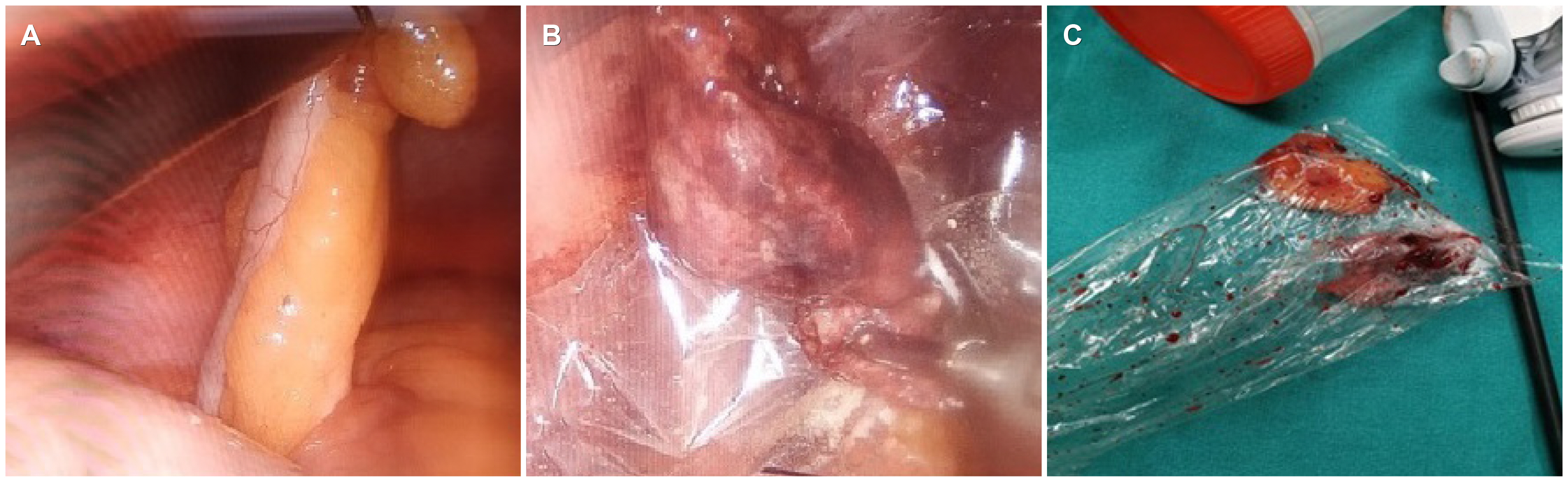
Fig. 2 Surgical specimens: (A) the vermiform appendix appears normal, while (B) a twisted, necrotic epiploic appendage originating from the antimesenteric border of the mid ascending colon is identified. (C) The two surgical specimens are placed alongside.
Reference
-
1. Sand M, Gelos M, Bechara FG, et al. 2007; Epiploic appendagitis--clinical characteristics of an uncommon surgical diagnosis. BMC Surg. 7:11. DOI: 10.1186/1471-2482-7-11. PMID: 17603914. PMCID: PMC1925058.
Article2. Choi YU, Choi PW, Park YH, et al. 2011; Clinical characteristics of primary epiploic appendagitis. J Korean Soc Coloproctol. 27:114–121. DOI: 10.3393/jksc.2011.27.3.114. PMID: 21829765. PMCID: PMC3145881.
Article3. Son HJ, Lee SJ, Lee JH, et al. 2002; Clinical diagnosis of primary epiploic appendagitis: differentiation from acute diverticulitis. J Clin Gastroenterol. 34:435–438. DOI: 10.1097/00004836-200204000-00010. PMID: 11907356.4. Nadida D, Amal A, Ines M, et al. 2016; Acute epiploic appendagitis: radiologic and clinical features of 12 patients. Int J Surg Case Rep. 28:219–222. DOI: 10.1016/j.ijscr.2016.09.015. PMID: 27728879. PMCID: PMC5065630.
Article5. Rioux M, Langis P. 1994; Primary epiploic appendagitis: clinical, US, and CT findings in 14 cases. Radiology. 191:523–526. DOI: 10.1148/radiology.191.2.8153333. PMID: 8153333.
Article6. Jalaguier A, Zins M, Rodallec M, Nakache JP, Boulay-Coletta I, Jullès MC. 2010; Accuracy of multidetector computed tomography in differentiating primary epiploic appendagitis from left acute colonic diverticulitis associated with secondary epiploic appendagitis. Emerg Radiol. 17:51–56. DOI: 10.1007/s10140-009-0822-x. PMID: 19517148.
Article7. Lohle PN, Coerkamp EG, Puylaert JB. , Van Breda Vriesman AC. 1999; Infarction of omentum and epiploic appendage: diagnosis, epidemiology and natural history. Eur Radiol. 9:1886–1892. DOI: 10.1007/s003300050942. PMID: 10602970.8. Mollà E, Ripolés T, Martínez MJ, Morote V, Roselló-Sastre E. 1998; Primary epiploic appendagitis: US and CT findings. Eur Radiol. 8:435–438. DOI: 10.1007/s003300050408. PMID: 9510579.
Article9. Zissin R, Hertz M, Osadchy A, Kots E, Shapiro-Feinberg M, Paran H. 2002; Acute epiploic appendagitis: CT findings in 33 cases. Emerg Radiol. 9:262–265. DOI: 10.1007/s10140-002-0243-6. PMID: 15290551.
Article10. Hwang JA, Kim SM, Song HJ, et al. 2013; Differential diagnosis of left-sided abdominal pain: primary epiploic appendagitis vs colonic diverticulitis. World J Gastroenterol. 19:6842–6848. DOI: 10.3748/wjg.v19.i40.6842. PMID: 24187459. PMCID: PMC3812483.11. Chen JH, Wu CC, Wu PH. 2011; Epiploic appendagitis: an uncommon and easily misdiagnosed disease. J Dig Dis. 12:448–452. DOI: 10.1111/j.1751-2980.2011.00543.x. PMID: 22118694.
Article12. Legome EL, Belton AL, Murray RE, Rao PM, Novelline RA. 2002; Epiploic appendagitis: the emergency department presentation. J Emerg Med. 22:9–13. DOI: 10.1016/S0736-4679(01)00430-9. PMID: 11809550.
Article13. Ozdemir S, Gulpinar K, Leventoglu S, et al. 2010; Torsion of the primary epiploic appendagitis: a case series and review of the literature. Am J Surg. 199:453–458. DOI: 10.1016/j.amjsurg.2009.02.004. PMID: 19520357.
Article14. Ergelen R, Asadov R, Özdemir B, Tureli D, Demirbaş BT, Tuney D. 2017; Computed tomography findings of primary epiploic appendagitis as an easily misdiagnosed entity: case series and review of literature. Ulus Travma Acil Cerrahi Derg. 23:489–494. DOI: 10.5505/tjtes.2017.99894. PMID: 29115651.15. Hasbahceci M, Erol C, Seker M. 2012; Epiploic appendagitis: is there need for surgery to confirm diagnosis in spite of clinical and radiological findings? World J Surg. 36:441–446. DOI: 10.1007/s00268-011-1382-2. PMID: 22167263.
Article16. Ng KS, Tan AG, Chen KK, Wong SK, Tan HM. 2006; CT features of primary epiploic appendagitis. Eur J Radiol. 59:284–288. DOI: 10.1016/j.ejrad.2006.02.008. PMID: 16580806.
Article17. Nugent JP, Ouellette HA, O'Leary DP, Khosa F, Nicolaou S, McLaughlin PD. 2018; Epiploic appendagitis: 7-year experience and relationship with visceral obesity. Abdom Radiol (NY). 43:1552–1557. DOI: 10.1007/s00261-017-1355-5. PMID: 29043404.
Article18. Almeida RR, Singh AK, Mansouri M, Spilberg G, Alkasab T, Lev MH. 2019; Impact of radiology report wording on care of patients with acute epiploic appendagitis. AJR Am J Roentgenol. 212:1265–1270. DOI: 10.2214/AJR.18.20747. PMID: 30860892.
Article19. Sirvanci M, Balci NC, Karaman K, Duran C, Karakaş E. 2002; Primary epiploic appendagitis: MRI findings. Magn Reson Imaging. 20:137–139. DOI: 10.1016/S0730-725X(02)00478-2. PMID: 11973040.20. Saad J, Mustafa HA, Elsani AM, Alharbi F, Alghamdi S. 2014; Primary epiploic appendagitis: reconciling CT and clinical challenges. Indian J Gastroenterol. 33:420–426. DOI: 10.1007/s12664-014-0466-y. PMID: 24934824.
Article
- Full Text Links
-
- Actions
-
Cited
- CITED
-
- Close
- Share
-
- Similar articles
-
- A Case of Epiploic Appendagitis after COVID-19
- A Case of Epiploic Appendagitis Presented with Right Lower Quadrant Pain of Abdomen
- Seven Cases of Epiploic Appendagitis: Retrograde Analysis of Clinical Characteristics
- A Case of Primary Epiploic Appendagitis
- A Case of Epiploic Appendagitis with Acute Gastroenteritis
