Comparison of Clinical and Echocardiographic Characteristics between Cardiac Myxomas and Masses Mimicking Myxoma
- Affiliations
-
- 1Division of Cardiology, Department of Internal Medicine, Jeonbuk National University Medical School and Research Institute of Clinical Medicine of Jeonbuk National University-Biomedical Research Institute of Jeonbuk National University Hospital, Jeonju, Korea
- 2Department of Cardiovascular Medicine, Chonnam National University Hospital, Gwangju, Korea
- 3Department of Cardiology in Internal Medicine, School of Medicine, Chungnam National University and Regional Cardiocerebrovascular Center, Chungnam National University Hospital, Daejeon, Korea
- 4Cardiovascular Center and Division of Cardiology, Department of Internal Medicine, Eulji University Hospital, College of Medicine, Eulji University, Daejeon, Korea
- KMID: 2505738
- DOI: http://doi.org/10.4070/kcj.2020.0024
Abstract
- Background and Objectives
Cardiac myxoma is the most frequent benign cardiac tumor that can result in cardiac and systemic symptoms. We investigated clinical and echocardiographic characteristics of patients with cardiac masses suggesting myxoma.
Methods
We investigated 265 consecutive patients with an echocardiographic diagnosis of cardiac myxomas in 4 teaching hospitals in Korea.
Results
The mean age was 61±16 years and 169 patients (63.8%) were female. The most frequent referral reason for echocardiography was an evaluation of cardiac symptoms (43.4%). Tumors were incidentally detected in 82 patients (30.9%). Left atrium (LA) was the most frequently involved site (84.5%) and 19 patients (7.2%) had non-atrial tumors. The mean tumor size was 38.7×26.0 mm (range, 4–96 mm). Of 186 patients (70.2%) who had pathological diagnosis, 174 (93.5%) were confirmed with myxoma, 8 (4.3%) with other tumors and 4 (2.2%) with thrombi. Compared to myxoma, smaller size (20.4×12.6 mm vs. 41.4×27.6 mm, p<0.01) and non-LA location (87.5% vs. 10.5%, p<0.001) were associated with non-myxoma tumors, and more frequent atrial fibrillation (AF, 75.0% vs. 7.0%, p<0.001) and larger LA diameter (55.0±14.6 mm vs. 41.3±7.7 mm, p=0.001) were related to thrombi.
Conclusions
Of 265 patients with an echocardiographic diagnosis with cardiac myxomas, 174 (65.7%) were surgically confirmed with myxomas. Compared with cardiac myxoma, other tumors were smaller and more frequently found in non-atrial sites. Thrombi were associated with AF and larger LA diameter.
Keyword
Figure
-
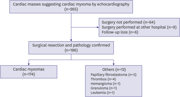
Figure 1 A flow diagram showing the number of patients diagnosed as cardiac myxomas by transthoracic echocardiography.
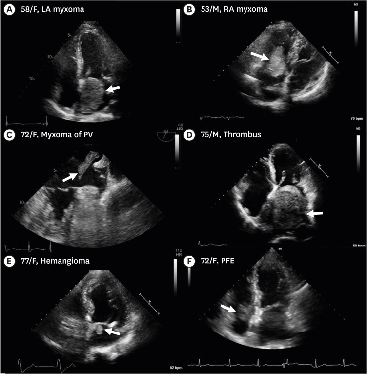
Figure 2 Representative cases. (A) An incidentally detected a 64×49 mm sized myxoma with smooth surface in the LA in a 58-year old woman. (B) A myxoma with polypoid surface in the RA prolapsing through the TV in a 53-year old man. (C) A transesophageal echocardiographic image showing an irregular shaped myxoma measured 34×9 mm in size originating from the left upper PV in the LA cavity in a 72-year old woman with a history of AF and cerebral infarction 3 years ago. (D) An 84×67 mm-sized mass-like thrombus in the LA in a 75-year old man with a history of AF presenting with abdominal pain. (E) A 14×13 mm-sized round echogenic mass below the medial mitral annulus in the LA in a 77-year old woman presenting with chest pain diagnosed as hemangioma. (F) A 22×21 mm-sized round mass proven to be a PFE below the septal leaflet of the TV in the RA in a 72-year old woman presenting with chest pain. Arrows indicate myxomas and masses mimicking myxoma.AF = atrial fibrillation; LA = left atrium; PFE = papillary fibroelastoma; PV = pulmonary vein; RA = right atrium; TV = tricuspid valve.
Cited by 1 articles
-
A Comprehensive Perspective of Clinical and Echocardiographic Features in the Differential Diagnosis of Cardiac Myxomas and Myxoma-Like Masses
Dong-Soo Kim
Korean Circ J. 2020;50(9):833-835. doi: 10.4070/kcj.2020.0309.
Reference
-
1. Burke AP, Virmani R. Cardiac myxoma. A clinicopathologic study. Am J Clin Pathol. 1993; 100:671–680. PMID: 8249916.2. Colin GC, Gerber BL, Amzulescu M, Bogaert J. Cardiac myxoma: a contemporary multimodality imaging review. Int J Cardiovasc Imaging. 2018; 34:1789–1808. PMID: 29974293.
Article4. Fang BR, Chiang CW, Hung JS, Lee YS, Chang CS. Cardiac myxoma--clinical experience in 24 patients. Int J Cardiol. 1990; 29:335–341. PMID: 2283191.5. Basso C, Valente M, Poletti A, Casarotto D, Thiene G. Surgical pathology of primary cardiac and pericardial tumors. Eur J Cardiothorac Surg. 1997; 12:730–738. PMID: 9458144.
Article6. Poterucha TJ, Kochav J, O'Connor DS, Rosner GF. Cardiac tumors: clinical presentation, diagnosis, and management. Curr Treat Options Oncol. 2019; 20:66. PMID: 31250250.
Article7. Pacini D, Careddu L, Pantaleo A, et al. Primary benign cardiac tumours: long-term results. Eur J Cardiothorac Surg. 2012; 41:812–819. PMID: 22219403.
Article8. Cianciulli TF, Cozzarin A, Soumoulou JB, et al. Twenty years of clinical experience with cardiac myxomas: diagnosis, treatment, and follow up. J Cardiovasc Imaging. 2019; 27:37–47. PMID: 30701715.
Article9. Karabinis A, Samanidis G, Khoury M, Stavridis G, Perreas K. Clinical presentation and treatment of cardiac myxoma in 153 patients. Medicine (Baltimore). 2018; 97:e12397. PMID: 30213011.
Article10. Yu SH, Lim SH, Hong YS, Yoo KJ, Chang BC, Kang MS. Clinical experiences of cardiac myxoma. Yonsei Med J. 2006; 47:367–371. PMID: 16807986.
Article11. Bullock-Palmer RP, Tak V, Mitchell JE. Mitral valve annular bacterial vegetative mass masquerading as a left atrial myxoma. Echocardiography. 2010; 27:E62–E64. PMID: 20545987.
Article12. Erdoes G, Reineke D, Basciani R, Carrel T, Eberle B. Left atrial myxoma attached to the anterior mitral leaflet with symptoms suggestive of infective endocarditis. Eur J Echocardiogr. 2010; 11:E8. PMID: 19959532.
Article13. El Sabbagh A, Al-Hijji MA, Thaden JJ, et al. Cardiac myxoma: the great mimicker. JACC Cardiovasc Imaging. 2017; 10:203–206. PMID: 28183439.14. Kodali S, Yamrozik J, Biederman RW. Left atrial thrombus masquerading as a myxoma in a patient with mitral stenosis. Echocardiography. 2010; 27:E98–101. PMID: 21039808.
Article15. Singh Y, Kharge J, Ramegowda RT, Nanjappa MC. Large atrial thrombus at unusual site: masquerading atrial myxoma. J Thromb Thrombolysis. 2013; 36:74–76. PMID: 23354968.
Article16. Demirel M, Acar E, Toprak C, İzci S, Öcal L. Papillary fibroelastoma of the mitral valve: an unusual cause of mitral valve obstruction. Korean Circ J. 2017; 47:286–287. PMID: 28382087.
Article
- Full Text Links
-
- Actions
-
Cited
- CITED
-
- Close
- Share
-
- Similar articles
-
- A Comprehensive Perspective of Clinical and Echocardiographic Features in the Differential Diagnosis of Cardiac Myxomas and Myxoma-Like Masses
- Multi Modality Imaging Features of Cardiac Myxoma
- A Case of Multiple Right Atrial Myxomas with Pulmonary Embolism
- Familial Atrial Myxoma with Carney's Complex: 1 Case
- A Case of Carney Complex: Diagnosed 11 Years after Resection of Recurrent Cardiac Myxomas
