Value of the Frontal Assessment Battery Tool for Assessing the Frontal Lobe Function in Stroke Patients
- Affiliations
-
- 1Department of Rehabilitation Medicine, Incheon St. Mary’s Hospital, College of Medicine, The Catholic University of Korea, Incheon, Korea
- 2Department of Rehabilitation Medicine, Incheon Hospital of Korea Workers’ Compensation and Welfare Service, Incheon, Korea
- KMID: 2505386
- DOI: http://doi.org/10.5535/arm.19111
Abstract
Objective
To examine the correlation between the Frontal Assessment Battery (FAB) test, which is used to assess the frontal lobe function, and anatomical lesions as well as the ability of the test to detect frontal lobe dysfunction.
Methods
Records of stroke patients undergoing a FAB test and Mini-Mental State Examination (MMSE) were retrospectively reviewed. The patients were divided into three groups according to the lesions determined by an imaging study: frontal lobe cortex lesions, frontal subcortical circuit lesions, and other lesions. The FAB scores of the three groups were compared using the Kruskal-Wallis test. The validity of the FAB test to detect frontal lobe dysfunction was assessed by a comparison with the Computerized Neuropsychological Function Test (CNT) using the Spearman correlation coefficient. The correlation coefficients between the FAB test and MMSE were analyzed further based on the MMSE cutoff score.
Results
Patients with frontal cortex lesions had significantly lower total and subtest scores according to the FAB test than the other patients. The FAB test correlated better with the CNT than the MMSE, particularly in the executive function and memory domains. A high MMSE score (r=0.435) indicated a lower correlation with the FAB test score than a low MMSE score (r=0.714).
Conclusion
The FAB test could differentiate frontal lobe lesions from others in stroke patients and showed a good correlation with the CNT. Moreover, the FAB test can be used in patients with high MMSE scores to detect frontal lobe dysfunction and determine the treatment strategies for stroke patients.
Figure
-

Fig. 1. Frontal Assessment Battery (FAB) and Mini-Mental State Examination (MMSE) scores of patients according to brain lesions. (A) The mean FAB scores were significantly different in the FL group (7.3±5.0), SC group (9.5±5.3), and OL group (10.5±4.8). (B) The mean MMSE scores were significantly different in the FL group (16.94±9.04), SC group (20.68±8.38), and OL group (23.36±6.67). FL group, with lesions involving the frontal lobe cortex; SC group, with lesions related to the frontal subcortical circuit without frontal cortex involvement; OL group, with other lesions that were unrelated to the frontal subcortical circuit. *p<0.05 indicate a significant difference among three groups.
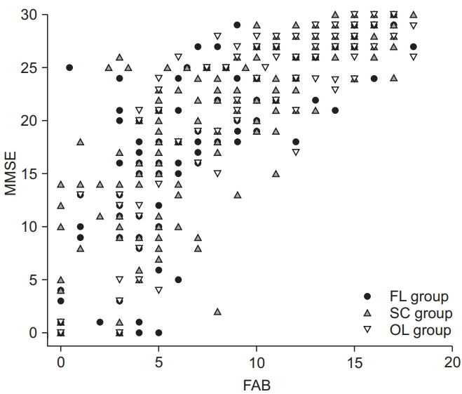
Fig. 2. Scatterplots for the Frontal Assessment Battery (FAB) and Mini-Mental State Examination (MMSE) scores of patients according to brain lesions. Scatterplots showed a positive correlation between the FAB and MMSE tests when divided among the three lesion groups (FL group: r=0.839, p<0.001; SC group: r=0.864, p<0.001; OL group: r=0.765, p<0.001). When the MMSE scores were divided according to the cutoff value that (MMSE=24), a high MMSE score (r=0.488, p<0.001) had a lower correlation with the FAB score than did a low MMSE score (r=0.704, p<0.001). FL group, with lesions involving the frontal lobe cortex; SC group, with lesions related to the frontal subcortical circuit without frontal cortex involvement; OL group, with other lesions that were unrelated to the frontal subcortical circuit.
Reference
-
1. Hoffmann M. The human frontal lobes and frontal network systems: an evolutionary, clinical, and treatment perspective. ISRN Neurol. 2013; 2013:892459.
Article2. Stuss DT, Alexander MP. Executive functions and the frontal lobes: a conceptual view. Psychol Res. 2000; 63:289–98.
Article3. Estevez-Gonzalez A, Garcia-Sanchez C, Barraquer-Bordas L. Frontal lobes: the executive brain. Rev Neurol. 2000; 31:566–77.4. Kokubo K, Suzuki K, Hattori N, Miyai I, Mori E. Executive dysfunction in patients with putaminal hemorrhage. J Stroke Cerebrovasc Dis. 2015; 24:1978–85.
Article5. Laplane D, Levasseur M, Pillon B, Dubois B, Baulac M, Mazoyer B, et al. Obsessive-compulsive and other behavioural changes with bilateral basal ganglia lesions: a neuropsychological, magnetic resonance imaging and positron tomography study. Brain. 1989; 112(Pt 3):699–725.6. Bhatia KP, Marsden CD. The behavioural and motor consequences of focal lesions of the basal ganglia in man. Brain. 1994; 117(Pt 4):859–76.
Article7. Cohen OS, Vakil E, Tanne D, Molshatzki N, Nitsan Z, Hassin-Baer S. The frontal assessment battery as a tool for evaluation of frontal lobe dysfunction in patients with Parkinson disease. J Geriatr Psychiatry Neurol. 2012; 25:71–7.
Article8. Zucchella C, Federico A, Martini A, Tinazzi M, Bartolo M, Tamburin S. Neuropsychological testing. Pract Neurol. 2018; 18:227–37.
Article9. Lima CF, Meireles LP, Fonseca R, Castro SL, Garrett C. The Frontal Assessment Battery (FAB) in Parkinson’s disease and correlations with formal measures of executive functioning. J Neurol. 2008; 255:1756–61.
Article10. Takagi R, Kajimoto Y, Kamiyoshi S, Miwa H, Kondo T. The frontal assessment battery at bed side (FAB) in patients with Parkinson’s disease. No To Shinkei. 2002; 54:897–902.11. Biundo R, Weis L, Pilleri M, Facchini S, Formento-Dojot P, Vallelunga A, et al. Diagnostic and screening power of neuropsychological testing in detecting mild cognitive impairment in Parkinson’s disease. J Neural Transm (Vienna). 2013; 120:627–33.
Article12. Slachevsky A, Villalpando JM, Sarazin M, Hahn-Barma V, Pillon B, Dubois B. Frontal assessment battery and differential diagnosis of frontotemporal dementia and Alzheimer disease. Arch Neurol. 2004; 61:1104–7.
Article13. Castiglioni S, Pelati O, Zuffi M, Somalvico F, Marino L, Tentorio T, et al. The frontal assessment battery does not differentiate frontotemporal dementia from Alzheimer’s disease. Dement Geriatr Cogn Disord. 2006; 22:125–31.
Article14. Cunha PJ, Nicastri S, de Andrade AG, Bolla KI. The frontal assessment battery (FAB) reveals neurocognitive dysfunction in substance-dependent individuals in distinct executive domains: abstract reasoning, motor programming, and cognitive flexibility. Addict Behav. 2010; 35:875–81.
Article15. Paviour DC, Winterburn D, Simmonds S, Burgess G, Wilkinson L, Fox NC, et al. Can the frontal assessment battery (FAB) differentiate bradykinetic rigid syndromes? Relation of the FAB to formal neuropsychological testing. Neurocase. 2005; 11:274–82.
Article16. Kopp B, Rosser N, Tabeling S, Sturenburg HJ, de Haan B, Karnath HO, et al. Performance on the Frontal Assessment Battery is sensitive to frontal lobe damage in stroke patients. BMC Neurol. 2013; 13:179.
Article17. Fu C, Jin X, Chen B, Xue F, Niu H, Guo R, et al. Comparison of the Mini-Mental State Examination and Montreal Cognitive Assessment executive subtests in detecting post-stroke cognitive impairment. Geriatr Gerontol Int. 2017; 17:2329–35.
Article18. Trzepacz PT, Hochstetler H, Wang S, Walker B, Saykin AJ; Alzheimer’s Disease Neuroimaging. Relationship between the Montreal Cognitive Assessment and Mini-Mental State Examination for assessment of mild cognitive impairment in older adults. BMC Geriatr. 2015; 15:107.
Article19. Dubois B, Slachevsky A, Litvan I, Pillon B. The FAB: a Frontal Assessment Battery at bedside. Neurology. 2000; 55:1621–6.
Article20. Gualtieri CT, Johnson LG. Reliability and validity of a computerized neurocognitive test battery, CNS Vital Signs. Arch Clin Neuropsychol. 2006; 21:623–43.
Article21. Kim YH, Shin SH, Park SH, Ko MH. Cognitive assessment for patient with brain injury by computerized neuropsychological test. J Korean Acad Rehabil Med. 2001; 25:209–16.22. Kang CG, Chun MH, Kang JA, Do KH, Choi SJ. Neurocognitive dysfunction according to hypoperfusion territory in patients with moyamoya disease. Ann Rehabil Med. 2017; 41:1–8.
Article23. Cho HY, Kim KT, Jung JH. Effects of computer assisted cognitive rehabilitation on brain wave, memory and attention of stroke patients: a randomized control trial. J Phys Ther Sci. 2015; 27:1029–32.
Article24. Kim JY. Analysis of cognitive factors affecting stroke patient’s activity of daily living performance: using the computerized neurocognitive function test. J Korea Acad Ind Coop Soc. 2011; 12:5715–21.
Article25. de Oliveira MO, Brucki SMD. Computerized Neurocognitive Test (CNT) in mild cognitive impairment and Alzheimer’s disease. Dement Neuropsychol. 2014; 8:112–6.26. Mataix-Cols D, Bartres-Faz D. Is the use of the wooden and computerized versions of the Tower of Hanoi puzzle equivalent? Appl Neuropsychol. 2002; 9:117–20.
Article27. Parsey CM, Schmitter-Edgecombe M. Applications of technology in neuropsychological assessment. Clin Neuropsychol. 2013; 27:1328–61.
Article28. Kwon JS, Lyoo IK, Hong KS, Yeon BK, Ha KS. Development and standardization of the computerized memory assessment for Korean adults. J Korean Neuropsychiatr Assoc. 2002; 41:347–58.29. Ha KS, Kwon JS, Lyoo IK, Kong SW, Lee DW, Youn T. Development and standardization process, and factor analysis of the computerized cognitive function test system for Korea adults. J Korean Neuropsychiatr Assoc. 2002; 41:551–62.30. Lyoo IK, Kwon JS, Ha KS. Development and standardization of the computerized higher cortical function assessment for Korean adults. J Korean Neuropsychiatr Assoc. 2002; 41:538–50.31. Appollonio I, Leone M, Isella V, Piamarta F, Consoli T, Villa ML, et al. The Frontal Assessment Battery (FAB): normative values in an Italian population sample. Neurol Sci. 2005; 26:108–16.
Article32. Lipton AM, Ohman KA, Womack KB, Hynan LS, Ninman ET, Lacritz LH. Subscores of the FAB differentiate frontotemporal lobar degeneration from AD. Neurology. 2005; 65:726–31.
Article33. Dong Y, Venketasubramanian N, Chan BP, Sharma VK, Slavin MJ, Collinson SL, et al. Brief screening tests during acute admission in patients with mild stroke are predictive of vascular cognitive impairment 3-6 months after stroke. J Neurol Neurosurg Psychiatry. 2012; 83:580–5.34. Nys GM, van Zandvoort MJ, de Kort PL, Jansen BP, Kappelle LJ, de Haan EH. Restrictions of the Mini-Mental State Examination in acute stroke. Arch Clin Neuropsychol. 2005; 20:623–9.
Article35. Sundar U, Adwani S. Post-stroke cognitive impairment at 3 months. Ann Indian Acad Neurol. 2010; 13:42–6.
Article36. Oshima E, Terada S, Sato S, Ikeda C, Nagao S, Takeda N, et al. Frontal assessment battery and brain perfusion imaging in Alzheimer’s disease. Int Psychogeriatr. 2012; 24:994–1001.
Article37. Lee JH, Byun MS, Sohn BK, Choe YM, Yi D, Han JY, et al. Functional neuroanatomical correlates of the frontal assessment battery performance in Alzheimer disease: a FDG-PET study. J Geriatr Psychiatry Neurol. 2015; 28:184–92.38. Brugger F, Abela E, Hagele-Link S, Bohlhalter S, Galovic M, Kagi G. Do executive dysfunction and freezing of gait in Parkinson’s disease share the same neuroanatomical correlates? J Neurol Sci. 2015; 356:184–7.
Article39. Nakamura-Palacios EM, Souza RS, Zago-Gomes MP, de Melo AM, Braga FS, Kubo TT, et al. Gray matter volume in left rostral middle frontal and left cerebellar cortices predicts frontal executive performance in alcoholic subjects. Alcohol Clin Exp Res. 2014; 38:1126–33.
Article40. Demakis GJ. Frontal lobe damage and tests of executive processing: a meta-analysis of the category test, stroop test, and trail-making test. J Clin Exp Neuropsychol. 2004; 26:441–50.
Article
- Full Text Links
-
- Actions
-
Cited
- CITED
-
- Close
- Share
-
- Similar articles
-
- Depression and the Frontal Lobe
- A Case of Giant Mucocele at Frontal Sinus
- Brain F - 18 FDG PET for localization of epileptogenic zones in frontal lobe epilepsy ; visual assessment and statistical parametric mapping analysis
- Hypersexuality and Obsessive-Compulsive Behaviors in a Stroke Patient with the Left Mesial Frontal Cortex and Both Basal Ganglia Lesion
- The function of frontal lobe of schizophrenics on the neuropsychological test
