Recurrent pancreatitis in the setting of gallbladder agenesis,ansa pancreatica, Santorinicoele and eventual intraductal papillary mucinous neoplasia (IPMN)
- Lee SW
 1,2
1,2 - Davidson CJ2,3
- Kia Y2,4
- Devereaux B2,4
- Godinho S2,5
- Appleyard M4
- O’Rourke N2,6
- Chandrasegaram MD2,3
- Affiliations
-
- 1Princess Alexandra Hospital Brisbane, Queensland, Australia
- 2School of Medicine The University of Brisbane, Queensland, Australia
- 3Department of General Surgery, The Prince Charles Hospital Brisbane, Queensland, Australia
- 4Department of Gastroenterology, Royal Brisbane and Woman’s Hospital Brisbane, Queensland, Australia
- 5Department of Radiology, The Prince Charles Hospital Brisbane, Queensland, Australia
- 6Department of HPB Surgery, Royal Brisbane and Woman’s Hospital, Brisbane, Queensland, Australia
- KMID: 2505355
- DOI: http://doi.org/10.14701/ahbps.2020.24.3.381
Abstract
- Gallbladder agenesis is a rare condition. Patients with gallbladder agenesis can present with biliary type symptoms and rarely pancreatitis. We present the case of a 35-year-old gentleman who was admitted and treated for recurrent pancreatitis on a background of gallbladder agenesis, ansa pancreatica and Santorinicoele. He has had several admissions with pancreatitis and has had multiple imaging modalities during these admissions which we delineate. We discuss this rare anatomical variant and describe the course and management of his illness leading up to his eventual diagnosis of intraductal papillary neoplasia (IPMN).
Keyword
Figure
-

Fig. 1 Magnetic Resonance Cholangiopancreatography (MRCP) image confirming an absent gallbladder and defining the biliary anatomy. The main pancreatic duct (hollow red arrow) is shown. There is a dilated duct of Santorini draining into the minor papilla (solid red arrow) and an ansa pancreatica (yellow solid arrow) connecting the main pancreatic duct and duct of Santorini.
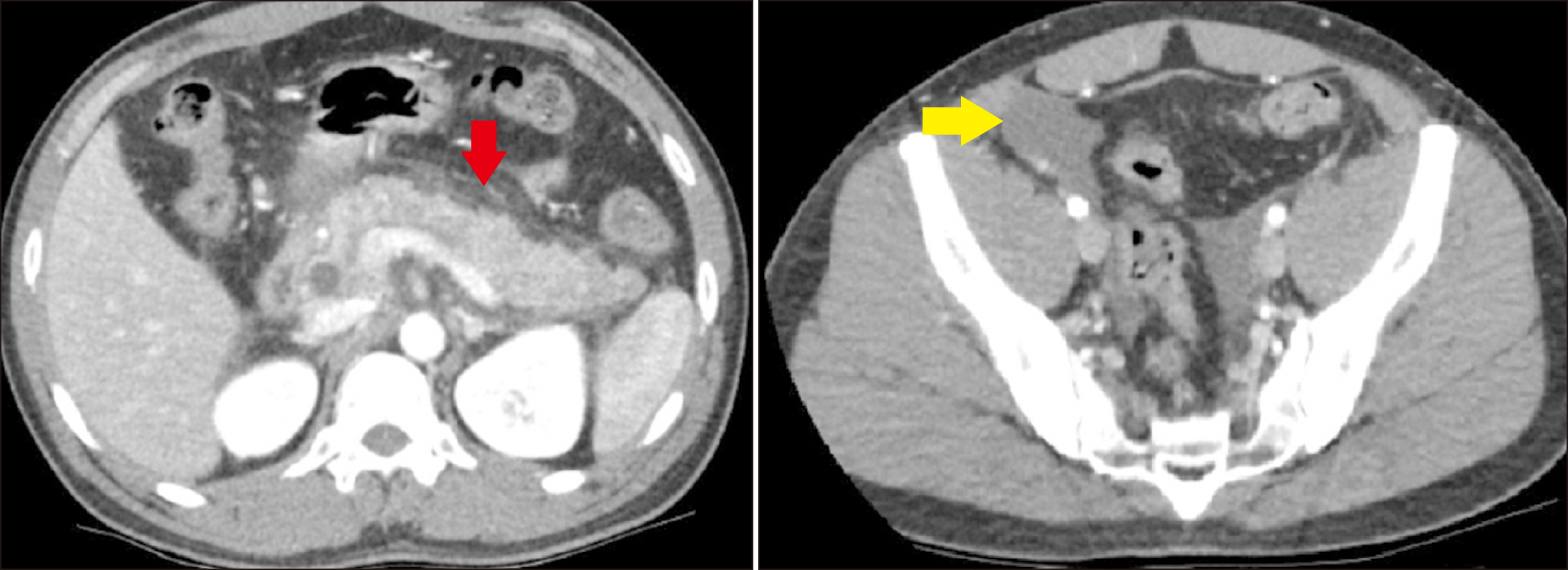
Fig. 2 Computed tomography (CT) of the abdomen showing pancreatitis with peripancreatic fat stranding (red arrow) with free fluid extending to the pelvis (yellow arrow).

Fig. 3 Magnetic Resonance Cholangiopancreatography (MRCP) image showing mild intrahepatic duct dilatation and a dilated common bile duct which measures 8 mm at the porta hepatis and 15 mm at the mid CBD with smooth distal tapering. The main pancreatic duct (hollow red arrow) is prominent at 3 mm, there is a Santorinicoele (solid red arrow) draining into minor papilla and an ansa pancreatica (yellow solid arrow) between the main pancreatic duct and Santorinicoele which is more prominent and dilated compared to Fig. 1 which was an MRCP done the month prior.
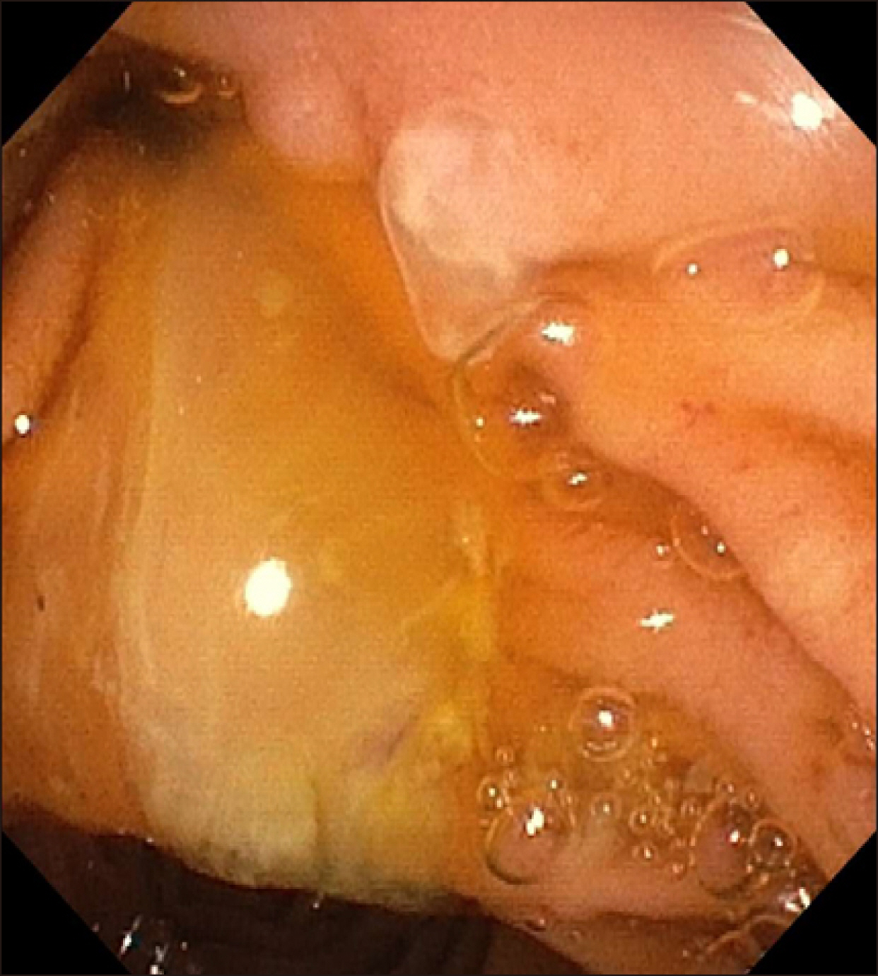
Fig. 4 Mucin seen exuding the minor papilla at endoscopy.
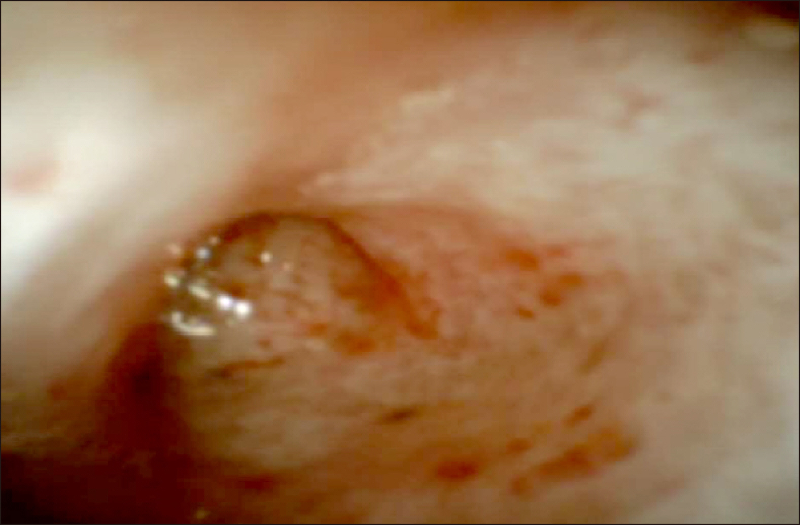
Fig. 5 Spyglass view of main pancreatic duct.
Reference
-
1. Bani-Hani KE. 2005; Agenesis of the gallbladder: difficulties in management. J Gastroenterol Hepatol. 20:671–675. DOI: 10.1111/j.1440-1746.2005.03740.x. PMID: 15853977.
Article2. Bedi N, Bond-Smith G, Kumar S, Hutchins R. 2013; Gallbladder agenesis with choledochal cyst--a rare association: a case report and review of possible genetic or embryological links. BMJ Case Rep. 2013:bcr2012006786. DOI: 10.1136/bcr-2012-006786. PMID: 23307450. PMCID: PMC3604316.
Article3. Yoldas O, Yazıcı P, Ozsan I, Karabuga T, Alpdogan O, Sahin E, et al. 2014; Coexistence of gallbladder agenesis and cholangiocarcinoma: report of a case. J Gastrointest Surg. 18:1373–1376. DOI: 10.1007/s11605-014-2472-x. PMID: 24519037.
Article4. Malde S. 2010; Gallbladder agenesis diagnosed intra-operatively: a case report. J Med Case Rep. 4:285. DOI: 10.1186/1752-1947-4-285. PMID: 20731843. PMCID: PMC2933638.
Article5. Rajkumar A, Piya A. 2017; Gall bladder agenesis: a rare embryonic cause of recurrent biliary colic. Am J Case Rep. 18:334–338. DOI: 10.12659/AJCR.903176. PMID: 28365715. PMCID: PMC5386428.
Article6. Kosmidis CS, Koimtzis GD, Kosmidou MS, Ieridou F, Koletsa T, Zarampouka KT, et al. 2017; Gallbladder hypoplasia, a congenital abnormality of the gallbladder: a case report. Am J Case Rep. 18:1320–1324. DOI: 10.12659/AJCR.905963. PMID: 29225328. PMCID: PMC5737095.
Article7. Bennion RS, Thompson JE Jr, Tompkins RK. 1988; Agenesis of the gallbladder without extrahepatic biliary atresia. Arch Surg. 123:1257–1260. DOI: 10.1001/archsurg.1988.01400340083014. PMID: 3052366.
Article8. Eisen G, Schutz S, Metzler D, Baillie J, Cotton PB. 1994; Santorinicele: new evidence for obstruction in pancreas divisum. Gastrointest Endosc. 40:73–76. DOI: 10.1016/S0016-5107(94)70015-X. PMID: 8163142.
Article9. Costamagna G, Ingrosso M, Tringali A, Mutignani M, Manfredi R. 2000; Santorinicele and recurrent acute pancreatitis in pancreas divisum: diagnosis with dynamic secretin-stimulated magnetic resonance pancreatography and endoscopic treatment. Gastrointest Endosc. 52:262–267. DOI: 10.1067/mge.2000.107711. PMID: 10922107.
Article10. Seibert DG, Matulis SR. 1995; Santorinicele as a cause of chronic pancreatic pain. Am J Gastroenterol. 90:121–123. PMID: 7801911.11. Khan SA, Chawla T, Azami R. 2009; Recurrent acute pancreatitis due to a santorinicele in a young patient. Singapore Med J. 50:e163–e165. PMID: 19495498.12. Tang H, Kay CL, Devonshire DA, Tagge E, Cotton PB. 1999; Recurrent pancreatitis in a child with pancreas divisum. Endoscopic therapy of a Santorinicele. Surg Endosc. 13:1040–1043. DOI: 10.1007/s004649901165. PMID: 10526045.13. Peterson MS, Slivka A. 2001; Santorinicele in pancreas divisum: diagnosis with secretin-stimulated magnetic resonance pancreatography. Abdom Imaging. 26:260–263. DOI: 10.1007/s002610000156. PMID: 11429949.
Article14. Manfredi R, Costamagna G, Brizi MG, Spina S, Maresca G, Vecchioli A, et al. 2000; Pancreas divisum and "santorinicele": diagnosis with dynamic MR cholangiopancreatography with secretin stimulation. Radiology. 217:403–408. DOI: 10.1148/radiology.217.2.r00nv29403. PMID: 11058635.
Article15. Joo KR, Bang SJ, Shin JW, Kim DH, Park NH. 2004; Santorinicele containing a pancreatic duct stone in a patient with incomplete pancreas divisum. Yonsei Med J. 45:952–955. DOI: 10.3349/ymj.2004.45.5.952. PMID: 15515212.
Article16. Byeon JS, Kim MH, Lee SK, Yang DH, Bae JS, Kim HJ, et al. 2003; Santorinicele without pancreas divisum. Gastrointest Endosc. 58:800–803. DOI: 10.1016/S0016-5107(03)01887-X. PMID: 14595329.
Article17. Gonoi W, Akai H, Hagiwara K, Akahane M, Hayashi N, Maeda E, et al. 2013; Santorinicele without pancreas divisum pathophysiology: initial clinical and radiographic investigations. BMC Gastroenterol. 13:62. DOI: 10.1186/1471-230X-13-62. PMID: 23570616. PMCID: PMC3637151.
Article18. Nam KD, Joo KR, Jang JY, Kim NH, Lee SK, Dong SH, et al. 2006; A case of santorinicele without pancreas divisum: diagnosis with multi-detector row computed tomography. J Korean Med Sci. 21:358–360. DOI: 10.3346/jkms.2006.21.2.358. PMID: 16614530. PMCID: PMC2734020.
Article
- Full Text Links
-
- Actions
-
Cited
- CITED
-
- Close
- Share
-
- Similar articles
-
- Ansa Pancreatica-Type Anatomic Variation of the Pancreatic Duct in Patients with Recurrent Acute Pancreatitis and Chronic Localized Pancreatitis
- Ansa Pancreatica: A Case Report of a Type of Ductal Variation in a Patient with Idiopathic Acute Recurrent Pancreatitis
- A Case of Branch Duct Intraductal Papillary Mucinous Neoplasm Mimicking Pseudocysts Complicated by Recurrent Pancreatitis
- Pathologic View of Intraductal Papillary Mucinous Neoplasm
- A Case of Acute Recurrent Pancreatitis Caused by Branch Duct Type IPMN and Ampulla of Vater Adenoma with High Grade Dysplasia
