Effects of the long-term use of maxillary protraction facemasks with skeletal anchorage on pharyngeal airway dimensions in growing patients with cleft lip and palate
- Affiliations
-
- 1Department of Dentistry, Graduate School, Kyung Hee University, Seoul, Korea
- 2Department of Dentistry, Graduate School, Seoul National University, Seoul, Korea
- 3Department of Oral and Maxillofacial Surgery, School of Dentistry, Seoul National University, Seoul, Korea
- 4Department of Plastic and Reconstructive Surgery, College of Medicine, Seoul National University, Seoul, Korea
- 5Department of Orthodontics, School of Dentistry, Kyung Hee University, Seoul, Korea
- 6Department of Orthodontics, School of Dentistry, Seoul National University, Seoul, Korea
- KMID: 2504141
- DOI: http://doi.org/10.4041/kjod.2020.50.4.238
Abstract
Objective
To investigate the effects of the long-term use of a maxillary protraction facemask with miniplate (FM-MP) on pharyngeal airway dimensions in growing patients with cleft lip and palate (CLP).
Methods
The study included 24 boys with CLP (mean age, 12.2 years; mean duration of FM-MP therapy, 4.9 years), divided into two groups according to the amount of A point advancement to the vertical reference plane (VRP): Group 1, > 4 mm; Group 2, < 2 mm; n = 12/group. After evaluating the skeletodental and airway variables using lateral cephalograms acquired before and after FM-MP therapy, statistical analyses were performed.
Results
Group 1 showed greater forward and downward displacements of the posterior maxilla (posterior nasal spine [PNS]-horizontal reference plane [HRP]; PNSVRP), greater increase in ANB, more forward tongue position (tongue tip-Pt vertical line to Frankfort horizontal plane), and greater increase in the oropharynx (superior posterior airway space [SPAS]; middle airway space [MAS]) and upper nasopharynx (PNS-adenoid2) than did Group 2. While maxillary advancement (A-VRP and PNS-VRP) correlated with increases in SPAS, MAS, and PNS-adenoid2, downward displacement of the PNS (PNS-HRP) correlated with increases in SPAS, MAS, PNSadenoid1, and PNS-adenoid2, and with a decrease in vertical airway length (VAL). Mandibular forward displacement and decrease in mandibular plane correlated with increases in MAS.
Conclusions
FM-MP therapy had positive effects on the oropharyngeal and nasopharyngeal airway spaces without increases in VAL in Group 1 rather than in Group 2. However, further validation using an untreated control group is necessary.
Keyword
Figure
-
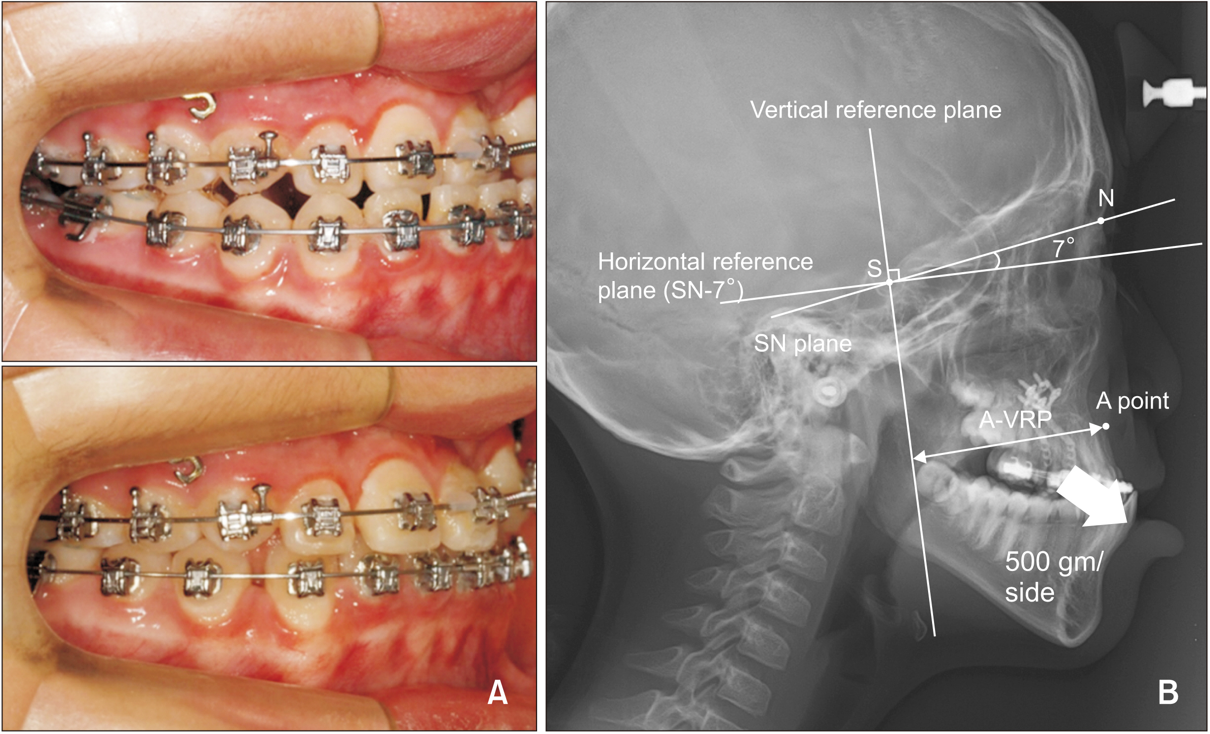
Figure 1 A, An example of facemask with miniplate (FM-MP) therapy. B, Reference lines. The horizontal reference plane (HRP) passes the Sella (S) at an angle of 7 degree clockwise to the Sella-Nasion (SN) plane. The vertical reference plane (VRP) is perpendicular to the HRP passing through the S. A-VRP is the horizontal distance from the VRP to A point.
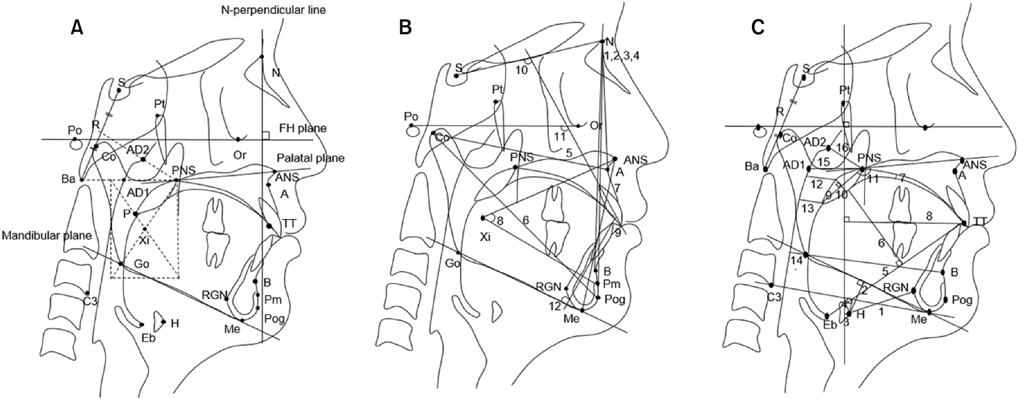
Figure 2 Cephalometric landmarks and measurements. A, Landmarks. S, Sella; N, nasion; Ba, basion; Or, orbitale; Po, porion; Pt, pterygoid point; R, midpoint between sella and basion; AD1, adenoid point 1 (adenoid tissue on the PNS-Ba line); AD2, adenoid point 2 (adenoid tissue on the R-PNS line); Co, condylion; Go, gonion; ANS, anterior nasal spine; PNS, posterior nasal spine; A, point A; B, point B; TT, tongue tip; Pog, pogonion; Me, menton; P, tip of the soft palate; Eb, base of the epiglottic fold; H, hyoidale (the most anterosuperior point on the body of the hyoid bone); RGN, retrognathion; Pm, protuberance menti; Xi, geometric center of the ramus; FH, Frankfort horizontal; C3, the third vertebrae. B, Skeletodental variables. 1, SNA; 2, SNB; 3, SN-Pog; 4, ANB; 5, effective maxillary length (EMxL); 6, effective mandibular length (EMnL); 7, ANS-Me; 8, lower facial height (ANS-Xi-Pm); 9, overjet; 10, U1-SN; 11, U1-FH; 12, L1-MP. C, Airway-related variables. hyoid bone 1, H-RGN; 2, MP-H; 3, H-PTV; 4, H-C3Me; tongue 5, TGL; 6, TGH; 7, Td-PP; 8, TT-PTV; soft palate 9, SPL; 10, SPT; 11, SPA; pharyngeal airway 12, SPAS; 13, MAS; 14, IAS; 15, PNS-AD1; 16, PNS-AD2. See Table 2 for definitions of each landmark or measurement.
Reference
-
1. Celikoglu M, Buyuk SK, Sekerci AE, Ucar FI, Cantekin K. 2014; Three-dimensional evaluation of the pharyngeal airway volumes in patients affected by unilateral cleft lip and palate. Am J Orthod Dentofacial Orthop. 145:780–6. DOI: 10.1016/j.ajodo.2014.02.008. PMID: 24880849.
Article2. Satoh K, Wada T, Tachimura T, Sakoda S, Shiba R. 1998; A cephalometric study by multivariate analysis of growth of the bony nasopharynx in patients with clefts and non-cleft controls. J Craniomaxillofac Surg. 26:394–9. DOI: 10.1016/S1010-5182(98)80074-7.
Article3. Jolleys A. 1954; A review of the results of operations on cleft palates with reference to maxillary growth and speech function. Br J Plast Surg. 7:229–41. DOI: 10.1016/S0007-1226(54)80027-0.
Article4. Shibasaki Y, Ross RB. 1969; Facial growth in children with isolated cleft palate. Cleft Palate J. 6:290–302. PMID: 5261874.5. Hermann NV, Jensen BL, Dahl E, Bolund S, Kreiborg S. 1999; A comparison of the craniofacial morphology in 2-month-old unoperated infants with unilateral complete cleft lip and palate, and unilateral incomplete cleft lip. J Craniofac Genet Dev Biol. 19:80–93.6. Karia H, Shrivastav S, Karia AK. 2017; Three-dimensional evaluation of the airway spaces in patients with and without cleft lip and palate: a digital volume tomographic study. Am J Orthod Dentofacial Orthop. 152:371–81. DOI: 10.1016/j.ajodo.2016.12.026. PMID: 28863918.
Article7. Akarsu-Guven B, Karakaya J, Ozgur F, Aksu M. 2015; Growth-related changes of skeletal and upper-airway features in bilateral cleft lip and palate patients. Am J Orthod Dentofacial Orthop. 148:576–86. DOI: 10.1016/j.ajodo.2015.02.031. PMID: 26432313.
Article8. Kunkel M, Wahlmann U, Wagner W. 1997; Nasal airway in cleft-palate patients: acoustic rhinometric data. J Craniomaxillofac Surg. 25:270–4. DOI: 10.1016/S1010-5182(97)80065-0. PMID: 9368863.
Article9. Muntz H, Wilson M, Park A, Smith M, Grimmer JF. 2008; Sleep disordered breathing and obstructive sleep apnea in the cleft population. Laryngoscope. 118:348–53. DOI: 10.1097/MLG.0b013e318158195e. PMID: 18025949.
Article10. Robison JG, Otteson TD. 2011; Increased prevalence of obstructive sleep apnea in patients with cleft palate. Arch Otolaryngol Head Neck Surg. 137:269–74. DOI: 10.1001/archoto.2011.8. PMID: 21422312.
Article11. Cordasco G, Matarese G, Rustico L, Fastuca S, Caprioglio A, Lindauer SJ, et al. 2014; Efficacy of orthopedic treatment with protraction facemask on skeletal Class III malocclusion: a systematic review and meta-analysis. Orthod Craniofac Res. 17:133–43. DOI: 10.1111/ocr.12040. PMID: 24725349.
Article12. Baccetti T, McGill JS, Franchi L, McNamara JA Jr, Tollaro I. 1998; Skeletal effects of early treatment of Class III malocclusion with maxillary expansion and face-mask therapy. Am J Orthod Dentofacial Orthop. 113:333–43. DOI: 10.1016/S0889-5406(98)70306-3. PMID: 9517727.
Article13. Kaygisiz E, Tuncer BB, Yüksel S, Tuncer C, Yildiz C. 2009; Effects of maxillary protraction and fixed appliance therapy on the pharyngeal airway. Angle Orthod. 79:660–7. DOI: 10.2319/072408-391.1. PMID: 19537871.14. Oktay H, Ulukaya E. 2008; Maxillary protraction appliance effect on the size of the upper airway passage. Angle Orthod. 78:209–14. DOI: 10.2319/122806-535.1. PMID: 18251620.
Article15. Sayinsu K, Isik F, Arun T. 2006; Sagittal airway dimensions following maxillary protraction: a pilot study. Eur J Orthod. 28:184–9. DOI: 10.1093/ejo/cji095. PMID: 16464873.16. Cevidanes L, Baccetti T, Franchi L, McNamara JA Jr, De Clerck H. 2010; Comparison of two protocols for maxillary protraction: bone anchors versus face mask with rapid maxillary expansion. Angle Orthod. 80:799–806. DOI: 10.2319/111709-651.1. PMID: 20578848. PMCID: PMC2930261.
Article17. Baek SH, Kim KW, Choi JY. 2010; New treatment modality for maxillary hypoplasia in cleft patients. Protraction facemask with miniplate anchorage. Angle Orthod. 80:783–91. DOI: 10.2319/073009-435.1. PMID: 20482368.18. Ahn HW, Kim KW, Yang IH, Choi JY, Baek SH. 2012; Comparison of the effects of maxillary protraction using facemask and miniplate anchorage between unilateral and bilateral cleft lip and palate patients. Angle Orthod. 82:935–41. DOI: 10.2319/010112-1.1. PMID: 22380632.
Article19. On SW, Baek SH, Choi JY. 2018; Effect of long-term use of facemask with miniplate on maxillary protraction in patients with cleft lip and palate. J Craniofac Surg. 29:309–14. DOI: 10.1097/SCS.0000000000004122. PMID: 29135737.
Article20. Fishman LS. 1982; Radiographic evaluation of skeletal maturation. A clinically oriented method based on hand-wrist films. Angle Orthod. 52:88–112. DOI: 10.1043/0003-3219(1982)052<0088:REOSM>2.0.CO;2. PMID: 6980608.21. Zhang Y, Jia H, Fu Z, Huang Y, Wang Z, Guo R, et al. 2018; Dentoskeletal effects of facemask therapy in skeletal Class III cleft patients with or without bone graft. Am J Orthod Dentofacial Orthop. 153:542–9. DOI: 10.1016/j.ajodo.2017.07.024. PMID: 29602346.
Article22. Li HY, Chen NH, Wang CR, Shu YH, Wang PC. 2003; Use of 3-dimensional computed tomography scan to evaluate upper airway patency for patients undergoing sleep-disordered breathing surgery. Otolaryngol Head Neck Surg. 129:336–42. DOI: 10.1016/S0194-5998(03)00629-6. PMID: 14574286.
Article23. Ngan P, Wilmes B, Drescher D, Martin C, Weaver B, Gunel E. 2015; Comparison of two maxillary protraction protocols: tooth-borne versus bone-anchored protraction facemask treatment. Prog Orthod. 16:26. DOI: 10.1186/s40510-015-0096-7. PMID: 26303311. PMCID: PMC4547969.
Article24. Ozbek MM, Memikoglu TU, Gögen H, Lowe AA, Baspinar E. 1998; Oropharyngeal airway dimensions and functional-orthopedic treatment in skeletal Class II cases. Angle Orthod. 68:327–36. DOI: 10.1043/0003-3219(1998)068<0327:OADAFO>2.3.CO;2. PMID: 9709833.25. Akcam MO, Toygar TU, Wada T. 2002; Longitudinal investigation of soft palate and nasopharyngeal airway relations in different rotation types. Angle Orthod. 72:521–6. DOI: 10.1043/0003-3219(2002)072<0521:LIOSPA>2.0.CO;2. PMID: 12518943.26. van den Dungen GM, Ongkosuwito EM, Aartman IH, Prahl-Andersen B. 2008; Craniofacial morphology of Dutch patients with bilateral cleft lip and palate and noncleft controls at the age of 15 years. Cleft Palate Craniofac J. 45:661–6. DOI: 10.1597/07-166.1. PMID: 18956940.
Article27. Lee JW, Park KH, Kim SH, Park YG, Kim SJ. 2011; Correlation between skeletal changes by maxillary protraction and upper airway dimensions. Angle Orthod. 81:426–32. DOI: 10.2319/082610-499.1. PMID: 21299388.
Article28. Xu Y, Zhao S, Shi J, Wang Y, Shi B, Zheng Q, et al. 2013; 3-dimensional computed tomographic analysis of the pharynx in adult patients with unrepaired isolated cleft palate. J Oral Maxillofac Surg. 71:1424–34. DOI: 10.1016/j.joms.2013.01.022. PMID: 23541993.
Article29. McNamara JA Jr. 1987; An orthopedic approach to the treatment of Class III malocclusion in young patients. J Clin Orthod. 21:598–608.30. Kapust AJ Jr, Sinclair PM, Turley PK. 1998; Cephalometric effects of face mask/expansion therapy in Class III children: a comparison of three age groups. Am J Orthod Dentofacial Orthop. 113:204–12. DOI: 10.1016/S0889-5406(98)70141-6. PMID: 9484212.
Article
- Full Text Links
-
- Actions
-
Cited
- CITED
-
- Close
- Share
-
- Similar articles
-
- Does surgically assisted maxillary protraction with skeletal anchorage and Class III elastics affect the pharyngeal airway? A retrospective, long-term study
- Comparison of the effects on the pharyngeal airway space of maxillary protraction appliances according to the methods of anchorage
- Maxillary protraction treatment of skeletal Class III children using miniplate anchorage
- Long-term follow-up of early cleft maxillary distraction
- A study on the maxillary dental arch and palate of unilateral cleft lip and palate individuals
