Choroidal fissure and choroidal fissure cysts: a comprehensive review
- Affiliations
-
- 1Department of Neurosurgery, Hospital Santo Tomas, Panama City, Panama, USA
- 2Department of Anatomical Sciences, St. George’s University, St. George’s, Grenada, USA
- 3Aiken Neurosciences, Aiken Regional Medical Centers, Aiken, SC, USA
- 4Division of Gross and Clinical Anatomy, Department of Anatomy, Kurume,University School of Medicine, Kurume, Japan
- 5Department of Neurosurgery, Tulane Center for Clinical Neurosciences, Tulane University School of Medicine, New Orleans, LA, USA
- 6Department of Structural & Cellular Biology, Tulane University School of Medicine, New Orleans, LA, USA
- KMID: 2503440
- DOI: http://doi.org/10.5115/acb.20.040
Abstract
- In this paper, the authors discuss the embryology and anatomy of the choroidal fissure, as well as the pathophysiology and treatment of cerebrospinal fluid cysts of this structure. Understanding its anatomical relations to nearby structures plays an essential role during brain surgeries. With the advancement and availability of imaging techniques, lesions of the choroidal fissure are often found incidentally. Patients are usually asymptomatic or exhibit symptoms that do not correlate with anatomical location or do not require surgical treatment. The choroidal fissure is a key landmark used during brain surgery. Therefore, a comprehensive understanding of it and nearby anatomical structures is essential. Choroidal fissure cysts can be found incidentally, and well-known key features will allow one to differentiate them from other lesions. Surgical treatment should be reserved for symptomatic patients while asymptomatic patients should be monitored.
Figure
-
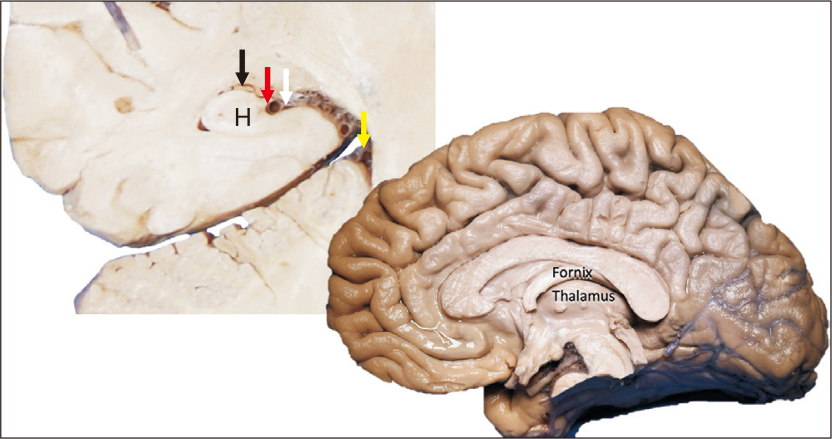
Fig. 1 Coronal brain slice (left) noting the ambient cistern (yellow arrow), the choroidal fissure (white arrow), fimbria (red arrow), hippocampus (H) and choroid plexus in the temporal horn (black arrow); The right sagittal image of the brain notes the relationship between the fornix and thalamus from a medial view.

Fig. 2 Schematic drawing of the developing neural tube. As the telencephalic vesicles form from the prosencephalon, note the groove on the dorsal tube at the base of the vesicles, which is the primitive choroidal fissure. Observe that the choroidal vessels are entering and leaving the ventricle via this fissure. ACA, anterior cerebral artery; ICA, internal carotid artery; PCom, posterior communicating artery.
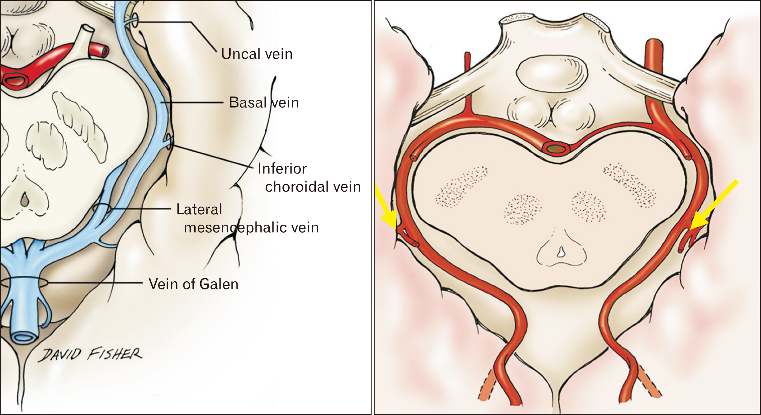
Fig. 3 Axial views of the inferior choroidal vein (left) and posterior lateral choroidal arteries (yellow arrows; right) at the choroidal fissure.
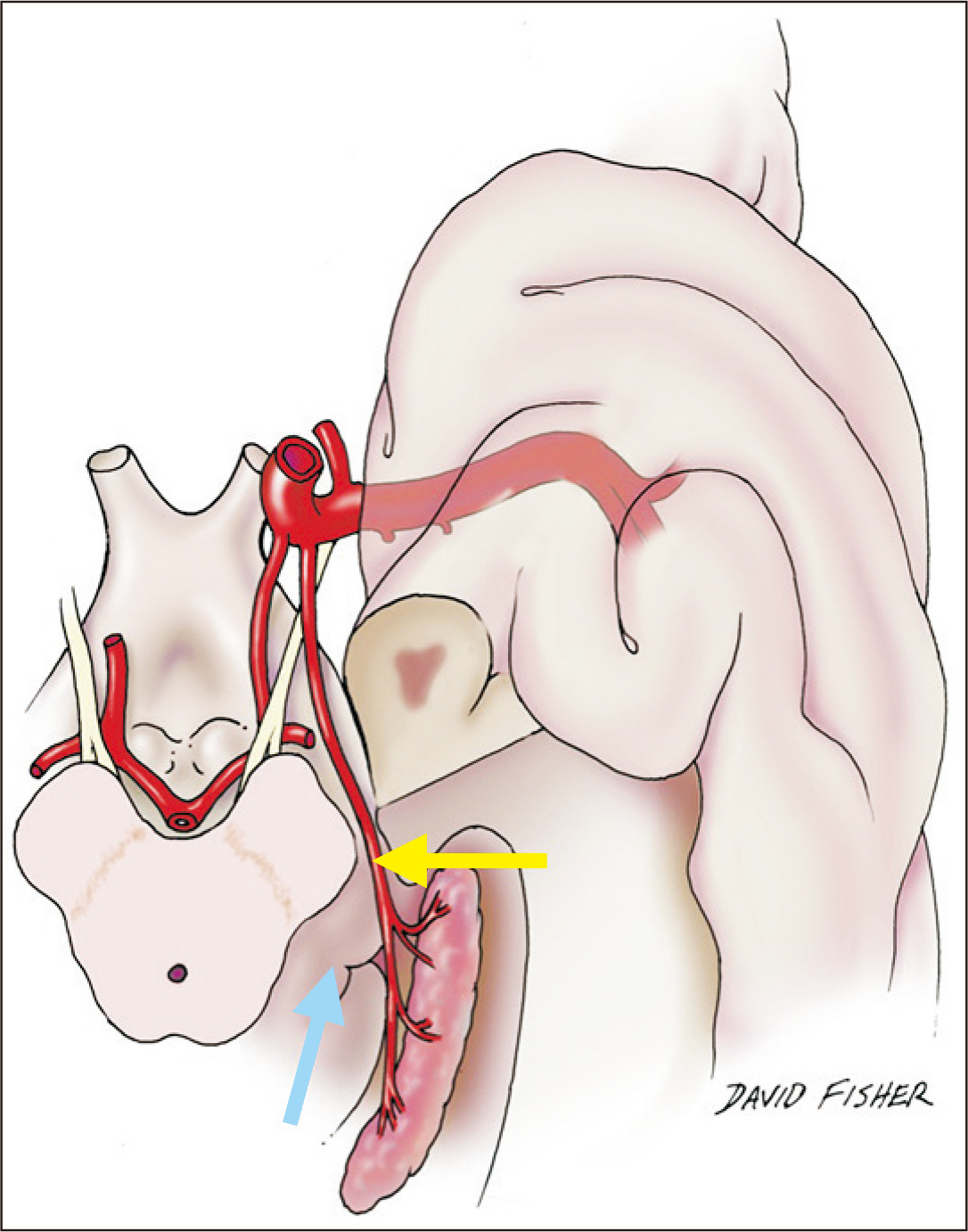
Fig. 4 Schematic drawing of a basal view of the brain (left) noting the choroidal fissure at the point of entrance (inferior choroidal point) of the anterior choroidal artery (yellow arrow) and for reference, the thalamus (blue arrow).
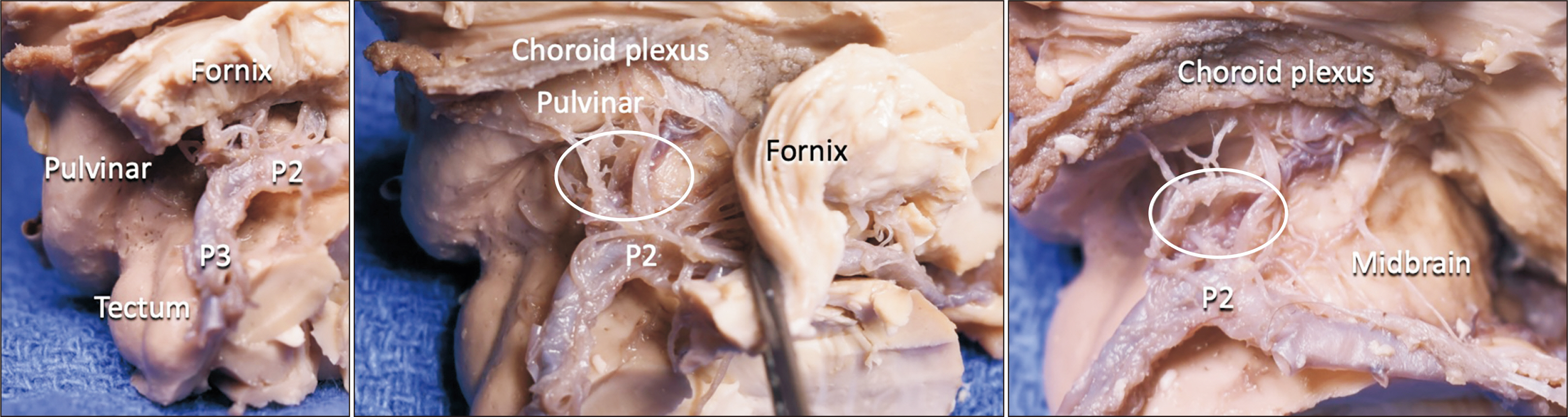
Fig. 5 Sequential dissection of the brain noting the relationship between the pulvinar and choroid plexus and posterior lateral choroidal arteries (white circle). Note that there are usually multiple (average 4) posterior lateral choroidal arteries as seen in this specimen. The P2 and P3 parts of the posterior cerebral artery are labeled.

Fig. 6 Magnetic resonance imaging noting large right and left choroidal fissure cysts.
Reference
-
1. de Jong L, Thewissen L, van Loon J, Van Calenbergh F. 2011; Choroidal fissure cerebrospinal fluid-containing cysts: case series, anatomical consideration, and review of the literature. World Neurosurg. 75:704–8. DOI: 10.1016/j.wneu.2010.12.056. PMID: 21704940.
Article2. Zemmoura I, Velut S, François P. 2012; The choroidal fissure: anatomy and surgical implications. Adv Tech Stand Neurosurg. 38:97–113. DOI: 10.1007/978-3-7091-0676-1_5. PMID: 22592413.
Article3. Yamada S, Ishikawa M, Iwamuro Y, Yamamoto K. 2016; Choroidal fissure acts as an overflow device in cerebrospinal fluid drainage: morphological comparison between idiopathic and secondary normal-pressure hydrocephalus. Sci Rep. 6:39070. DOI: 10.1038/srep39070. PMID: 27941913. PMCID: PMC5150242.
Article4. da Costa MDS, Santos BFO, Bouchabki de Almeida Guardini F, Chaddad-Neto F. 2017; Microsurgical treatment for arteriovenous malformation of the corpus callosum and choroidal fissure. Neurosurg Focus. 43(VideoSuppl1):V12. DOI: 10.3171/2017.7.FocusVid.1733. PMID: 28669263.
Article5. Jean WC. 2018; Transcallosal, transchoroidal resection of a recurrent craniopharyngioma. J Neurol Surg B Skull Base. 79(Suppl 3):S259–60. DOI: 10.1055/s-0038-1624585. PMID: 29588891. PMCID: PMC5868919.
Article6. Isolan GR, Oliveira Ed, Recalde R. 2005; [Microanatomical study of the choroidal fissure in ventricular and cisternal approaches]. Arq Neuropsiquiatr. 63:801–6. Portuguese. DOI: 10.1590/S0004-282X2005000500015. PMID: 16258659.7. Wen HT, Rhoton AL Jr, de Oliveira E. 1998; Transchoroidal approach to the third ventricle: an anatomic study of the choroidal fissure and its clinical application. Neurosurgery. 42:1205–17. discussion 1217–9. DOI: 10.1097/00006123-199806000-00001. PMID: 9632178.
Article8. Siwanuwatn R, Deshmukh P, Zabramski JM, Preul MC, Spetzler RF. 2005; Microsurgical anatomy and quantitative analysis of the transtemporal-transchoroidal fissure approach to the ambient cistern. Neurosurgery. 57(4 Suppl):228–35. discussion 228–35. DOI: 10.1227/01.NEU.0000176407.35946.88. PMID: 16234669.
Article9. Wu A, Chang SW, Deshmukh P, Spetzler RF, Preul MC. 2010; Through the choroidal fissure: a quantitative anatomic comparison of 2 incisions and trajectories (transsylvian transchoroidal and lateral transtemporal). Neurosurgery. 66(6 Suppl Operative):221–8. discussion 228–9. DOI: 10.1227/01.NEU.0000369920.68166.6C. PMID: 20489509.10. Ikeda K, Shoin K, Mohri M, Kijima T, Someya S, Yamashita J. 2002; Surgical indications and microsurgical anatomy of the transchoroidal fissure approach for lesions in and around the ambient cistern. Neurosurgery. 50:1114–9. discussion 1120DOI: 10.1227/00006123-200205000-00030. PMID: 11950415.
Article11. Nagata S, Rhoton AL Jr, Barry M. 1988; Microsurgical anatomy of the choroidal fissure. Surg Neurol. 30:3–59. DOI: 10.1016/0090-3019(88)90180-2.
Article12. Catala M, Poirier J. 1998; [Arachnoid cysts: histologic, embryologic and physiopathologic review]. Rev Neurol (Paris). 154:489–501. French. PMID: 9773082.13. Maher CO, Piatt JH Jr. 2015; Incidental findings on brain and spine imaging in children. Pediatrics. 135:e1084–96. DOI: 10.1542/peds.2015-0071. PMID: 25825535.
Article14. Tubbs RS, Muhleman M, McClugage SG, Loukas M, Miller JH, Chern JJ, Rozzelle CJ, Oakes WJ, Cohen-Gadol AA. 2012; Progressive symptomatic increase in the size of choroidal fissure cysts. J Neurosurg Pediatr. 10:306–9. DOI: 10.3171/2012.7.PEDS11515. PMID: 22900488.
Article15. Morioka T, Nishio S, Suzuki S, Fukui M, Nishiyama T. 1994; Choroidal fissure cyst in the temporal horn associated with complex partial seizure. Clin Neurol Neurosurg. 96:164–7. DOI: 10.1016/0303-8467(94)90054-X.
Article16. Morioka T, Nishio S, Ishibashi H, Fukui M. 1998; Relationship between arachnoid cysts and seizure foci. Epilepsia. 39:804–5. DOI: 10.1111/j.1528-1157.1998.tb01168.x. PMID: 9670911.17. Chitkara R, Rajani A, Bernstein J, Shah S, Hahn JS, Barnes P, Hintz SR. 2011; Newborn with prenatally diagnosed choroidal fissure cyst and panhypopituitarism and review of the literature. AJP Rep. 1:111–4. DOI: 10.1055/s-0031-1293512. PMID: 23705098. PMCID: PMC3653523.
Article18. Tas E, Tracy M, Sarco DP, Eksioglu YZ, Prabhu SP, Loddenkemper T. 2011; Septo-optic dysplasia complicated by infantile spasms and bilateral choroidal fissure arachnoid cysts. J Neuroimaging. 21:89–91. DOI: 10.1111/j.1552-6569.2009.00453.x. PMID: 20002969.
Article19. Sherman JL, Camponovo E, Citrin CM. 1990; MR imaging of CSF-like choroidal fissure and parenchymal cysts of the brain. AJR Am J Roentgenol. 155:1069–75. DOI: 10.2214/ajr.155.5.2120937. PMID: 2120937.
Article20. Reimão S, Sousa R, Morgado C. 2006; [Intraparenchymal neuroepithelial cysts--imaging findings in four clinical cases]. Acta Med Port. 19:509–12. Portuguese.21. Osborn AG, Preece MT. 2006; Intracranial cysts: radiologic-pathologic correlation and imaging approach. Radiology. 239:650–64. DOI: 10.1148/radiol.2393050823. PMID: 16714456.
Article22. Aprile I, Iaiza F, Lavaroni A, Budai R, Dolso P, Scott CA, Beltrami CA, Fabris G. 1999; Analysis of cystic intracranial lesions performed with fluid-attenuated inversion recovery MR imaging. AJNR Am J Neuroradiol. 20:1259–67. PMID: 10472983.23. Kjos BO, Brant-Zawadzki M, Kucharczyk W, Kelly WM, Norman D, Newton TH. 1985; Cystic intracranial lesions: magnetic resonance imaging. Radiology. 155:363–9. DOI: 10.1148/radiology.155.2.3983386. PMID: 3983386.
Article24. Park SW, Yoon SH, Cho KH, Shin YS. 2006; A large arachnoid cyst of the lateral ventricle extending from the supracerebellar cistern--case report. Surg Neurol. 65:611–4. DOI: 10.1016/j.surneu.2005.07.069. PMID: 16720186.
Article25. Becker T, Wagner M, Hofmann E, Warmuth-Metz M, Nadjmi M. 1991; Do arachnoid cysts grow? A retrospective CT volumetric study. Neuroradiology. 33:341–5. DOI: 10.1007/BF00587820. PMID: 1922751.26. Eymann R, Kiefer M. 2018; [Relevance and therapy of intracranial arachnoidal cysts]. Radiologe. 58:135–41. German. DOI: 10.1007/s00117-017-0340-x. PMID: 29255958.27. Parrent AG. 2000; Endoscopically guided fenestration of the choroidal fissure for treatment of trapped temporal horn. J Neurosurg. 93:891–4. DOI: 10.3171/jns.2000.93.5.0891. PMID: 11059675.
Article
