Echogenic Mass Lesion within the Main Pulmonary Artery in a Neonate
- Affiliations
-
- 1Department of Pediatrics, Samsung Medical Center, Sungkyunkwan University School of Medicine, Seoul, Korea
- 2Department of Thoracic and Cardiovascular Surgery, Samsung Medical Center, Sungkyunkwan University School of Medicine, Seoul, Korea
- KMID: 2503164
- DOI: http://doi.org/10.5385/nm.2020.27.2.89
Abstract
- Here we present a rare case of pulmonary arterial thrombosis associated with a ductus arteriosus aneurysm that caused severe pulmonary stenosis. A 5-day-old newborn was admitted to our hospital for the evaluation of an intracardiac mass-like lesion found after the detection of a cardiac murmur. Echocardiography and heart computed tomography revealed a mass-like lesion measuring 8.1 mm in diameter across the distal main pulmonary artery to the proximal left pulmonary artery resulting in localized severe stenosis of the left pulmonary artery. Left pulmonary artery angioplasty for surgical resection of the thrombus revealed that the mass was adherent to the proximal part of the left pulmonary artery anterior wall and extended to the ductus arteriosus. Histological examination of the mass showed an old thrombus with dystrophic calcification. Five months after surgery, follow-up echocardiography showed that the left pulmonary artery peak pressure gradient had decreased but the proximal left pulmonary artery stenosis remained. Cardiac catheterization and balloon angioplasty suc cessfully relieved the pulmonary stenosis.
Keyword
Figure
-
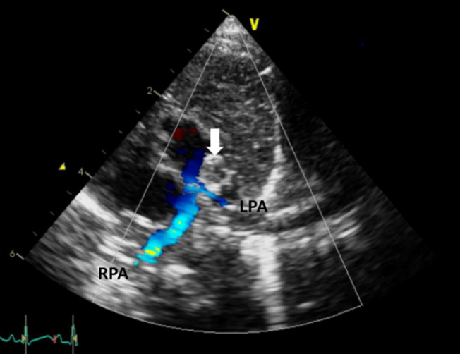
Figure 1. The parasternal short-axis view on transthoracic echocardiography revealed an oval-shaped mass (arrow) protruding from the lumen of the distal main pulmonary artery (MPA) to the proximal left pulmonary artery (LPA) causing LPA stenosis. Abbreviation: RPA, right pulmonary artery.

Figure 2. Left pulmonary artery flow velocity estimated by Doppler echocardiography revealed an increased left pulmonary artery pressure.
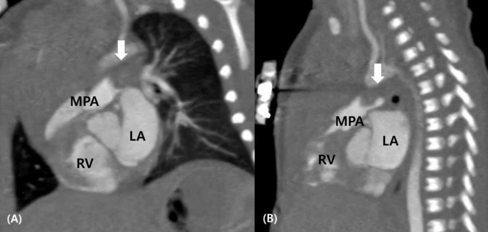
Figure 3. Sagittal (A) and left pulmonary artery (B) views of cardiac computed tomography showing the mass (arrows) in the lumen of the main pulmonary artery. Abbreviations: LA, left atrium; MPA, main pulmonary artery; RV, right ventricle.
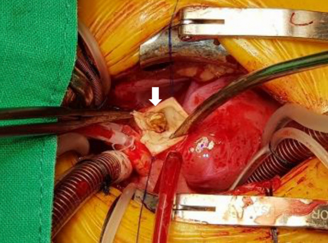
Figure 4. Intraoperative image showing that the mass (arrow) was tightly attached to the left pulmonary artery anterior wall just below the main pulmonary artery bifurcation. It seemed to extend from the main pulmonary artery bifurcation to the patent ductus arteriosus.

Figure 5. Histologic examination of the tissue removed from the left pulmonary artery showing an old thrombus (arrows) with dystrophic calcifications (arrowheads). (A) H&E, ×100; (B) H&E, ×200.
Reference
-
1. van Schendel MP, Visser DH, Rammeloo LA, Hazekamp MG, Hruda J. Left pulmonary artery thrombosis in a neonate with left lung hypoplasia. Case Rep Pediatr. 2012; 2012:314256.2. Sawyer T, Antle A, Studer M, Thompson M, Perry S, Mahnke CB. Neonatal pulmonary artery thrombosis presenting as persistent pulmonary hypertension of the newborn. Pediatr Cardiol. 2009; 30:520–2.3. Dijk FN, Curtin J, Lord D, Fitzgerald DA. Pulmonary embolism in children. Paediatr Respir Rev. 2012; 13:112–22.4. McArdle DJ, Paterson FL, Morris LL. Ductus arteriosus aneurysm thrombosis with mass effect causing pulmonary hypertension in the first week of life. J Pediatr. 2017; 180:289.5. Hornberger LK. Congenital ductus arteriosus aneurysm. J Am Coll Cardiol. 2002; 39:348–50.6. Jan SL, Hwang B, Fu YC, Chai JW, Chi CS. Isolated neonatal ductus arteriosus aneurysm. J Am Coll Cardiol. 2002; 39:342–7.7. Lund JT, Jensen MB, Hjelms E. Aneurysm of the ductus arteriosus. A review of the literature and the surgical implications. Eur J Cardiothorac Surg. 1991; 5:566–70.8. Dyamenahalli U, Smallhorn JF, Geva T, Fouron JC, Cairns P, Jutras L, et al. Isolated ductus arteriosus aneurysm in the fetus and infant: a multi-institutional experience. J Am Coll Cardiol. 2000; 36:262–9.9. Deeg KH, Ruffer A. Thrombus formation and arterial dissection of the wall of the left pulmonary artery caused by an aneurysm of the ductus arteriosus. Ultraschall Med. 2019; 40:85–6.10. Doreswamy SM, Twiss J, Predescu D, Shivananda SKP. Extending thrombus within the PDA in an infant with tetralogy of Fallot and pulmonary atresia: an averted disaster. J Clin Neonatol. 2015; 4:42–5.11. Sattar P, Ehrensperger J, Ducommun JC. Thrombosed aneurysmal nonpatent ductus arteriosus: a case report. Pediatr Radiol. 1996; 26:207–9.
- Full Text Links
-
- Actions
-
Cited
- CITED
-
- Close
- Share
-
- Similar articles
-
- Main Pulmonary Artery Stenosis Caused by Fibrocalcified Mass in a Young Infant
- RVOTO Caused by Pulmonary Artery Sarcoma Originating from Pulmonary Valve: One case report
- Contrast Echo-A Simple Diagnostic Tool for a Coronary Artery Fistula
- Congenital Giant Aneurysm of Pulmonary Artery-Associated with Ventricular Septal Defect and Pulmonary Stenosis : A Case Report
- Anomalous origin of the left coronary artery from the pulmonary artery
