Operative Treatment for Osteochondral Lesions of the Talus: Bone Marrow Aspirate Concentrate and Matrix-induced Chondrogenesis
- Affiliations
-
- 1Department of Orthopaedic Surgery, Inha University College of Medicine, Korea
- 2Department of Orthopaedic Surgery, Inha University Hospital, Incheon, Korea
- KMID: 2502911
- DOI: http://doi.org/10.14193/jkfas.2020.24.2.61
Abstract
- Bone marrow aspirate concentrate and matrix-induced chondrogenesis (BMIC) is an interesting treatment option for osteochondral lesions of the talus with promising short- to mid-term results. The various terminologies used to describe this surgical method need to be addressed. These include bone marrow-derived cell transplantation, matrix-induced bone marrow aspirate concentrate, and matrixassociated stem cell transplantation. BMIC is a one-stage, minimally invasive surgery performed arthroscopically or using a mini-open arthrotomy approach without a malleolar osteotomy in most cases. The lesion is replaced with hyaline-like cartilage, and treatmentrelated complications are rare. BMIC is a safe and effective treatment option and should be considered in large lesions or lesions with a prior treatment history.
Figure
-
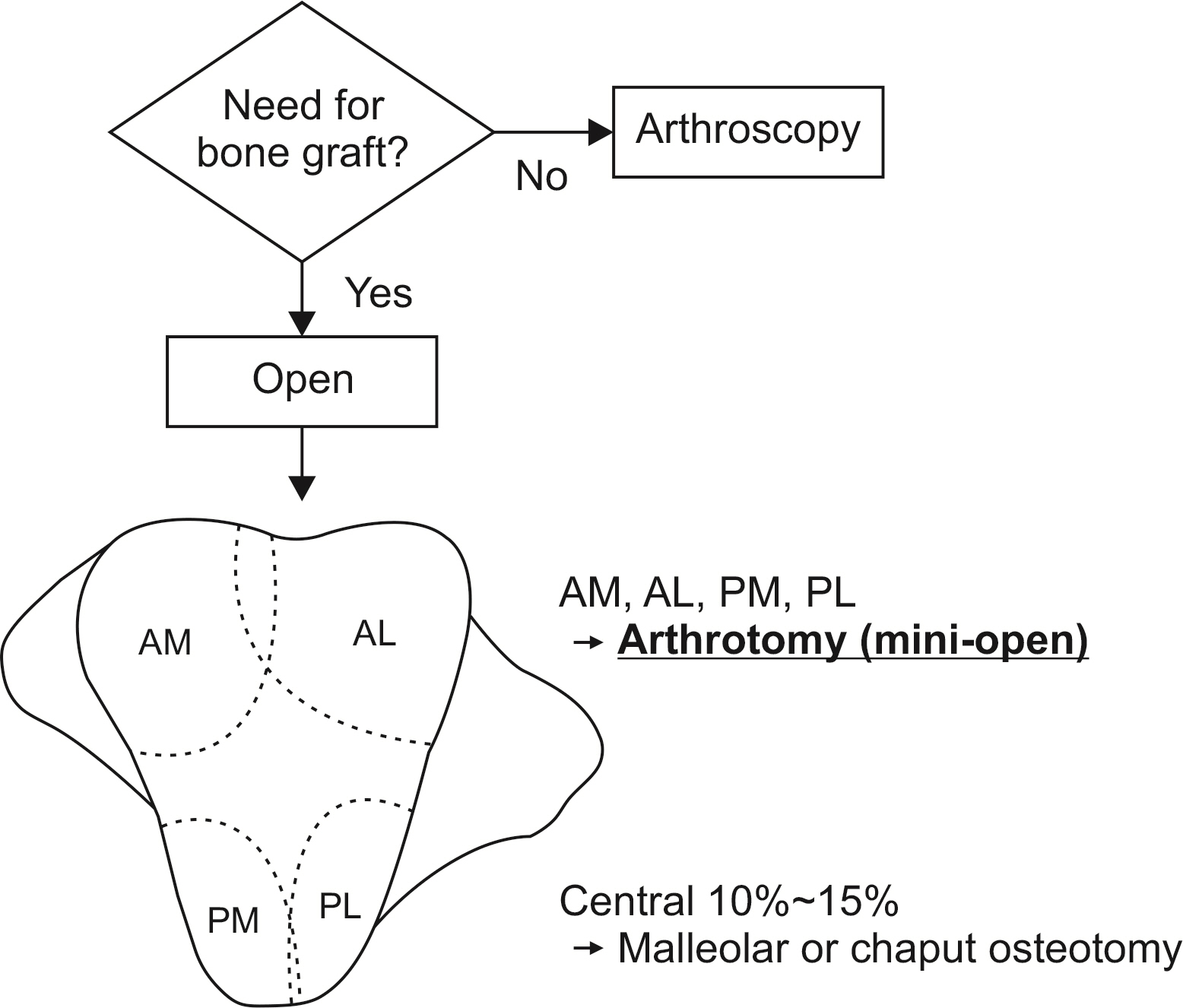
Figure. 1 Suggested algorithm for the selection of surgical approach to treat osteochondral lesions of the talus. The surgical approach should be determined based on the necessity of bone graft and the location of the lesion. AM: anteromedial, AL: anterolateral, PM: posteromedial, PL: posterolateral.

Figure. 2 Example of a bone marrow aspirate concentrate and matrix-induced chondrogenesis (BMIC) surgery performed through a posteromedial arthrotomy approach (A) without malleolar osteotomy. The lesion is clearly debrided and multiple holes have been made using a 1.2 mm Kirschner wire (B). (C) The subchondral bone defect has been filled with autologous iliac cancellous bone. (D) The lesion is covered with a matrix soaked with bone marrow aspirate concentrate.

Figure. 3 Preparation of the matrix. After removal of the damaged cartilage (top), an aluminum template is designed to fit the lesion, in about 10% smaller in size (middle). The matrix is trimmed in accordance with the template (bottom). (A) Hyalofast (Anika Therapeutics S.r.l, Padova, Italy), (B) Chondro-gide (Geistlich Pharma AG, Wolhusen, Switzerland).
Cited by 2 articles
-
Supramalleolar Osteotomy Combined with Redo Arthroscopy for a Patient with Persistent Pain after Primary Arthroscopic Microfracture for Medial Osteochondral Lesion of the Talus: A Case Report
Tae Hun Song, Jin Soo Suh, Jun Young Choi
J Korean Foot Ankle Soc. 2023;27(2):71-74. doi: 10.14193/jkfas.2023.27.2.71.Bony Union of Osteochondral Lesion of the Talus after Bone Marrow Aspirate Concentrate and Matrix-Induced Chondrogenesis: A Case Report
Tae Hun Song, Jin Soo Suh, Jun Young Choi
J Korean Foot Ankle Soc. 2023;27(4):148-153. doi: 10.14193/jkfas.2023.27.4.148.
Reference
-
1. Richter M, Zech S, Andreas Meissner S. 2017; Matrix-associated stem cell transplantation (MAST) in chondral defects of the 1st metatarsophalangeal joint is safe and effective-2-year-follow-up in 20 patients. Foot Ankle Surg. 23:195–200. doi: 10.1016/j.fas.2016.05.318. DOI: 10.1016/j.fas.2016.05.318. PMID: 28865590.
Article2. Giannini S, Buda R, Battaglia M, Cavallo M, Ruffilli A, Ramponi L, et al. 2013; One-step repair in talar osteochondral lesions: 4-year clinical results and t2-mapping capability in outcome prediction. Am J Sports Med. 41:511–8. doi: 10.1177/0363546512467622. DOI: 10.1177/0363546512467622. PMID: 23221772.3. Yontar NS, Aslan L, Can A, Ogut T. 2019; One step treatment of talus osteochondral lesions with microfracture and cell free hyaluronic acid based scaffold combination. Acta Orthop Traumatol Turc. 53:372–5. doi: 10.1016/j.aott.2019.04.002. DOI: 10.1016/j.aott.2019.04.002. PMID: 31126702. PMCID: PMC6819796.
Article4. Weigelt L, Hartmann R, Pfirrmann C, Espinosa N, Wirth SH. 2019; Autologous matrix-induced chondrogenesis for osteochondral lesions of the talus: a clinical and radiological 2- to 8-year follow-up study. Am J Sports Med. 47:1679–86. doi: 10.1177/0363546519841574. DOI: 10.1177/0363546519841574. PMID: 31084491.
Article5. Galla M, Duensing I, Kahn TL, Barg A. 2019; Open reconstruction with autologous spongiosa grafts and matrix-induced chondrogenesis for osteochondral lesions of the talus can be performed without medial malleolar osteotomy. Knee Surg Sports Traumatol Arthrosc. 27:2789–95. doi: 10.1007/s00167-018-5063-7. DOI: 10.1007/s00167-018-5063-7. PMID: 30019075.
Article6. Usuelli FG, D'Ambrosi R, Maccario C, Boga M, de Girolamo L. 2018; All-arthroscopic AMIC® (AT-AMIC®) technique with autologous bone graft for talar osteochondral defects: clinical and radiological results. Knee Surg Sports Traumatol Arthrosc. 26:875–81. doi: 10.1007/s00167-016-4318-4. DOI: 10.1007/s00167-016-4318-4. PMID: 27620469. PMCID: PMC5847209.7. Hannon CP, Ross KA, Murawski CD, Deyer TW, Smyth NA, Hogan MV, et al. 2016; Arthroscopic bone marrow stimulation and concentrated bone marrow aspirate for osteochondral lesions of the talus: a case-control study of functional and magnetic resonance observation of cartilage repair tissue outcomes. Arthroscopy. 32:339–47. doi: 10.1016/j.arthro.2015.07.012. DOI: 10.1016/j.arthro.2015.07.012. PMID: 26395409.
Article8. Murphy EP, McGoldrick NP, Curtin M, Kearns SR. 2019; A prospective evaluation of bone marrow aspirate concentrate and microfracture in the treatment of osteochondral lesions of the talus. Foot Ankle Surg. 25:441–8. doi: 10.1016/j.fas.2018.02.011. DOI: 10.1016/j.fas.2018.02.011. PMID: 30321966.
Article9. Giannini S, Buda R, Vannini F, Cavallo M, Grigolo B. 2009; One-step bone marrow-derived cell transplantation in talar osteochondral lesions. Clin Orthop Relat Res. 467:3307–20. doi: 10.1007/s11999-009-0885-8. DOI: 10.1007/s11999-009-0885-8. PMID: 19449082. PMCID: PMC2772930.
Article10. Sadlik B, Kolodziej L, Puszkarz M, Laprus H, Mojzesz M, Whyte GP. 2019; Surgical repair of osteochondral lesions of the talus using biologic inlay osteochondral reconstruction: clinical outcomes after treatment using a medial malleolar osteotomy approach compared to an arthroscopically-assisted approach. Foot Ankle Surg. 25:449–56. doi: 10.1016/j.fas.2018.02.010. DOI: 10.1016/j.fas.2018.02.010. PMID: 30321967.
Article11. Eren TK, Ataoğlu MB, Eren A, Geylan DE, Öner AY, Kanatlı U. 2019; Comparison of arthroscopic microfracture and cell-free scaffold implantation techniques in the treatment of talar osteochondral lesions. Eklem Hastalik Cerrahisi. 30:97–105. doi: 10.5606/ehc.2019.64401. DOI: 10.5606/ehc.2019.64401. PMID: 31291856.
Article12. Vannini F, Cavallo M, Ramponi L, Castagnini F, Massimi S, Giannini S, et al. 2017; Return to sports after bone marrow-derived cell transplantation for osteochondral lesions of the talus. Cartilage. 8:80–7. doi: 10.1177/1947603516642574. DOI: 10.1177/1947603516642574. PMID: 27994723. PMCID: PMC5154421.
Article13. DʼAmbrosi R, Villafañe JH, Indino C, Liuni FM, Berjano P, Usuelli FG. 2019; Return to sport after arthroscopic autologous matrix-induced chondrogenesis for patients with osteochondral lesion of the talus. Clin J Sport Med. 29:470–5. doi: 10.1097/JSM.0000000000000560. DOI: 10.1097/JSM.0000000000000560. PMID: 31688177.14. Richter M, Zech S. 2013; Matrix-associated stem cell transplantation (MAST) in chondral defects of foot and ankle is effective. Foot Ankle Surg. 19:84–90. doi: 10.1016/j.fas.2012.11.005. DOI: 10.1016/j.fas.2012.11.005. PMID: 23548448.
Article15. Murphy EP, Fenelon C, Egan C, Kearns SR. 2019; Matrix-associated stem cell transplantation is successful in treating talar osteochondral lesions. Knee Surg Sports Traumatol Arthrosc. 27:2737–43. doi: 10.1007/s00167-019-05452-z. DOI: 10.1007/s00167-019-05452-z. PMID: 30888452.
Article16. Benthien JP, Behrens P. 2010; Autologous matrix-induced chondrogenesis (AMIC): combining microfracturing and a collagen I/III matrix for articular cartilage resurfacing. Cartilage. 1:65–8. doi: 10.1177/1947603509360044. DOI: 10.1177/1947603509360044. PMID: 26069536. PMCID: PMC4440611.17. Volz M, Schaumburger J, Frick H, Grifka J, Anders S. 2017; A randomized controlled trial demonstrating sustained benefit of Autologous Matrix-Induced Chondrogenesis over microfracture at five years. Int Orthop. 41:797–804. doi: 10.1007/s00264-016-3391-0. DOI: 10.1007/s00264-016-3391-0. PMID: 28108777.18. Buda R, Vannini F, Castagnini F, Cavallo M, Ruffilli A, Ramponi L, et al. 2015; Regenerative treatment in osteochondral lesions of the talus: autologous chondrocyte implantation versus one-step bone marrow derived cells transplantation. Int Orthop. 39:893–900. doi: 10.1007/s00264-015-2685-y. DOI: 10.1007/s00264-015-2685-y. PMID: 25662594.
Article19. Buda R, Castagnini F, Cavallo M, Ramponi L, Vannini F, Giannini S. 2016; "One-step" bone marrow-derived cells transplantation and joint debridement for osteochondral lesions of the talus in ankle osteoarthritis: clinical and radiological outcomes at 36 months. Arch Orthop Trauma Surg. 136:107–16. doi: 10.1007/s00402-015-2344-1. DOI: 10.1007/s00402-015-2344-1. PMID: 26471987.20. Desando G, Bartolotti I, Vannini F, Cavallo C, Castagnini F, Buda R, et al. 2017; Repair potential of matrix-induced bone marrow aspirate concentrate and matrix-induced autologous chondrocyte implantation for talar osteochondral repair: patterns of some catabolic, inflammatory, and pain mediators. Cartilage. 8:50–60. doi: 10.1177/1947603516642573. DOI: 10.1177/1947603516642573. PMID: 27994720. PMCID: PMC5154420.21. Richter M, Zech S. 2019; Matrix-associated stem cell transplantation (MAST) in chondral lesions at the ankle as part of a complex surgical approach- 5-year-follow-up in 100 patients. Foot Ankle Surg. 25:264–71. doi: 10.1016/j.fas.2017.11.004. DOI: 10.1016/j.fas.2017.11.004. PMID: 29409182.
Article22. Sadlik B, Kolodziej L, Blasiak A, Szymczak M, Warchal B. 2017; Biological reconstruction of large osteochondral lesions of the talar dome with a modified "sandwich" technique-Midterm results. Foot Ankle Surg. 23:290–5. doi: 10.1016/j.fas.2016.09.001. DOI: 10.1016/j.fas.2016.09.001. PMID: 29202990.
Article23. Chen H, Hoemann CD, Sun J, Chevrier A, McKee MD, Shive MS, et al. 2011; Depth of subchondral perforation influences the outcome of bone marrow stimulation cartilage repair. J Orthop Res. 29:1178–84. doi: 10.1002/jor.21386. DOI: 10.1002/jor.21386. PMID: 21671261.
Article24. Wang D, Shen Z, Fang X, Jiao C, Guo Q, Hu Y, et al. 2019; Vascular compromising effect of drilling for osteochondral lesions of the talus: a three-dimensional micro-computed tomography study. Arthroscopy. 35:2930–7. doi: 10.1016/j.arthro.2019.05.021. DOI: 10.1016/j.arthro.2019.05.021. PMID: 31439459.
Article
- Full Text Links
-
- Actions
-
Cited
- CITED
-
- Close
- Share
-
- Similar articles
-
- Bony Union of Osteochondral Lesion of the Talus after Bone Marrow Aspirate Concentrate and Matrix-Induced Chondrogenesis: A Case Report
- Corrigendum: Operative Treatment for Osteochondral Lesions of the Talus: Bone Marrow Aspirate Concentrate and Matrix-induced Chondrogenesis
- Arthroscopic Treatment for an Osteochondral Lesion of the Talus
- Osteochondral Lesions of the Talus: Autologous Osteochondral Transplantation
- Osteochondral Lesions of the Talus
