A Case of Intraosseous Hemangioma in the Nasal Cavity
- Affiliations
-
- 1Department of Otorhinolaryngology-Head and Neck Surgery, Samsung Medical Center, Sungkyunkwan University School of Medicine, Seoul, Korea
- KMID: 2502798
- DOI: http://doi.org/10.18787/jr.2020.00315
Abstract
- Osseous hemangioma typically occurs in the vertebral column or skull bones. It it is extremely rare in the nasal bone. Only nine cases originating in the turbinate and maxillary bone have been reported in the English and Korean literature. Herein, we present the case of a 51-year-old women with a dorsum mass to share our experience with intraosseous hemangioma successfully removed and reconstructed by an endonasal approach.
Keyword
Figure
-

Fig. 1. Preoperative endoscopic finding. Submucosal mass (white arrow) was seen protruding from lateral nasal wall (W) medially to the septum (S).
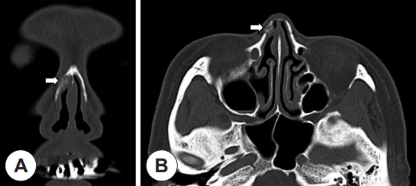
Fig. 2. Preoperative CT scan (A: coronal, B: axial). CT images present about 0.9×0.9 cm sized mass originated from nasal bone (white arrow).
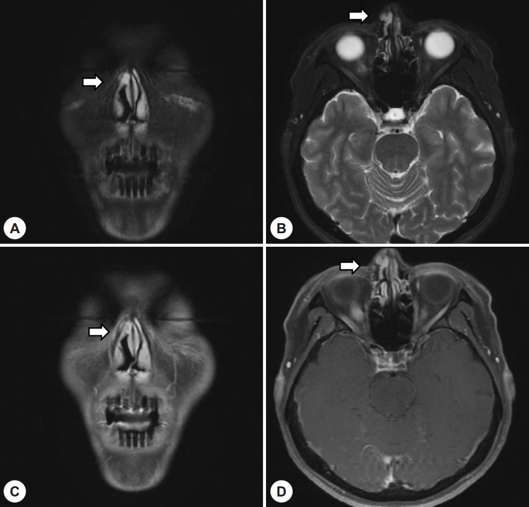
Fig. 3. Preoperative MRI scan (A: T2WI coronal, B: T2WI axial, C: T1WI coronal, D: T1WI axial). Heterogenous nasal bone mass (white arrow) presenting high signal intensity at T2WI and moderate homogeneous enhancement (A, B) and displaying low signal intensity at T1WI (C, D).
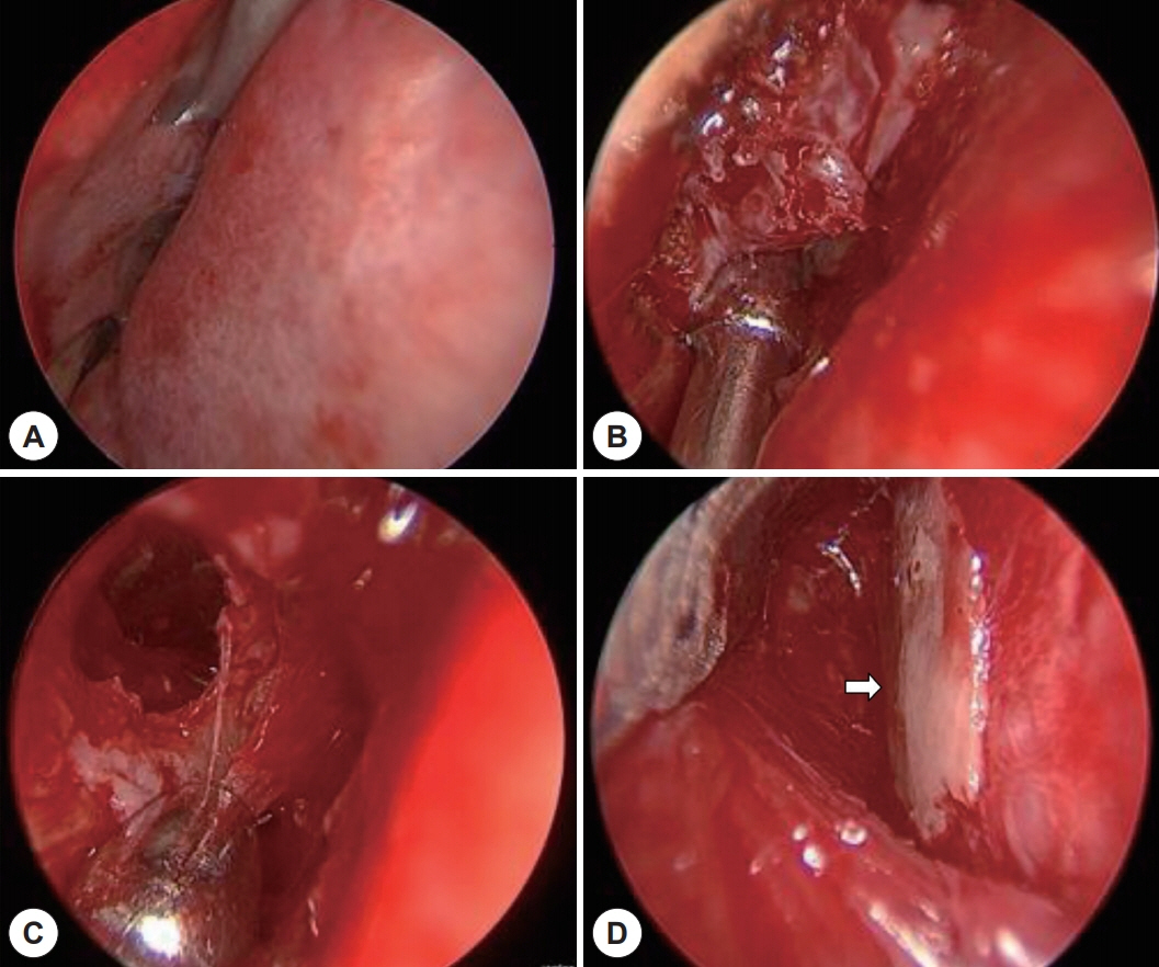
Fig. 4. Intraoperative findings (A-D). A: Intercartilaginous incision around mass was done with a canal knife and subperiosteal flap was elevated. B: The margin and base of the mass was carefully inspected. The origin of the mass was the lateral wall of the nasal cavity. C: Bone drilling was done via diamond burr to remove mass. D: The size of the defect was about 10 mm and was reconstructed with using a harvested nasal septal cartilage (white arrow).

Fig. 5. Pathologic finding (Hematoxylin and Eosin). Pathological examination showing endothelial lined blood vessel in the mature bone trabecula (hematoxylin and eosin staining; ×40).
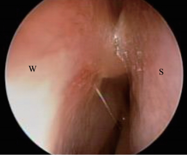
Fig. 6. Sinus endoscopy (6 month follow up). Mild adhesion between lateral nasal wall (W) and septum (S) is observed, but there are no evidence of recurrence.
Reference
-
References
1. Koybasi S, Saydam L, Kutluay L. Intraosseous hemangioma of the zygoma. Am J Otolaryngol. 2003; 24(3):194–7.2. Marshak G. Hemangioma of the zygomatic bone. Arch Otolaryngol. 1980; 106(9):581–2.3. Moore SL, Chun JK, Mitre SA, Som PM. Intraosseous hemangioma of the zygoma: CT and MR findings. AJNR Am J Neuroradiol. 2001; 22(7):1383–5.4. Taylor BG, Etheredge SN. Hemangiomas of the Mandible and Maxilla Presenting as Surgical Emergencies. Am J Surg. 1964; 108:574–7.5. Valentini V, Nicolai G, Lore B, Aboh IV. Intraosseous hemangiomas. J Craniofac Surg. 2008; 19(6):1459–64.6. Bridger MW. Haemangioma of the nasal bones. J Laryngol Otol. 1976; 90(2):191–200.7. Zins JE, Turegun MC, Hosn W, Bauer TW. Reconstruction of intraosseous hemangiomas of the midface using split calvarial bone grafts. Plast Reconstr Surg. 2006; 117(3):948–53; discussion 54.8. Peterson DL, Murk SE, Story JL. Multifocal cavernous hemangioma of the skull: report of a case and review of the literature. Neurosurgery. 1992; 30(5):778–81; discussion 82.
- Full Text Links
-
- Actions
-
Cited
- CITED
-
- Close
- Share
-
- Similar articles
-
- Intraosseous Hemangioma of the Nasal Septum: A Case Report
- A Case of 2-Month-Old Infant with Lobular Capillary Hemangioma
- A Case of Intraosseous Cavernous Hemangioma of the Inferior Turbinate
- A Case of Reconstruction of Surgical Defect after Removal of Intraosseous Hemangiomas on Nasal Dorsum
- Intraosseous Hemangioma of Frontal Bone: Report of Two Cases
