J Rhinol.
2020 May;27(1):50-53. 10.18787/jr.2019.00292.
Case Series of Antrovestibular Polyp: An Unusual Growth of Antral Polyp Toward the Nasal Vestibule Through the Anterior Fontanelle
- Affiliations
-
- 1Department of Otorhinolaryngology-Head and Neck Surgery, Seoul National University College of Medicine, Seoul National University Bundang Hospital, Seongnam, Korea
- 2Department of Otorhinolaryngology-Head and Neck Surgery, University of Santo Tomas Hospital, Manila, Philippines
- KMID: 2502795
- DOI: http://doi.org/10.18787/jr.2019.00292
Abstract
- Background and Objectives
This case series is aimed to introduce a new term, antrovestibular polyp (AVP), for an antral polyp herniating anteriorly toward the nasal vestibule and to describe an antral polyp direction of growth through the anterior and posterior fontanelles. Materials and Method: This is a retrospective study involving review of patients who underwent surgery due to maxillary sinus polyp herniating anteriorly toward the nasal vestibular area or posteriorly toward the choana at a tertiary training hospital from January 2007 through July 2016. Their demographic data, computed tomography scan findings, and endoscopic evaluations were analyzed.
Results
This study included 49 subjects; 8 (16.33%, 6 males) with AVP and 41 (83.67%, 24 males) with antrochoanal polyps (ACP). The mean ages of AVP and ACP patients were 9 and 14.4 years, respectively (p=0.006). The subjects were identified as AVP when computed tomography scan showed an antral polyp directed anteriorly toward the nasal vestibular area, while polyps growing toward the choana were identified as ACP. Endoscopic review showed that AVP grew out through an accessory ostium located anterior to the uncinate process at the area of the anterior fontanelle, while ACP started from an accessory ostium of the posterior fontanelle or a widened maxillary natural ostium.
Figure
-
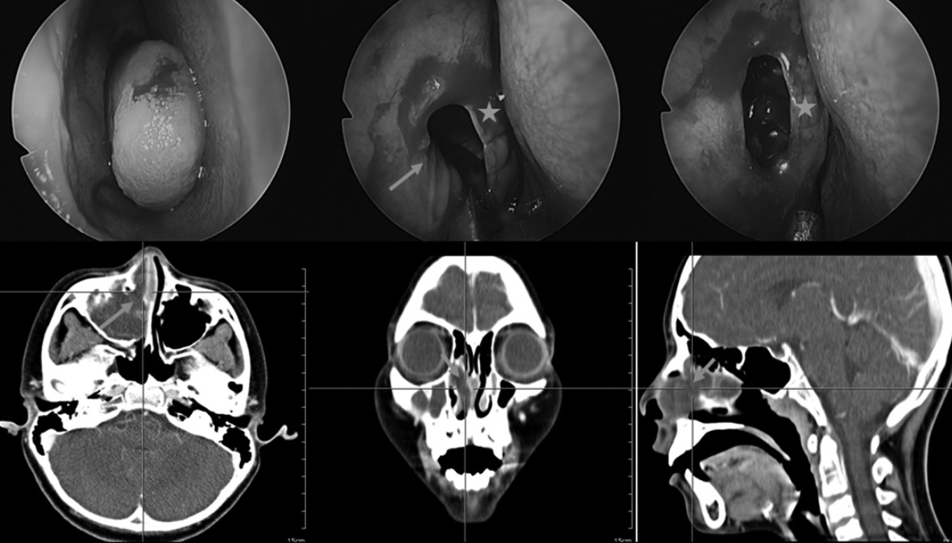
Fig. 1. Antrovestibular polyp. The crosshairs on the radiographs points to the antrovestibular polyp in different cross-sectional planes. In the endoscopic photographs, the arrow indicates the defect in the anterior fontanelle and the star marks indicate the uncinate process shown after antrochoanal polyp was removed.
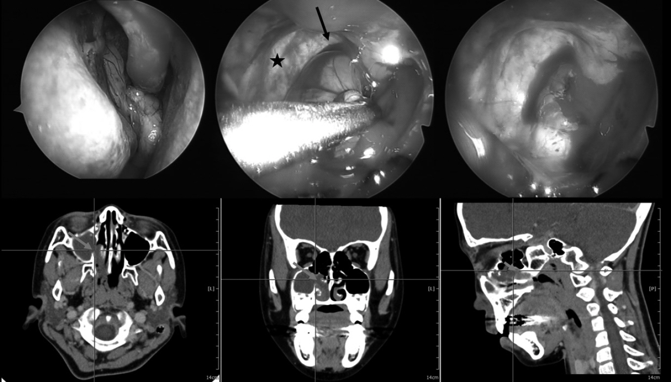
Fig. 2. Antrochoanal polyp. In the computed tomography scans, the crosshairs and arrows indicate the antrochoanal polyp. In the endoscopic photographs, the star mark indicates the large defect in the posterior fontanelle and the arrow indicates the antrochoanal polyp.
Reference
-
1. Frosini P, Picarella G, de Camporora E. Antrochoanal polyp analysis of 200 cases. ACTA Otorhinolaryngologica Italica. 2009; 29:21–6.2. Ruysch F. Observation um anatomica chirurgicaram anturca. 1691.3. Zuckerkandl E. Normale und pathologische Anatomie der Nasenholme. Vienna: 1892.4. Killian G. The origin of choanal polypi. 1906. p. 81–2.5. Stammberger HR, Kennedy DW, Anatomic Terminology G. Paranasal sinuses:anatomic terminology and nomenclature. Ann Otol Rhinol Laryngol Suppl. 1995; 167:7–16.6. Ashraf N, Bhattacharyya N. Determination of the incidental Lund-Mackay score for the staging of chronic rhinosinusitis. Otolaryngol Head Neck Surg. 2001; 125:483–6.7. Berg O, Carenfelt C, Silfversward C, Sobin A. Origin of the choanal polyp. Arch Otolaryngol Head Neck Surg. 1988; 114(11):1270–1.8. Chen JM, Schloss MD, Azouz ME. Antro-choanal polyp: a 10-year retrospective study in the pediatric population with a review of the literature. J Otolaryngol. 1989; 18(4):168–72.9. Berg O, Carenfelt C, Sobin A. On the diagnosis and pathogenesis of intramural maxillary cysts. Acta Otolaryngol. 1989; 108(5-6):464–8.10. Singhal M, Singhal D. Anatomy of accessory maxillary sinus ostium with clinical application. International Journal of Medical Science and Public Health. 2014; 3(3):327.11. Levine H, Mark M, Rontal M, Rontal E. Complex anatomy of lateral nasal wall simplified for endoscopic sinus surgery. New York: Thieme Medical Publishers;1993.12. Kumar H, Choudhry R, Kakar S. Osteomeatal complex obstruction is not associated with.pdf. 2011.13. Lund V. Anatomy of the nose and paranasal sinuses. 6th ed. Oxford: Butterworth Heinemann;1997.14. Jog M, McGarry GW. How frequent are accessory ostia? The Journal of Laryngology and Otology. 2003; 117:270–2.15. Mladina R, Skitarelic N, Casale M. Two holes syndrome (THS) is present in more than half of the postnasal drip patients? Acta Oto-Laryngolica. 2010; 130:1274–77.16. Mahajan A, Mahajan A, Gupta K, Verma P, Lalit M. Anatomical variations of accessory maxillary sinus ostium: an endoscopic study. Int J Anat Res. 2017; 5(1):3485–90.17. Bharathi D, Komala B, Sharath R. Anatomical Study of Accessory Maxillary Ostia and Its Surgical Importance. International Journal of Healthcare Sciences. 2015; 2(2):176–9.18. Patil M, Manjunath KY. Ostium maxillare accessarium-a morphologic study. National Journal of Clinical Anatomy. 2012; 1:171–5.
