Reconstruction of Medial Malleolar Defect Using Posterior Tibial Artery Perforator Based Deep Fascial Flap after Malignant Melanoma Ablation
- Affiliations
-
- 1Department of Plastic and Reconstructive Surgery, Yonsei University Wonju College of Medicine, Wonju, Korea
- KMID: 2502713
- DOI: http://doi.org/10.12790/ahm.20.0003
Abstract
- The soft tissue structure near the medial malleolus is an area where the bone is covered only with thin skin and subcutaneous tissue without structures like ligaments or tendons. In addition to the above anatomical features, wearing normal footwear should be considered when reconstructing the soft tissues in these areas. Therefore, it may be desirable to restore the original thickness of the soft tissue when performing a reconstruction. The authors reconstructed defects on the medial malleolus of a 50-year-old woman, after wide excision of a malignant melanoma, using only a deep fascial flap based on the posterior tibial artery-based perforator. This technique is thought to be a good option for reconstructing soft tissue defects in this area.
Keyword
Figure
-

Fig. 1. A 0.8 cm×0.7 cm sized dark pigmented mole was noted on right ankle. Reconstruction using a posterior tibial artery-based perforator flap was planned. The running of the posterior tibial artery on the skin was marked as photo with Doppler.
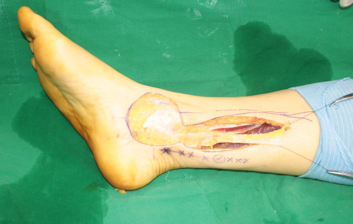
Fig. 2. After wide excision, a 2-cm proximal point from the medial malleolus was set as the pivot point, and the fascia was cut at the proximal 7 cm from the pivot point and then dissected toward the subfascial plane.
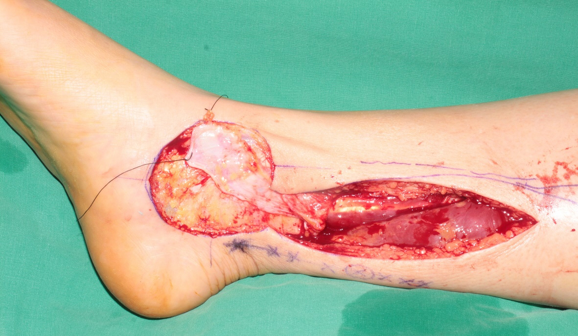
Fig. 3. After the flap was turned over to the defect, it was folded once into two layers for reinforcement, fixed on the exposed bone of the medial malleolus.

Fig. 4. Schematic illustration of the deep fascial flap. It is shown that the perforator vessels based on posterior tibial artery are segmented and branch out to supply deep fascia (left). The deep fascial flap is turned over based on the pivot point, and the distal perforator is feeding the whole flap (right).

Fig. 5. Eleven months after the operation, it was confirmed that the surgical site was maintained stably.
Reference
-
1. Schaverien MV, Hamilton SA, Fairburn N, Rao P, Quaba AA. Lower limb reconstruction using the islanded posterior tibial artery perforator flap. Plast Reconstr Surg. 2010; 125:1735–43.
Article2. Ciofu RN, Zamfirescu DG, Popescu SA, Lascar I. Reverse sural flap for ankle and heel soft tissues reconstruction. J Med Life. 2017; 10:94–8.3. Stecco C, Tiengo C, Stecco A, et al. Fascia redefined: anatomical features and technical relevance in fascial flap surgery. Surg Radiol Anat. 2013; 35:369–76.
Article4. Drimouras G, Kostopoulos E, Agiannidis C, et al. Redefining vascular anatomy of posterior tibial artery perforators: a cadaveric study and review of the literature. Ann Plast Surg. 2016; 76:705–12.5. Heymans O, Verhelle N, Peters S. The medial adiposofascial flap of the leg: anatomical basis and clinical applications. Plast Reconstr Surg. 2005; 115:793–801.
Article6. Ward KL, Romano A, Rodriguez-Collazo ER. Cadaveric atlas for orthoplastic lower limb and foot reconstruction of soft tissue defects. Clin Surg. 2018; 3:2001.7. Galumbeck M, Colen LB. Soft tissue reconstruction: coverage of lower leg: rotational flap. Orthop Clin North Am. 1993; 24:473–80.8. Kyung H, Choi JI, Song SH, Oh SH. Posterior tibial artery perforator-based adipofascial flap for skin necrosis and exposed extensor tendon after fixation of distal tibiofibular fracture. J Wound Manag Res. 2018; 14:54–7.
Article9. Bhadkamkar MA, Wolfswinkel EM, Hatef DA, et al. The ultra-thin, fascia-only anterolateral thigh flap. J Reconstr Microsurg. 2014; 30:599–606.
Article10. Fox P, Endress R, Sen S, Chang J. Fascia-only anterolateral thigh flap for extremity reconstruction. Ann Plast Surg. 2014; 72:S9–13.
Article
- Full Text Links
-
- Actions
-
Cited
- CITED
-
- Close
- Share
-
- Similar articles
-
- Perforator-Based Propeller Flap for Lower Extremity Reconstruction
- Posterior Tibial Artery Perforator Flap: An Anatomical Study and Clinical Applications
- Close-by Islanded Posterior Tibial Artery Perforator Flap: For Coverage of the Ankle Defect
- Soft Tissue Coverage Using a Combined Gastrocnemius-medial Sural Artery Perforator Flap
- Lower Extremity Reconstruction of Soft Tissue Defects with Perforator Island Flap
