Clinical Usefulness of Dual Red Imaging in Gastric Endoscopic Submucosal Dissection: A Pilot Study
- Yorita N1
- Oka S
 2
2 - Tanaka S1
- Kotachi T1
- Nagasaki N2
- Hata K2
- Kuroki K2
- Masuda K2
- Kurihara M2
- Kiso M2
- Boda T1
- Ito M2
- Chayama K2
- Affiliations
-
- 1Department of Endoscopy, Hiroshima University Hospital, Hiroshima, Japan
- 2Department of Gastroenterology and Metabolism, Hiroshima University Hospital, Hiroshima, Japan
- KMID: 2500905
- DOI: http://doi.org/10.5946/ce.2019.065
Abstract
- Background/Aims
Dual red imaging (DRI) is a new, image-enhanced endoscopy technique. There are few reports about the usefulness of DRI during gastric endoscopic submucosal dissection (ESD). We aimed to examine the usefulness of DRI in endoscopic hemostasis during gastric ESD.
Methods
We enrolled a total of 20 consecutive patients who underwent gastric ESD. Five endoscopists compared DRI with white light imaging (WLI) for the visibility of blood vessels and bleeding points while performing endoscopic hemostasis.
Results
The visibility of blood vessels was increased in 56% (19/34) of the cases, and the visibility of bleeding points was improved in 55% (11/20) of the cases with the use of DRI compared with the use of WLI.
Conclusions
DRI improved the visibility of blood vessels and bleeding points in cases with oozing bleeding, blood pooling around the bleeding points, and multiple bleeding points.
Keyword
Figure
-
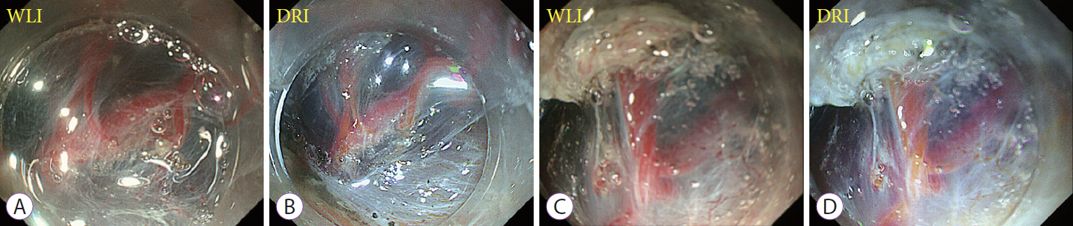
Fig. 1. Representative cases showing the usefulness of dual red imaging (DRI) in improving the visibility of blood vessels. Images from white light imaging (WLI) (A, C) and their corresponding DRI images (B, D). DRI enhanced the color tone of arteries and veins and improved visibility.

Fig. 2. Representative cases showing the usefulness of dual red imaging (DRI) in improving the visibility of bleeding points. Images from white light imaging (WLI) (A, C) and their corresponding DRI images (B, D). DRI emphasized the blood flow from the bleeding points in orange color, which made it easier to accurately detect the bleeding points (yellow and blue arrows).

Fig. 3. Mechanism of the dual red imaging (DRI) system. (A) In low concentration of blood, most of the 600-nm wavelength (orange color) is not attenuated, whereas the same wavelength is attenuated in the presence of a high concentration of blood. (B) Because yellow light of 600 nm is attenuated more in high-concentrated blood than in low-concentrated blood, the reflected image becomes reddish and contrast occurs. DRI was used at the 2 different blood concentrations. (C) A small amount of high-density blood was injected backward into the low-concentrated blood. DRI can distinguish between the different concentrations of blood. HCB, high concentration blood; LCB, low concentration blood.
Cited by 2 articles
-
Editors' Choice of Noteworthy Clinical Endoscopy Publications in the First Decade
Gwang Ha Kim, Kwang An Kwon, Do Hyun Park, Jimin Han
Clin Endosc. 2021;54(5):633-640. doi: 10.5946/ce.2021.216.The Role of Dual Red Imaging in Gastric Endoscopic Submucosal Dissection
In Kyung Yoo, Joo Young Cho
Clin Endosc. 2020;53(1):1-2. doi: 10.5946/ce.2020.018.
Reference
-
1. Oka S, Tanaka S, Kaneko I, et al. Advantage of endoscopic submucosal dissection compared with EMR for early gastric cancer. Gastrointest Endosc. 2006; 64:877–883.
Article2. Oka S, Tanaka S, Kaneko I, et al. Endoscopic submucosal dissection for residual/local recurrence of early gastric cancer after endoscopic mucosal resection. Endoscopy. 2006; 38:996–1000.
Article3. Higashimaya M, Oka S, Tanaka S, et al. Outcome of endoscopic submucosal dissection for gastric neoplasm in relationship to endoscopic classification of submucosal fibrosis. Gastric Cancer. 2013; 16:404–410.
Article4. Sanomura Y, Oka S, Tanaka S, et al. Clinical validity of endoscopic submucosal dissection for submucosal invasive gastric cancer: a single-center study. Gastric Cancer. 2012; 15:97–105.
Article5. Nagata S, Kimura S, Ogoshi H, Hidaka T. Endoscopic hemostasis of gastric ulcer bleeding by hemostatic forceps coagulation. Dig Endosc. 2010; 22 Suppl 1:S22–S25.
Article6. Ninomiya Y, Oka S, Tanaka S, et al. Clinical impact of dual red imaging in colorectal endoscopic submucosal dissection: a pilot study. Therap Adv Gastroenterol. 2016; 9:679–683.
Article7. Imagawa H, Oka S, Tanaka S, et al. Improved visibility of lesions of the small intestine via capsule endoscopy with computed virtual chromoendoscopy. Gastrointest Endosc. 2011; 73:299–306.
Article8. Yoshifuku Y, Sanomura Y, Oka S, et al. Evaluation of the visibility of early gastric cancer using linked color imaging and blue laser imaging. BMC Gastroenterol. 2017; 17:150.
Article9. Matsumoto A, Tanaka S, Oba S, et al. Outcome of endoscopic submucosal dissection for colorectal tumors accompanied by fibrosis. Scand J Gastroenterol. 2010; 45:1329–1337.
Article10. Higashiyama M, Oka S, Tanaka S, et al. Risk factors for bleeding after endoscopic submucosal dissection of gastric epithelial neoplasm. Dig Endosc. 2011; 23:290–295.
Article11. Jeon SW, Jung MK, Cho CM, et al. Predictors of immediate bleeding during endoscopic submucosal dissection in gastric lesions. Surg Endosc. 2009; 23:1974–1979.
Article12. Sanomura Y, Oka S, Tanaka S, et al. Taking warfarin with heparin replacement and direct oral anticoagulant is a risk factor for bleeding after endoscopic submucosal dissection for early gastric cancer. Digestion. 2018; 97:240–249.
Article13. Tajiri H, Kitano S. Complications associated with endoscopic mucosal resection: definition of bleeding that can be viewed as accidental. Dig Endosc. 2004; 16(Suppl 1):S134–S136.
Article14. Yahagi N, Horii J, Goto O, et al. Dual red imaging; a new endoscopic imaging technology for clear visualization of bleeding points in endoscopic submucosal dissection. Gastrointest Endosc. 2014; 79(5 Suppl):AB464.15. Tanaka H, Oka S, Tanaka S. Endoscopic hemostasis for spurting duodenal bleeding using dual red imaging. Dig Endosc. 2017; 29:816–817.
Article16. Naganuma M, Yahagi N, Bessho R, et al. Evaluation of the severity of ulcerative colitis using endoscopic dual red imaging targeting deep vessels. Endosc Int Open. 2017; 5:E76–E82.
Article17. Furuichi Y, Gotoda T, Kasai Y, et al. Role of dual red imaging to guide intravariceal sclerotherapy injection of esophageal varices (with videos). Gastrointest Endosc. 2018; 87:360–369.
Article18. Furuichi Y, Gotoda T, Moriyasu F, et al. Dual red imaging (novel advanced endoscopy) can increase visibility and can predict the depth in diagnosing esophageal varices. J Gastroenterol. 2017; 52:568–576.
Article19. Tanaka H, Oka S, Tanaka S, et al. Dual red imaging maintains clear visibility during colorectal endoscopic submucosal dissection. Dig Dis Sci. 2019; 64:224–231.
Article
- Full Text Links
-
- Actions
-
Cited
- CITED
-
- Close
- Share
-
- Similar articles
-
- The Role of Dual Red Imaging in Gastric Endoscopic Submucosal Dissection
- Usefulness of Narrow-Band Imaging in Endoscopic Submucosal Dissection of the Stomach
- Recent advances in endoscopic diagnosis and treatment of gastric cancer
- The Clinical Accuracy of Endoscopic Ultrasonography and White Light Imaging in Gastric Endoscopic Submucosal Dissection
- Pathological Interpretation of Gastric Tumors in Endoscopic Submucosal Dissection
