Korean J Orthod.
2020 Mar;50(2):129-135. 10.4041/kjod.2020.50.2.129.
Immediate effects of mandibular posterior displacement on the pharyngeal airway space: A preliminary study
- Affiliations
-
- 1Department of Orthodontics, Pusan National University Dental Hospital, Dental Research Institute, Yangsan, Korea. kmule@hanmail.net
- KMID: 2471873
- DOI: http://doi.org/10.4041/kjod.2020.50.2.129
Abstract
OBJECTIVE
This study aimed to evaluate the immediate effects of mandibular posterior displacement on the pharyngeal airway space (PAS) by using cephalometric evaluations and to investigate how the surrounding structures are schematically involved.
METHODS
In this retrospective study, 38 subjects with functional Class III malocclusion and two lateral cephalograms were selected. The first lateral cephalogram was taken with the mandible in the habitual occlusal position, and the second in anterior edge-to-edge bite. Paired t-test was used to analyze changes in the PAS, hyoid bone, tongue, and soft palate, followed by mandibular posterior displacement. Pearson's correlation analysis was used to determine the relationship between the amount of mandibular posterior displacement and other variables.
RESULTS
A statistically significant decrease was observed in the PAS following mandibular posterior displacement. Along with mandibular posterior displacement, the tongue decreased in length (p < 0.001) and increased in height (p < 0.05), while the soft palate increased in length, decreased in thickness, and was posteriorly displaced (p < 0.001). The hyoid bone was also posteriorly displaced (p < 0.05). There was no correlation between the amount of mandibular posterior displacement and the measured variables.
CONCLUSIONS
The PAS showed a statistically significant decrease following mandibular posterior displacement, which was a consequence of retraction of the surrounding structures. However, there were individual variances between the amount of mandibular posterior displacement and the measured variables.
Figure
-
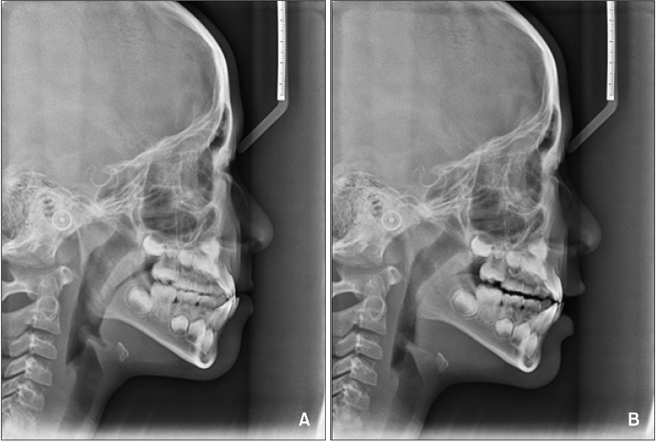
Figure 1 Two lateral cephalograms. A, In the habitual occlusal position. B, In the anterior edge-to-edge bite.
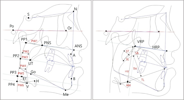
Figure 2 Cephalometric landmarks.S, Sella; N, nasion; Po, porion; Or, orbitale; PNS, posterior nasal spine; ANS, anterior nasal spine; A, point A; B, point B; Go, gonion; Me, menton.See Table 2 for definitions of the other landmarks.
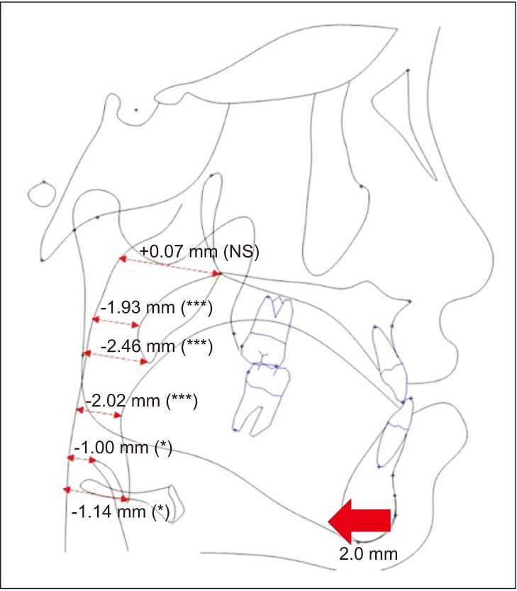
Figure 3 Changes in the pharyngeal airway space after mandibular posterior displacement.NS, Not significant.*p < 0.05, ***p < 0.001.
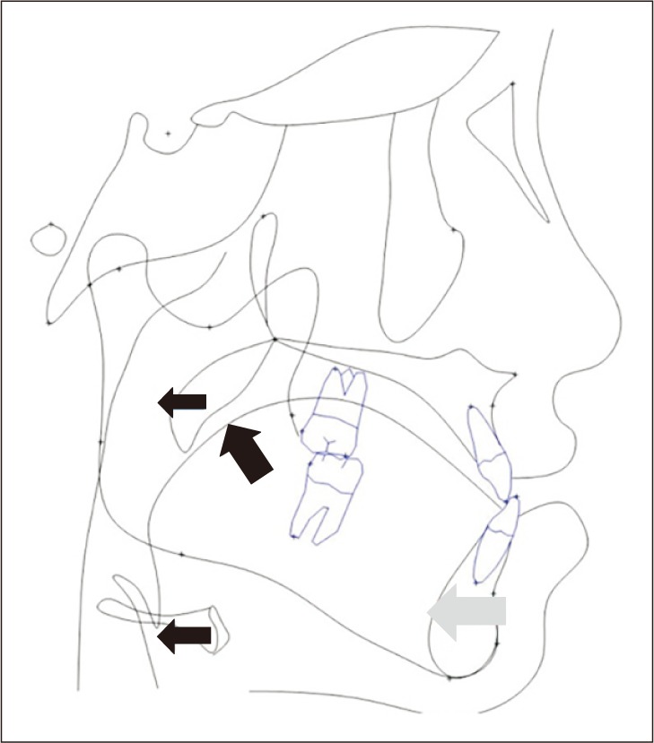
Figure 4 Changes in the surrounding structures after mandibular posterior displacement. Gray arrow, Changes in the mandible; Black arrows, changes in the tongue, soft palate, and hyoid bone.
Reference
-
1. Donner MW, Bosma JF, Robertson DL. Anatomy and physiology of the pharynx. Gastrointest Radiol. 1985; 10:196–212. PMID: 4029536.
Article2. El H, Palomo JM. Airway volume for different dentofacial skeletal patterns. Am J Orthod Dentofacial Orthop. 2011; 139:e511–e521. PMID: 21640863.
Article3. McNamara JA. Influence of respiratory pattern on craniofacial growth. Angle Orthod. 1981; 51:269–300. PMID: 6947703.
Article4. Dayyat E, Kheirandish-Gozal L, Sans Capdevila O, Maarafeya MMA, Gozal D. Obstructive sleep apnea in children: relative contributions of body mass index and adenotonsillar hypertrophy. Chest. 2009; 136:137–144. PMID: 19225059.5. Achilleos S, Krogstad O, Lyberg T. Surgical mandibular advancement and changes in uvuloglossopharyngeal morphology and head posture: a short- and long-term cephalometric study in males. Eur J Orthod. 2000; 22:367–381. PMID: 11029826.
Article6. Irani SK, Oliver DR, Movahed R, Kim YI, Thiesen G, Kim KB. Pharyngeal airway evaluation after isolated mandibular setback surgery using cone-beam computed tomography. Am J Orthod Dentofacial Orthop. 2018; 153:46–53. PMID: 29287649.
Article7. Han S, Choi YJ, Chung CJ, Kim JY, Kim KH. Long-term pharyngeal airway changes after bionator treatment in adolescents with skeletal Class II malocclusions. Korean J Orthod. 2014; 44:13–19. PMID: 24511511.
Article9. Jeans WD, Fernando DC, Maw AR, Leighton BC. A longitudinal study of the growth of the nasopharynx and its contents in normal children. Br J Radiol. 1981; 54:117–121. PMID: 7459548.
Article10. Athanasiou AE, Toutountzakis N, Mavreas D, Ritzau M, Wenzel A. Alterations of hyoid bone position and pharyngeal depth and their relationship after surgical correction of mandibular prognathism. Am J Orthod Dentofacial Orthop. 1991; 100:259–265. PMID: 1877552.
Article11. Panou E, Motro M, Ateş M, Acar A, Erverdi N. Dimensional changes of maxillary sinuses and pharyngeal airway in Class III patients undergoing bimaxillary orthognathic surgery. Angle Orthod. 2013; 83:824–831. PMID: 23438197.
Article12. Lamont J, Baldwin DR, Hay KD, Veale AG. Effect of two types of mandibular advancement splints on snoring and obstructive sleep apnoea. Eur J Orthod. 1998; 20:293–297. PMID: 9699407.
Article13. Ferguson KA, Ono T, Lowe AA, al-Majed S, Love LL, Fleetham JA. A short-term controlled trial of an adjustable oral appliance for the treatment of mild to moderate obstructive sleep apnoea. Thorax. 1997; 52:362–368. PMID: 9196520.
Article14. Pancherz H, Winnberg A, Westesson PL. Masticatory muscle activity and hyoid bone behavior during cyclic jaw movements in man. A synchronized electromyographic and videofluorographic study. Am J Orthod. 1986; 89:122–131. PMID: 3456206.15. Wright CR, Muyskens JH, Strong LH, Westerman KN, Kingery RH, Williams ST. A study of the tongue and its relation to denture stability. J Am Dent Assoc. 1949; 39:269–275. PMID: 18146829.
Article
- Full Text Links
-
- Actions
-
Cited
- CITED
-
- Close
- Share
-
- Similar articles
-
- Effects of bodily retraction of mandibular incisors versus mandibular setback surgery on pharyngeal airway space: A comparative study
- The effects of mandibular setback osteotomy on the oropharyngeal airway space in mandibular prognathic patients
- A Cephalometric Study on Changes in Pharyngeal Airway Space, Tongue and Hyoid Bone Positions Following the Surgical Correction of Mandibular Prognathism
- Changes in the hyoid bone, tongue, and oropharyngeal airway space after mandibular setback surgery evaluated by cone-beam computed tomography
- Retrospective study on change in pharyngeal airway space and hyoid bone position after mandibular setback surgery
