Changes in maximum lip-closing force after extraction and nonextraction orthodontic treatments
- Affiliations
-
- 1Department of Orthodontics, Seoul National University Bundang Hospital, Section of Dentistry, Seoul National University, Seongnam, Korea. ortho0328@naver.com
- 2Department of Oral Medicine and Diagnosis, College of Dentistry and Research Institute of Oral Science, Gangneung-Wonju National University, Gangneung, Korea.
- 3Department of Orthodontics, Seoul St. Mary's Hospital, The Catholic University of Korea, Seoul, Korea.
- 4Division of Orthodontics, School of Dentistry, University of Minnesota, Minneapolis, MN, USA.
- KMID: 2471872
- DOI: http://doi.org/10.4041/kjod.2020.50.2.120
Abstract
OBJECTIVE
The aims of the present study were to evaluate the changes in the maximum lip-closing force (MLF) after orthodontic treatment with or without premolar extractions and verify the correlation of these changes with dentoskeletal changes.
METHODS
In total, 17 women who underwent nonextraction orthodontic treatment and 15 women who underwent orthodontic treatment with extraction of all four first premolars were included in this retrospective study. For all patients, lateral cephalograms and dental models were measured before (T0) and after (T1) treatment. In addition, MLF was measured at both time points using the Lip De Cum LDC-110R® device. Statistical analyses were performed to evaluate changes in clinical variables and MLF and their correlations.
RESULTS
Both groups showed similar skeletal patterns, although the extraction group showed greater proclination of the maxillary and mandibular incisors and lip protrusion compared to the nonextraction group at T0. MLF at T0 was comparable between the two groups. The reduction in the arch width and depth and incisor retroclination from T0 to T1 were more pronounced in the extraction group than in the nonextraction group. MLF in the extraction group significantly increased during the treatment period, and this increase was significantly greater than that in the nonextraction group. The increase in MLF was found to be correlated with the increase in the interincisal angle and decrease in the intermolar width, arch depth, and incisor-mandibular plane angle.
CONCLUSIONS
This study suggests that MLF increases to a greater extent during extraction orthodontic treatment than during nonextraction orthodontic treatment.
Keyword
Figure
-
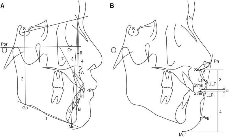
Figure 1 Landmarks, reference planes, and measurements in cephalometric analyses.A, Dentoskeletal variables. S, Sella; N, nasion; Por, porion; Or, orbitale; Me, menton; Go, gonion; A, A point; B, B point; U1, upper central incisor; L1, lower central incisor; SN plane (S-N); FH plane, Frankfort horizontal plane (Or-Por); mandibular plane (Go-Me); 1, angle between FH plane and mandibular plane (FMA); 2, S to Go (posterior facial height, PFH); 3, N to Me (anterior facial height, AFH); 4, angle between the SN plane and N-A line (SNA); 5, angle between the SN plane and N-B line (SNB); 6, angle between the N-A line and N-B line (ANB); 7, angle between the long axis of U1 and the FH plane (U1-FH); 8, angle between the long axis of L1 and the mandibular plane (IMPA); 9, angle between the long axis of U1 and the long axis of L1 (interincisal angle); 10, overbite; 11, overjet. B, Soft tissue variables. Pn, Pronasale; Cm, columella; Sn, subnasale; Ls, labrale superius; ULP, upper lip point; LLP, lower lip point; Stms, lowest point on the upper lip; Stmi, highest point on the lower lip; Pog', soft tissue pogonion; Me', soft tissue menton; 1, Rickett's E line (Pn-Pog'; EL) to ULP; 2, EL to LLP; 3, Sn to Stms; 4, Stmi to Me’; 5, interlabial gap (distance from Stms to Stmi); 6, nasolabial angle (angle between the Sn-Cm line and the Sn-Ls line).
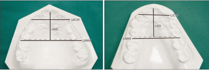
Figure 2 Measurements of the dental arch morphology on study models.UICW/LICW, Distance between both canine cusp tips in the maxillary/mandibular arch; UIMW/LIMW, distance between the mesiobuccal cusp tips of both first molars in the maxillary/mandibular arch; UAD/LAD, distance from the contact point between the central incisors perpendicular to a line connecting the mesial contact points of both first molars in the maxillary/mandibular arch.
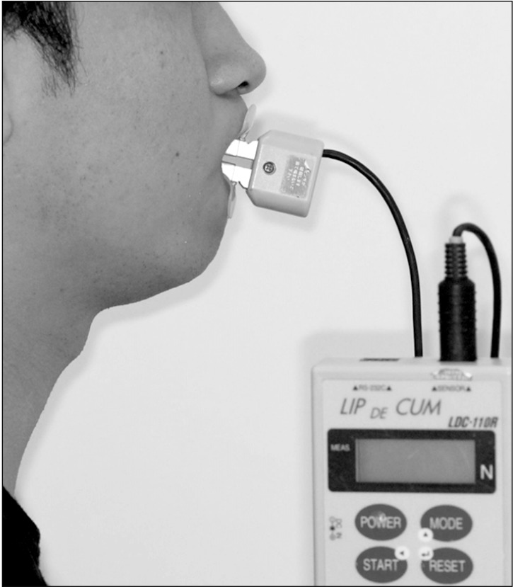
Figure 3 Measurement of the lip-closing force using a lip holder (Ducklings®) connected to a sensor in the Lip De Cum LDC-110R® device.
Reference
-
1. Nakatsuka K, Adachi T, Kato T, Oishi M, Murakami M, Okada Y, et al. Reliability of novel multidirectional lip-closing force measurement system. J Oral Rehabil. 2011; 38:18–26. PMID: 20722773.
Article2. Kaede K, Kato T, Yamaguchi M, Nakamura N, Yamada K, Masuda Y. Effects of lip-closing training on maximum voluntary lip-closing force during lip pursing in healthy young adults. J Oral Rehabil. 2016; 43:169–175. PMID: 26443933.
Article3. Weinstein S, Haack DC, Morris LY, Snyder BB, Attaway HE. On an equilibrium theory of tooth position. Angle Orthod. 1963; 33:1–26.4. Proffit WR. Equilibrium theory revisited: factors influencing position of the teeth. Angle Orthod. 1978; 48:175–186. PMID: 280125.5. Shaw WC, Addy M, Ray C. Dental and social effects of malocclusion and effectiveness of orthodontic treatment: a review. Community Dent Oral Epidemiol. 1980; 8:36–45. PMID: 6989548.
Article6. Chen S, Cai Y, Chen F. Lip closing force of Class III patients with mandibular prognathism: a case control study. Head Face Med. 2014; 10:33. PMID: 25159036.
Article7. Tomiyama N, Ichida T, Yamaguchi K. Electromyographic activity of lower lip muscles when chewing with the lips in contact and apart. Angle Orthod. 2004; 74:31–36. PMID: 15038488.8. Gamboa NA, Miralles R, Valenzuela S, Santander H, Cordova R, Bull R, et al. Comparison of muscle activity between subjects with or without lip competence: electromyographic activity of lips, supra- and infrahyoid muscles. Cranio. 2017; 35:385–391. PMID: 27997289.
Article9. Posen AL. The influence of maximum perioral and tongue force on the incisor teeth. Angle Orthod. 1972; 42:285–309. PMID: 4507148.10. Fındık Y, Baykul T, Aydın MA, Esenlik E, Ordu BN. Evaluation of lip force in patients with unilateral and bilateral cleft lip. Br J Oral Maxillofac Surg. 2017; 55:391–395. PMID: 28087210.
Article11. Kawabata A, Kobayashi T, Takagi A, Kuroyanagi F, Washino K, Sabashi K, et al. Multidirectional lip-closing force in adults with mandibular deviation. J Oral Rehabil. 2013; 40:664–669. PMID: 23855528.
Article12. Doto N, Yamada K. The relationship between maximum lip closing force and tongue pressure according to lateral craniofacial morphology. Orthod Waves. 2015; 74:69–75.
Article13. Takehana Y, Masuda Y, Kageyama T, Okazaki R, Murakami M, Yamada K. The relationship between lip-closing force and dental arch morphology in patient with Angle Class I malocclusion. J Oral Rehabil. 2017; 44:205–212. PMID: 27997984.
Article14. Young TM, Smith RJ. Effects of orthodontics on the facial profile: a comparison of changes during nonextraction and four premolar extraction treatment. Am J Orthod Dentofacial Orthop. 1993; 103:452–458. PMID: 8480714.
Article15. Kocadereli I. Changes in soft tissue profile after orthodontic treatment with and without extractions. Am J Orthod Dentofacial Orthop. 2002; 122:67–72. PMID: 12142899.
Article16. Ueki K, Mukozawa A, Okabe K, Miyazaki M, Moroi A, Marukawa K, et al. Changes in the lip closing force of patients with class III malocclusion before and after orthognathic surgery. Int J Oral Maxillofac Surg. 2012; 41:835–838. PMID: 22398020.
Article17. Ueki K, Moroi A, Sotobori M, Ishihara Y, Marukawa K, Iguchi R, et al. Evaluation of recovery in lip closing pressure and occlusal force and contact area after orthognathic surgery. J Craniomaxillofac Surg. 2014; 42:1148–1153. PMID: 24559719.
Article18. Yuce E, Baykul T, Findik Y, Alkis H. Evaluation of changes in lip closing force after surgically assisted rapid maxillary expansion. J Craniofac Surg. 2016; 27:649–653. PMID: 27054425.
Article19. Ono T, Hori K, Masuda Y, Hayashi T. Recent advances in sensing oropharyngeal swallowing function in Japan. Sensors (Basel). 2010; 10:176–202. PMID: 22315534.
Article20. Jung MH, Yang WS, Nahm DS. Effects of upper lip closing force on craniofacial structures. Am J Orthod Dentofacial Orthop. 2003; 123:58–63. PMID: 12532064.
Article21. Thüer U, Ingervall B. Pressure from the lips on the teeth and malocclusion. Am J Orthod Dentofacial Orthop. 1986; 90:234–242. PMID: 3463198.
Article22. Posen AL. The application of quantitative perioral assessment to orthodontic case analysis and treatment planning. Angle Orthod. 1976; 46:118–143. PMID: 1064341.23. Kato Y, Kuroda T, Togawa T. Perioral force measurement by a radiotelemetry device. Am J Orthod Dentofacial Orthop. 1989; 95:410–414. PMID: 2718971.
Article24. Jung MH, Yang WS, Nahm DS. Maximum closing force of mentolabial muscles and type of malocclusion. Angle Orthod. 2010; 80:72–79. PMID: 19852643.
Article25. Ingervall B, Janson T. The value of clinical lip strength measurements. Am J Orthod. 1981; 80:496–507. PMID: 6946706.
Article26. Partal I, Aksu M. Changes in lips, cheeks and tongue pressures after upper incisor protrusion in Class II division 2 malocclusion: a prospective study. Prog Orthod. 2017; 18:29. PMID: 28944417.
Article27. Lambrechts H, De Baets E, Fieuws S, Willems G. Lip and tongue pressure in orthodontic patients. Eur J Orthod. 2010; 32:466–471. PMID: 20089572.
Article28. Drobocky OB, Smith RJ. Changes in facial profile during orthodontic treatment with extraction of four first premolars. Am J Orthod Dentofacial Orthop. 1989; 95:220–230. PMID: 2923102.
Article29. Aksu M, Kocadereli I. Arch width changes in extraction and nonextraction treatment in class I patients. Angle Orthod. 2005; 75:948–952. PMID: 16448236.
- Full Text Links
-
- Actions
-
Cited
- CITED
-
- Close
- Share
-
- Similar articles
-
- Correction to: Changes in maximum lip-closing force after extraction and nonextraction orthodontic treatments
- Changes in occlusal force and occlusal contact area after orthodontic treatment
- Dentofacial Changes in Class I Cases Treated With and Without Extraction
- Evaluation of factors influencing the change of vertical dimension of face after orthodontic treatment
- Changes in buccal facial depth of female patients after extraction and nonextraction orthodontic treatments: A preliminary study
