Coronary Arteriovenous Fistulas Mimicking Coronary Perforation After Chronic Total Occlusion Recanalization
- Affiliations
-
- 1Division of Cardiology, Heart Institute, Asan Medical Center, University of Ulsan, Seoul, Korea. cheolwlee@amc.seoul.kr
- 2Department of Radiology and Research Institute of Radiology, Asan Medical Center, University of Ulsan College of Medicine, Seoul, Korea.
- KMID: 2471788
- DOI: http://doi.org/10.4070/kcj.2019.0300
Abstract
- No abstract available.
MeSH Terms
Figure
-
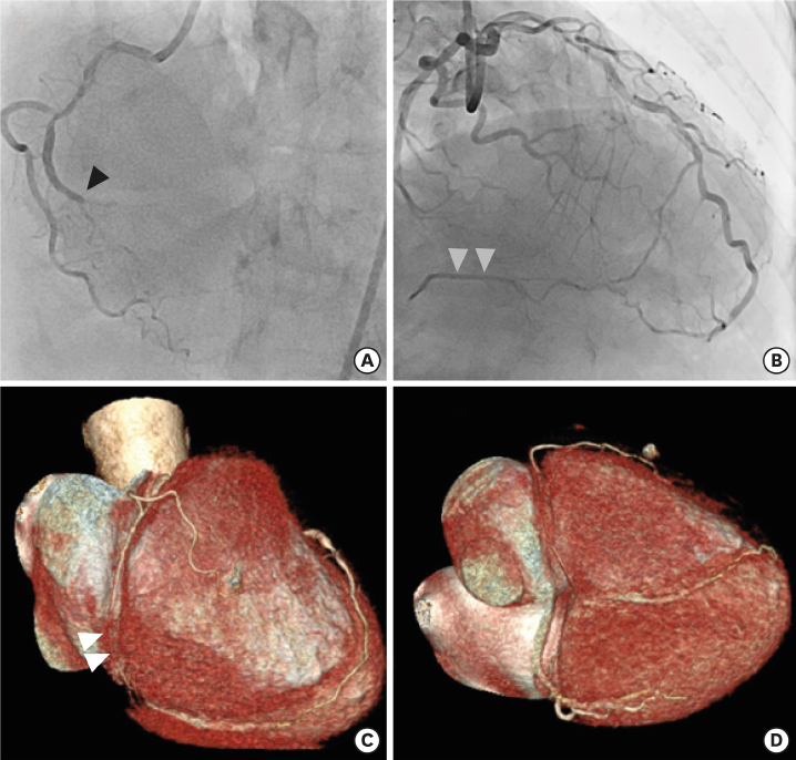
Figure 1 Diagnostic coronary angiography demonstrated total occlusion of the RCA (black arrow) (A) with good collaterals from the left anterior descending coronary artery (grey arrow) (B), and severe stenosis of the left anterior descending artery (B). Pre-procedure coronary computed tomography angiogram showed total occlusion of the distal RCA (white arrow) and there was no evidence of abnormal coronary artery drainage. (C, D). RCA = right coronary artery.
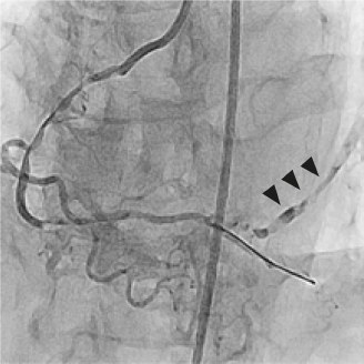
Figure 2 Coronary angiogram (left anterior oblique cranial view) showed contrast drainage through multiple channels after recanalization of chronic total occlusion, which mimicked coronary artery perforation (black arrow).

Figure 3 (A, B) Two drug-eluting stents (Orsiro, 2.5×40 and 2.5×40 mm) were implanted in the proximal to middle segment and distal segment of the right coronary artery (A: LAO view, B: AP cranial view). Communication between posterior descending coronary artery and coronary sinus was prominently noted after antegrade flow restoration (C: AP cranial view, D: AP caudal view). AP = anteroposterior; LAO = left anterior oblique.
Reference
-
1. Gowda RM, Vasavada BC, Khan IA. Coronary artery fistulas: clinical and therapeutic considerations. Int J Cardiol. 2006; 107:7–10.
Article
- Full Text Links
-
- Actions
-
Cited
- CITED
-
- Close
- Share
-
- Similar articles
-
- A Case of Dual Coronary Arteriovenous Fistulas Draining into the Coronary Sinus in a Patient with Acute Myocardial Infarction
- Bilateral Congenital Coronary Arteriovenous Fistulas
- A case of arteriovenous fistula with drainage into the coronary sinus during the percutaneous tranluminal coronary angioplasty of chronic total occlusion of circumflex coronary artery
- Changes in Coronary Perfusion after Occlusion of Coronary Arteries in Kawasaki Disease
- Iteration of Reverse Controlled Antegrade and Retrograde Tracking for Coronary Chronic Total Occlusion Intervention: a Current Appraisal
