The Experimental Study on Traumatic Epiphyseal Seperation
- Affiliations
-
- 1Department of Orthopaedics, Chonnam University Medical School, Kwangju, Korea.
- KMID: 2471466
- DOI: http://doi.org/10.4055/jkoa.1974.9.4.425
Abstract
- Traumatic epiphyseal separation was produced in the young rabbit weighing about 1 kg, and the type of injuries according to Salter and Harris method and location of the cleavage line within the epiphysis were studied radiologically and histologically, with the following result. 1. At the distal femur and the distal radius, the hyperextension force produced predominantly the Type 1 injury, in 95% and 75% respectively, while the rotation force resulted mainly in the Type I. in 65% and 90% respectively. The lateral bending produced almost the equal ratio of the Type 1 and 1. 2. Of all the injuries, the cleavage line within the epiphyseal growth plate could be located in the hypertrophying cartilage cell layer in 58.8%, in that it was 76% in the hyperextension group, 51% in the lateral bending group, and 49% in the rotation group. 3. The results of the above experiment have shown that the external violence of severe and complicated nature tended to produce the Type I injury, and the line of cleavage thus produced had the tendency to be located out of the hypertrophying cell layer, the common site of its separation.
Figure
-

Fig. 1 Radiograph of normal distal femur of the rabbit with its epiphyseal plate is shown.
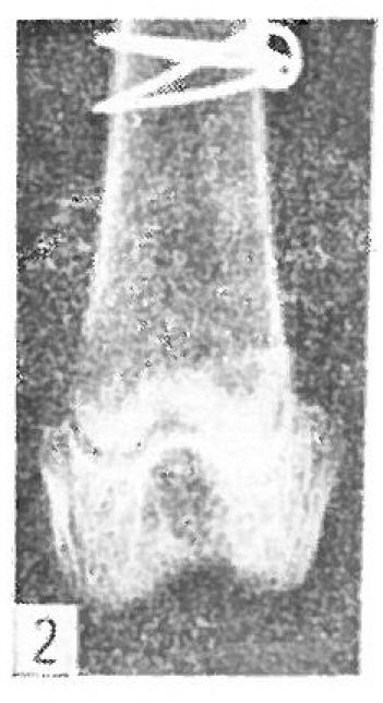
Fig. 2 Radiograph of Type I injury of the distal femur.
Fig. 3 Radiograph of Type II injury of the distal tibia.

Fig. 4 Radiograph of Type I injury of the proximal tibia.
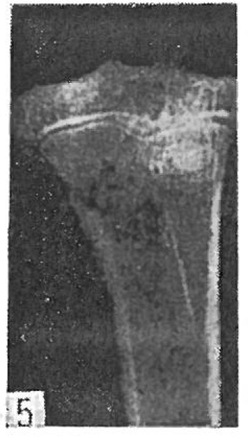
Fig. 5 Radiograph of Type II injury of the proximal tibia.
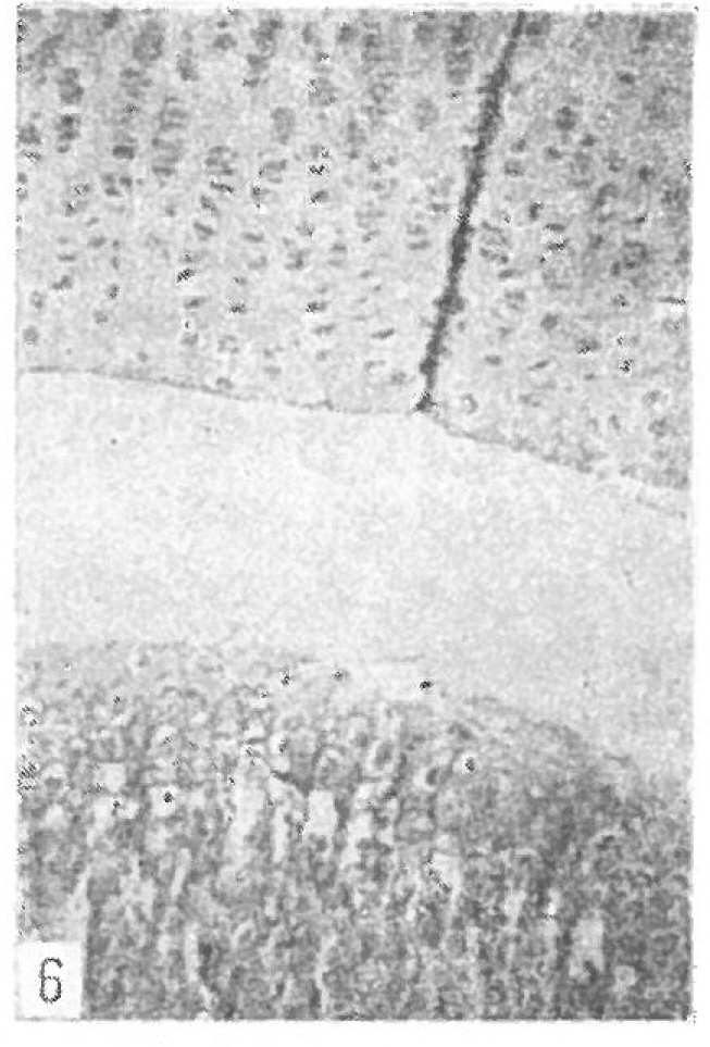
Fig. 6 The epiphyses of mormal rabbit are shown.
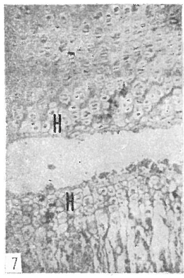
Fig. 7 The separation passes through the hypertrophying cartilage cell layer (H)
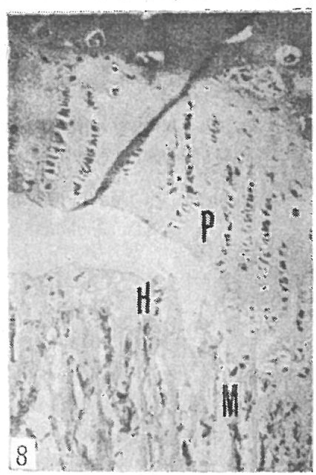
Fig. 8 Type II injury of the epiphysis is shown. The cleavage passes through the proliferating cartilage (P) and the hypertrophying cartilage cell layer (H) and then extend into the metaphysis(M)
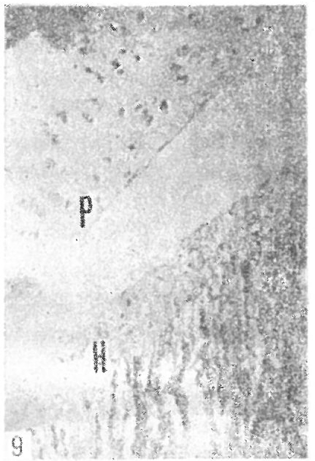
Fig. 9 The cleavage line of this separation passes between the proliferating cartilage and hypertrophying cartilage cell layer.
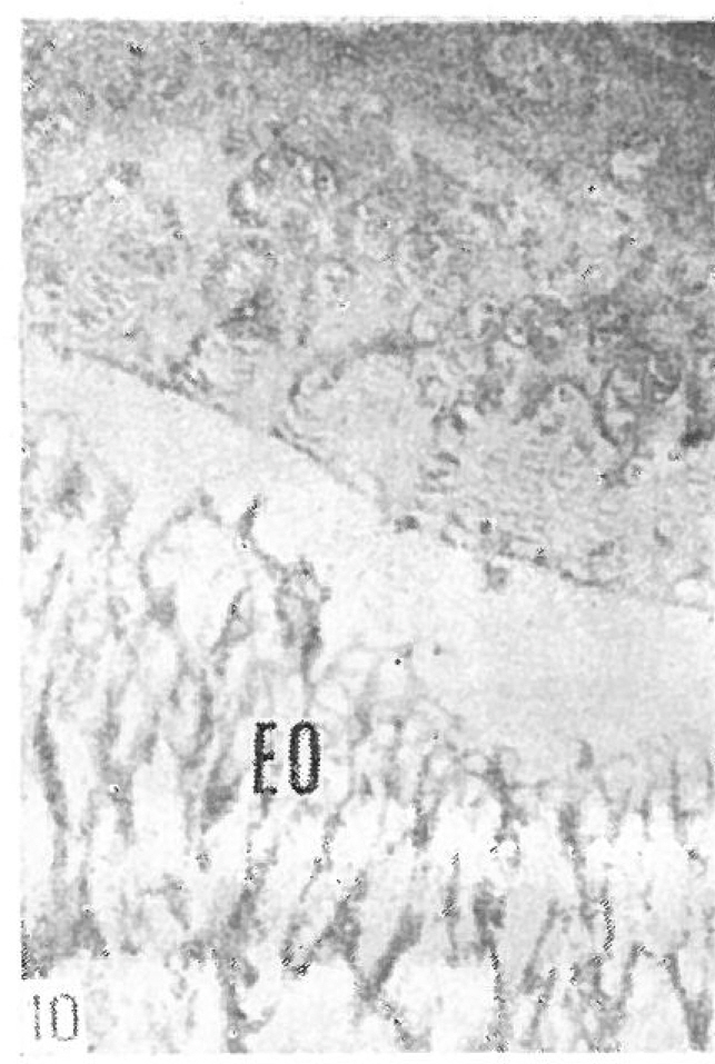
Fig. 10 In this Type I injury the separation occur between the hypertrophying cell layer and endochondral ossification zone (EO)
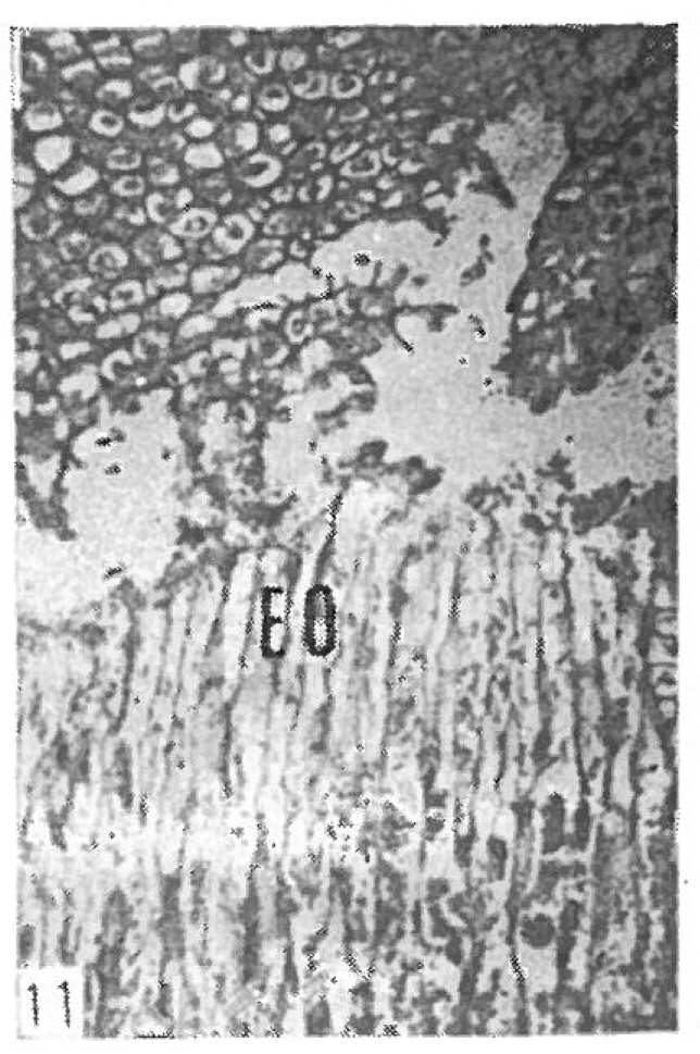
Fig. 11 In this Type I injury the separation occur between the hypertrophying cell layer and endochondral ossification zone (EO)
Reference
-
1. Aitken A.P.The and results of the fractured distal radial epiphysis. J. B. J. S. 17:302. 1935.2. Bergenfeldt E.Beiträge zur Kenntnis der traumatischen Epiphysenlösungen an den langen Röhrenknochen der Extremitäten. Acta Chir. Scand. 73(Suppl):28. 1933.3. Blount W.P.Fractures in children. Baltimore, The Williams and Wilkins Co.;1955.4. Brashear H.R.Epiphyseal fractures. A microscopic study of the healing process in rats. J. B. J. S. 41-A:1055. 1959.5. Campbell C.J., Grisolia A., Zanconato G.The effects produced in the cartilaginous epiphyseal plate of immature dogs by experimental surgical traumata. J. B. J. S. 41-A:1221. 1959.
Article6. Haas S.L.The localization of the growing point in the epiphyseal cartilage plate of bones. Amer. J. Orthop. Surg. 15:563. 1917.7. Hass S.L.Restriction of bone growth by pins through the epiphyseal cartilage plate. J. B. J. S. 32-A:338. 1950.8. Harris W.R., Hobson K.W.Histological changes in experimentally displaced upper femoral epiphysis in rabbits. J. B. J. S. 38-B:914. 1956.9. Oilier L.Quoted in Tachdjian, M. D.10. Poland J.Traumatic separation of the epiphyses in general. Historical. Clin. Orthop. 1965 (reprinted from Pediatrics. 4:49. 1897).11. Salter R.B., Harris W.R.Injuries involving the epiphyseal plate. J. B. J. S.,. 45-A:587. 1963.
Article12. Tachdjian M.D.Pediatric Orthopedics. p. 1534. Philadelphia, Saunders Co;1971.13. Trueta J., Trias A.The vascular contribution to osteogenesis. IV. The effect of pressure upon the epiphyseal cartilage of the rabbit. J. B. J. S. 43-B:800. 1961.14. Vogt P.Die traumatische Epiphysenntreinmung und deren Einfluss auf des Längenwachstum der Röhrenknochen. Arch. Klin. Chir. 22:343. 1878.
- Full Text Links
-
- Actions
-
Cited
- CITED
-
- Close
- Share
-
- Similar articles
-
- Clinical Significance of MRI for Assessment of Bony Bridge of Epiphyseal Plate: A Case Report
- A Study of Trauma to the Epiphyseal Plate in Immature Rabbits
- The Treatment of Acromioclavicular Seperation
- An Experimental Study of the Effect of Kirschner-wire and Screw insertion upon the Epiphyseal Plate of the Distal Femur in the Growing Rabbit
- A case of child seperation anxiety disorder with severe weight loss and school refusal
