Primary Cutaneous Solitary Fibrous Tumor on the Back
- Affiliations
-
- 1Department of Dermatology, Hanyang University Guri Hospital, Guri, Korea. tuentuen@hanyang.ac.kr
- KMID: 2471343
- DOI: http://doi.org/10.5021/ad.2020.32.2.155
Abstract
- Solitary fibrous tumors (SFT) are uncommon mesenchymal tumors. SFT have several synonyms including localized fibrous tumor, benign mesothelioma, localized fibrous mesothelioma, and submesothelial fibroma. SFT usually occur in the pleura or other serosal surfaces, but SFT can also develop in extrapleural areas including the nasal cavity, orbit, retroperitoneum, and pelvis. Cutaneous SFT is extremely rare, and more likely to occur in the head and neck region. Histologically, this tumor can mimic a variety of benign and malignant tumors such as dermatofibroma, dermatofibrosarcoma protuberans, spindle cell lipoma or other mesenchymal tumors. Most cases of SFT show non-aggressive clinical courses, with low recurrence rates. Herein, we describe a case of primary cutaneous SFT which presented with huge mass on the back.
Keyword
MeSH Terms
Figure
-
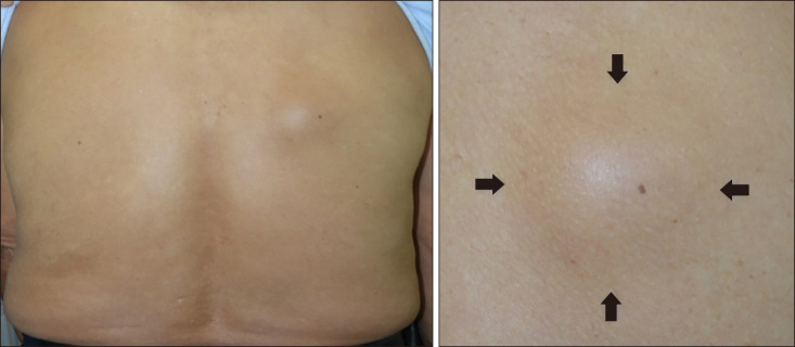
Fig. 1 Solitary, dome-shaped subcutaneous mass on patient's right back.
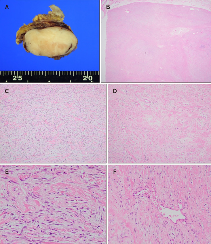
Fig. 2 (A) Cut section of the tumor showed an ovoid, well defined white-tan solid mass measuring 50×35×28 mm in size. (B) A spheroid, well-circumscribed tumor composed of alternating hypercellular and fibrous hypocellular areas was observed in the subcutis (H&E, ×40). (C) In the highly cellular areas, spindle-shaped cells were present in short interlacing fascicles, mixed with interstitial fibrous tissue (H&E, ×100). (D) In hypocellular foci, interspersed collagen fibers were mainly seen (H&E, ×100). (E) Many of the cells had enlarged vesicular nuclei with inconspicuous nucleoli (H&E, ×400). (F) Staghorn and ectatic blood vessels were found in some areas (H&E, ×200).
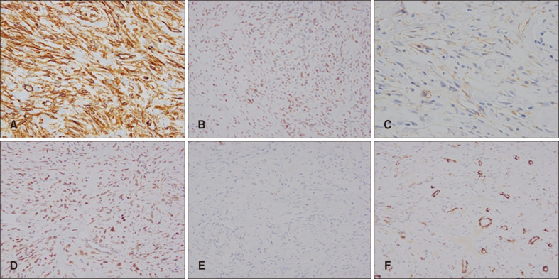
Fig. 3 Immunohistochemical staining was performed for smooth muscle actin (SMA), S-100, CD34, Bcl-2, CD99 and factor XIIIa. The tumor cells demonstrated positivity for CD34, factor XIIIa, CD99 and Bcl-2 (A: CD34, ×200; B: factor XIIIa, ×100; C: CD99, ×100; D: Bcl-2, ×100). However, S-100 and SMA staining were negative in tumor cell (E: S-100, ×100; F: SMA, ×100).
Reference
-
1. Erdag G, Qureshi HS, Patterson JW, Wick MR. Solitary fibrous tumors of the skin: a clinicopathologic study of 10 cases and review of the literature. J Cutan Pathol. 2007; 34:844–850. PMID: 17944724.
Article2. Kang TW, Kim HJ, Kim YC, Kim SC. A case of solitary fibrous tumor that developed on the scalp. Korean J Dermatol. 2009; 47:615–617.3. Moran CA, Suster S, Koss MN. The spectrum of histologic growth patterns in benign and malignant fibrous tumors of the pleura. Semin Diagn Pathol. 1992; 9:169–180. PMID: 1609159.4. Klemperer P, Rabin CB. Primary neoplasms of the pleura: a report of five cases. Arch Pathol. 1931; 11:385–412.
Article5. Okamura JM, Barr RJ, Battifora H. Solitary fibrous tumor of the skin. Am J Dermatopathol. 1997; 19:515–518. PMID: 9335244.
Article6. Soldano AC, Meehan SA. Cutaneous solitary fibrous tumor: a report of 2 cases and review of the literature. Am J Dermatopathol. 2008; 30:54–58. PMID: 18212546.
Article7. Terada T. Solitary fibrous tumor of the shoulder showing diverse distinct histologic patterns. Int J Dermatol. 2011; 50:208–211. PMID: 21244389.
Article8. Ali SZ, Hoon V, Hoda S, Heelan R, Zakowski MF. Solitary fibrous tumor. A cytologic-histologic study with clinical, radiologic, and immunohistochemical correlations. Cancer. 1997; 81:116–121. PMID: 9126139.
